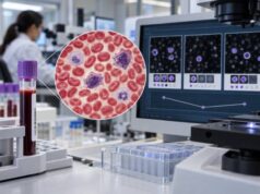
A liquid biopsy resistance mutation test looks for genetic changes in circulating tumor DNA (ctDNA) that can explain why a cancer has stopped responding—or may soon stop responding—to a targeted treatment. Because the test uses blood rather than a new tumor biopsy, it can sample tumor DNA repeatedly as cancer evolves under treatment pressure. This is especially useful in advanced cancers treated with targeted drugs, where new resistant clones can appear months or years after the original tissue biopsy. Examples include EGFR resistance alterations in lung cancer, ESR1 mutations during endocrine therapy for breast cancer, RAS or EGFR-pathway changes after anti-EGFR treatment in colorectal cancer, and secondary alterations affecting other targeted pathways. A detected resistance mutation can sometimes point directly to a different drug or treatment strategy, but not every resistance mechanism is detectable in plasma or has an approved therapy. Negative results therefore require careful interpretation and may need tissue re-biopsy.
- A positive resistance mutation result means a treatment-relevant alteration was detected in plasma, but the exact gene, variant, cancer type, and current therapy determine what it means.
- A negative blood test does not rule out resistance, because low ctDNA shedding, non-genetic resistance, or an alteration outside the assay’s coverage can produce a negative result.
- Testing is often most informative at clinical or radiographic progression, although some settings use serial testing before visible progression.
- No fasting is usually required; sample timing, tumor burden, and laboratory processing matter more than diet.
- Resistance testing complements rather than automatically replaces tissue biopsy, especially when histologic transformation or another non-DNA mechanism is suspected.
Table of Contents
- What Resistance Mutation Testing Detects
- How Cancer Develops Drug Resistance
- Common Resistance Markers by Cancer Type
- When Liquid Biopsy Is Most Useful
- How to Read a Resistance Report
- Limitations and False-Negative Results
- What Happens After a Resistance Mutation Is Found
What Resistance Mutation Testing Detects
Cancer treatment can create intense selection pressure. Drug-sensitive tumor cells shrink or disappear, while cells carrying a resistance mechanism may survive and expand. Those resistant cells can release DNA fragments into the bloodstream. A liquid biopsy can capture some of those fragments and reveal how the tumor’s molecular profile has changed.
Most resistance-focused blood tests analyze cell-free DNA in plasma, looking for the tumor-derived fraction known as ctDNA. Some tests use a broad ctDNA mutation panel, while others focus on predefined hotspots in one or a few genes. Broad next-generation sequencing (NGS) can identify multiple possible escape pathways at once. Highly sensitive digital PCR can be useful when clinicians are tracking a specific mutation at very low levels.
A resistance report may identify:
- A new point mutation that changes the drug-binding site
- Amplification of a gene that restores signaling despite treatment
- Activation of a bypass pathway
- A secondary fusion or rearrangement
- Reversion of a previously damaging DNA-repair mutation
- Multiple resistant clones at the same time
The phrase acquired resistance mutation means the alteration became detectable after treatment selection pressure or increased from a previously minor clone. It does not always prove that the mutation alone caused progression. Clinical interpretation depends on known biology, treatment timing, the mutation’s allele fraction, and whether the alteration has been validated as a resistance marker for that specific cancer and drug.
Blood testing is particularly helpful because resistant disease can be genetically heterogeneous. One metastatic lesion may develop one escape mechanism while another develops a different one. A tissue biopsy samples only one location. Plasma may collect ctDNA from several tumor sites and therefore reveal a broader picture—although it can still miss lesions that do not shed much DNA.
How Cancer Develops Drug Resistance
Drug resistance is not one biological event. It can arise through several routes, and only some are visible on a DNA-based liquid biopsy.
On-target resistance
The cancer changes the same protein that the drug was designed to inhibit. The alteration can reduce drug binding while preserving the cancer-driving signal. Classic examples have occurred in kinase-driven cancers, where secondary mutations alter the drug target after months of therapy.
Bypass pathway activation
Instead of changing the original target, tumor cells activate another signaling pathway. Amplification or mutation of a different gene can provide an alternate route for growth. Broad NGS is more likely than a single-gene test to find these parallel mechanisms.
Downstream reactivation
A cancer may reactivate signaling downstream from the inhibited target. RAS-MAPK pathway alterations are a well-known example in colorectal cancer after anti-EGFR therapy. These changes can make continued EGFR blockade ineffective even when the original tumor was RAS wild type.
Clonal selection
A resistant subclone may have been present at a very low level before therapy and become dominant only after sensitive cells are suppressed. Serial blood testing can sometimes show that clone rising over time.
Non-genetic resistance
Not all resistance is encoded by a detectable DNA mutation. Tumors can change cell state, protein expression, epigenetic regulation, immune environment, or histology. For example, a lung adenocarcinoma can occasionally transform into small-cell lung cancer during targeted therapy. A plasma DNA panel may suggest resistance but cannot diagnose that histologic transformation; tissue biopsy is usually needed.
This is why a liquid biopsy test is best viewed as one piece of a resistance workup, not a universal replacement for pathology and imaging.
Common Resistance Markers by Cancer Type
The clinically meaningful resistance markers depend strongly on the tumor and treatment. The same mutation can have very different implications in another cancer type.
| Cancer setting | Examples of resistance signals detectable in ctDNA | Why they matter |
|---|---|---|
| EGFR-mutant NSCLC | Secondary EGFR alterations, MET amplification, other bypass changes | Can explain progression on EGFR-targeted therapy and help guide re-biopsy or next treatment |
| ER-positive breast cancer | ESR1 mutations | Can indicate resistance to aromatase-inhibitor-based endocrine pressure and affect endocrine strategy |
| Metastatic colorectal cancer | KRAS, NRAS, BRAF, EGFR extracellular-domain and other pathway alterations | Can emerge during anti-EGFR therapy and affect eligibility for rechallenge |
| ALK- or other fusion-driven NSCLC | Secondary kinase-domain mutations and bypass alterations | May help match the next inhibitor or identify a need for tissue testing |
| BRCA1/2-associated tumors treated with PARP inhibitors | BRCA reversion mutations | Can restore DNA-repair function and contribute to PARP-inhibitor or platinum resistance |
In EGFR-mutant lung cancer, resistance testing has evolved as newer generations of EGFR tyrosine kinase inhibitors have replaced older drugs. The once-famous EGFR T790M liquid biopsy remains an important historical and biological example, but current progression patterns can involve many other alterations. A modern broad panel is often more informative than testing only T790M.
In hormone receptor-positive, HER2-negative advanced breast cancer, acquired ESR1 mutations in blood can emerge during aromatase-inhibitor exposure. The PADA-1 randomized trial provided a major proof of principle: patients were monitored for rising ESR1 mutations in blood before radiographic progression, and an early endocrine switch improved progression-free survival in the specific trial setting. That does not mean every emerging mutation should trigger an automatic treatment change, but it shows that ctDNA can sometimes move from a passive biomarker to a treatment-guiding tool.
In metastatic colorectal cancer, RAS, BRAF, and EGFR-pathway resistant clones can appear during anti-EGFR treatment. The CHRONOS trial used plasma screening to exclude patients who still had detectable resistance alterations before panitumumab rechallenge. This illustrates another use of resistance testing: not only finding a new target, but determining whether a previously effective therapy may be reasonable again after resistant clones decline.
When Liquid Biopsy Is Most Useful
Resistance testing is most useful when the result can answer a concrete treatment question. Common situations include:
- Radiographic or clinical progression on a targeted drug. A fresh blood sample can identify acquired mutations without delaying treatment for an invasive biopsy.
- Insufficient or inaccessible tissue. Some metastatic sites are unsafe or technically difficult to biopsy.
- Concern that old tissue is outdated. A biopsy from diagnosis may not reflect years of treatment-driven tumor evolution.
- Need to survey multiple resistant clones. Plasma can sometimes capture heterogeneity across metastatic sites better than a single-lesion biopsy.
- Serial molecular monitoring in a validated setting. Certain trials and clinical strategies use repeated ctDNA to detect emerging resistance before standard imaging shows progression.
Timing matters. Testing too early after starting therapy may simply document baseline disease. Testing after a clear progression event is often more likely to capture the dominant resistance clone. Conversely, some biomarkers are specifically useful before visible progression when prospective evidence supports an earlier intervention.
The amount of ctDNA also matters. Progressive, high-volume metastatic disease often sheds more tumor DNA than small-volume or compartmentalized disease. A patient with only small lung nodules, peritoneal disease, or isolated central nervous system progression may have a negative plasma result despite genuine molecular resistance.
Broader commercial platforms, including assays such as the Tempus xF liquid biopsy, can report multiple mutation classes and resistance-associated alterations in one specimen. Other platforms use different gene lists, sequencing depths, filtering strategies, and reporting thresholds. The best assay is therefore the one validated for the clinical question—not necessarily the test with the largest gene count.
How to Read a Resistance Report
A resistance report should be read in layers rather than as a list of mutations.
1. Confirm the clinical context
The first question is what treatment the cancer is receiving and whether the patient is actually progressing. A mutation labeled as “resistance associated” can be irrelevant if the corresponding drug was never used or if the cancer type does not rely on that pathway.
2. Identify the exact alteration
Gene names alone are not enough. EGFR C797S, for example, is not interchangeable with EGFR T790M. KRAS G12C, G12D, and amplification of KRAS also have different implications. Exact nomenclature matters.
3. Review variant allele frequency and tumor fraction
A resistance mutation at 0.2% VAF may represent a small emerging clone, while another alteration may dominate the plasma profile. But VAF is not a direct measurement of the percentage of tumor cells carrying the mutation. It is influenced by how much normal cell-free DNA is present and how much DNA different tumor sites release.
4. Look for multiple resistance mechanisms
Resistance is often polyclonal. Large ctDNA studies have found more than one emerging resistance alteration in some patients. A treatment directed against only one clone may therefore have limited durability.
5. Separate actionable findings from uncertain findings
Reports may classify variants as therapeutically actionable, biologically relevant, or of uncertain significance. Only some findings have evidence supporting a specific treatment. Clinical-trial options may exist even when no standard therapy is established.
A mutation result should also be distinguished from KRAS liquid biopsy testing or another single-gene test used for baseline profiling. The same gene can function as an initial driver biomarker in one context and an acquired resistance marker in another.
Limitations and False-Negative Results
The most important limitation is that absence of a resistance mutation in plasma does not prove absence of resistance. A negative result can occur because:
- The tumor releases little ctDNA.
- The resistant lesion is confined to the brain or another low-shedding site.
- The mutation is below the assay’s limit of detection.
- The resistance mechanism is a fusion, copy-number change, or structural event that the assay detects less sensitively.
- The mechanism is outside the panel’s gene list.
- Resistance is epigenetic, phenotypic, immune-mediated, or histologic rather than genomic.
Clonal hematopoiesis can create the opposite problem: a mutation may come from an expanded blood-cell clone rather than from the solid tumor. Some laboratories reduce this risk by sequencing matched white blood cells or applying specialized bioinformatic filters. Still, an unexpected low-level finding should be interpreted in context.
Tissue remains especially valuable when clinicians suspect transformation, when plasma is repeatedly non-informative, or when morphology and protein expression matter. Tissue can also provide RNA, immunohistochemistry, and histology that plasma DNA cannot.
There is another limitation: detecting a resistance marker does not automatically prove that changing therapy because of that marker improves outcomes. Some biomarkers have randomized or prospective evidence; others are biologically plausible but still investigational. This distinction is critical when serial ctDNA detects a change before conventional progression.
Pre-analytical factors also matter more than many patients realize. Cell-free DNA is present in small quantities and can be diluted by DNA released from normal white blood cells if specimen handling is delayed or the wrong collection tube is used. Laboratories use specialized tubes, rapid processing, and quality-control thresholds to reduce this problem. A technically adequate sample can still be biologically uninformative, however, if the cancer is not shedding measurable ctDNA. Those are different reasons for a negative report and can lead to different follow-up decisions.
It is also important to distinguish molecular progression from clinical progression. A resistant clone can sometimes become detectable in plasma before a scan shows growth. That early signal is scientifically important, but the right response depends on evidence for that particular biomarker and treatment. In some settings, prospective trials support acting on a rising mutation; in others, changing an effective therapy too early could remove a drug that is still controlling most of the cancer. Serial ctDNA is therefore most powerful when a testing schedule and action threshold have been defined in advance rather than improvised after every small fluctuation.
What Happens After a Resistance Mutation Is Found
A useful resistance result should lead to a structured next step. The oncology team typically asks whether the alteration is validated, targetable, and compatible with the patient’s prior treatments and current disease pattern.
Possible next actions include:
- Switching to another approved targeted therapy that addresses the resistance mechanism
- Combining therapies when dual pathway blockade is supported
- Reusing a previous drug after resistant clones become undetectable in a validated rechallenge strategy
- Ordering tissue biopsy to confirm a complex or unexpected result
- Looking for a clinical trial matched to the resistance alteration
- Continuing the current therapy if the molecular finding is not yet clinically actionable and the cancer remains controlled
A resistance mutation is therefore not inherently “bad news” or a command to stop treatment. It is molecular information about tumor evolution. Sometimes it explains why a drug failed. Sometimes it creates a new treatment option. Sometimes it is an early warning that still needs confirmation. And sometimes no genetic mechanism is found at all.
The most productive question to ask after testing is: What decision does this result change today? If the answer is unclear, the report may still be useful for future treatment or trial planning, but it should not be overinterpreted. Modern precision oncology works best when ctDNA findings are integrated with imaging, pathology, symptoms, treatment response, and the evolving evidence for each resistance marker.
References
- ESMO recommendations on the use of circulating tumour DNA assays for patients with cancer: a report from the ESMO Precision Medicine Working Group 2022 (Guideline)
- Liquid biopsy in cancer drug resistance: real-time monitoring, mechanistic insights, and translational applications 2026 (Review)
- Utilizing ctDNA to discover mechanisms of resistance to targeted therapies in patients with metastatic NSCLC: towards more informative trials 2025 (Review)
- Circulating tumor DNA landscape and prognostic impact of acquired resistance to targeted therapies in cancer patients: a national center for precision medicine (PRISM) study 2023
- Switch to fulvestrant and palbociclib versus no switch in advanced breast cancer with rising ESR1 mutation during aromatase inhibitor and palbociclib therapy (PADA-1): a randomised, open-label, multicentre, phase 3 trial 2022 (RCT)
- Circulating tumor DNA to guide rechallenge with panitumumab in metastatic colorectal cancer: the phase 2 CHRONOS trial 2022
Disclaimer
Liquid biopsy resistance findings must be interpreted by an oncology team using the patient’s cancer type, treatment history, imaging, pathology, and the specific assay’s validated capabilities. A negative plasma result does not rule out drug resistance, and treatment should not be changed solely on the basis of this article or an unreviewed molecular report.