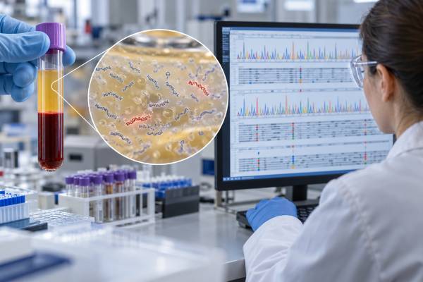
A liquid biopsy is a laboratory test that looks for cancer-related material in blood or another body fluid instead of removing a piece of tumor tissue. In everyday oncology practice, the term most often refers to plasma testing for circulating tumor DNA (ctDNA), the small fraction of cell-free DNA released by cancer cells. Depending on the assay, a liquid biopsy can identify tumor mutations, help match an advanced cancer to targeted therapy, reveal emerging drug resistance, estimate molecular disease burden, or monitor for residual disease after treatment. The major advantage is repeatability: a blood draw is usually easier and safer than another tissue biopsy. The major limitation is equally important—some cancers release very little ctDNA, so a negative blood result may miss an alteration that is present in the tumor. Liquid biopsy therefore complements tissue pathology and imaging rather than replacing them in every setting.
- Most cancer liquid biopsies analyze plasma ctDNA, although some tests examine circulating tumor cells, RNA, methylation patterns, proteins, or extracellular vesicles.
- A positive tumor mutation can guide treatment when the alteration is clinically validated, but the significance depends on the exact gene, variant, and cancer type.
- A negative result does not reliably exclude cancer or a tumor mutation, especially when tumor burden or ctDNA shedding is low.
- Fasting is generally not required; correct blood collection, handling, and timing are more important for preserving cell-free DNA.
- Liquid biopsy is established for selected molecular profiling uses in advanced cancer, while screening and many MRD applications remain more context dependent.
Table of Contents
- What a Liquid Biopsy Measures
- How Blood-Based Tumor Testing Works
- When Liquid Biopsy Is Used
- How to Interpret Liquid Biopsy Results
- Liquid Biopsy Versus Tissue Biopsy
- Limitations, False Results, and Clonal Hematopoiesis
- Preparation, Timing, and Follow-Up
What a Liquid Biopsy Measures
“Liquid biopsy” describes a family of tests rather than one single assay. The analyte most widely used in precision oncology is circulating tumor DNA, but other tumor-related material can also be measured.
Cell-free DNA and circulating tumor DNA
Cell-free DNA (cfDNA) consists of short DNA fragments released into the bloodstream as cells die and turn over. Most cfDNA comes from normal tissues and blood cells. In a person with cancer, a smaller subset may originate from tumor cells. That tumor-derived portion is ctDNA.
A cell-free DNA cancer test may therefore analyze a mixture in which only a tiny fraction of molecules are actually tumor-derived. Advanced assays use deep sequencing, molecular barcodes, digital PCR, or other error-suppression strategies to find rare cancer-associated variants within that background.
Other liquid-biopsy analytes
Some tests examine intact circulating tumor cells rather than DNA fragments. Research and emerging commercial platforms may also analyze circulating RNA, extracellular vesicles, DNA methylation, fragment size and end patterns, proteins, or combinations of several signals.
These approaches answer different questions. Mutation-focused ctDNA testing is especially useful for identifying actionable genomic changes. Methylation and fragmentomics may be used in early-detection research because they can carry tissue-of-origin information. CTCs can provide cellular features that DNA alone cannot.
The phrase “blood biopsy” can therefore be misleading if it suggests that blood reproduces everything a tissue biopsy provides. It does not. Plasma can reveal molecular signals, but it cannot show tumor architecture, cell morphology, invasion pattern, or many protein-expression features that pathologists assess under the microscope.
How Blood-Based Tumor Testing Works
Most ctDNA liquid biopsies follow the same broad workflow.
- Blood is collected into specialized tubes. These tubes help stabilize the specimen and limit contamination from DNA released by normal white blood cells.
- Plasma is separated from blood cells. Laboratories process the sample under controlled conditions because delays and poor handling can reduce test quality.
- Cell-free DNA is extracted. The amount may be small, particularly in early-stage or low-volume disease.
- The DNA is analyzed. Tests may use digital PCR for selected hotspot mutations or next-generation sequencing for broader genomic profiling.
- Bioinformatics separates likely tumor findings from noise. Modern assays use error correction and may incorporate matched white-blood-cell analysis or computational filters.
- A clinical report is generated. It may list pathogenic variants, potentially actionable alterations, variant allele frequencies, tumor fraction estimates, and relevant treatment or trial information.
A focused assay can be extremely sensitive for one known target. A broad ctDNA mutation panel can detect many genes and alteration classes but may have different limits of detection for single-nucleotide variants, insertions or deletions, copy-number changes, and gene fusions.
There is no single “normal range” for a liquid biopsy. Results are usually categorical—detected or not detected—or genomic, listing specific variants. Some reports also provide a variant allele frequency (VAF). A VAF of 2%, for example, means that about 2% of sequenced DNA molecules at that position carried the variant. It does not mean that 2% of the body is cancer or that the tumor occupies 2% of an organ.
When Liquid Biopsy Is Used
The strongest routine use of ctDNA is molecular profiling in patients with known advanced cancer, particularly when tissue is unavailable, insufficient, old, or difficult to obtain. Validated plasma assays can identify actionable alterations that help select targeted therapy.
Treatment selection in advanced cancer
Examples include detecting EGFR alterations in non-small cell lung cancer, RAS or BRAF alterations in colorectal cancer, ESR1 mutations in advanced breast cancer, or other genomic changes covered by a broad panel. When a clinically actionable mutation is found in plasma, treatment can often proceed without waiting for another invasive biopsy.
However, a negative plasma result is different from a negative tissue result. Professional recommendations emphasize reflex tissue testing after a non-informative ctDNA result when finding an actionable mutation would change care and tissue can be obtained.
Detecting acquired resistance
Tumors evolve during treatment. Serial blood samples can reveal new mutations that explain why a targeted therapy has stopped working. A liquid biopsy resistance mutation test may show a newly expanded clone that was absent or rare at diagnosis.
This ability to repeat testing is a major advantage over relying on a years-old tissue specimen. Plasma can provide a current molecular snapshot after several lines of therapy.
Molecular residual disease and recurrence monitoring
After surgery or curative-intent treatment, ctDNA may be present at levels far below the threshold of imaging. Highly sensitive assays can sometimes detect this molecular residual disease (MRD) and identify patients at higher risk of recurrence.
Two broad MRD strategies are used. A tumor-informed ctDNA test first sequences an individual patient’s tumor and then tracks selected tumor-specific variants. A tumor-naive ctDNA test uses a fixed or predefined blood-based approach without requiring that patient’s tumor tissue for assay design.
MRD positivity has strong prognostic value in several solid tumors, but whether treatment should be started, intensified, reduced, or stopped solely because of an MRD result depends on the cancer type, assay, timing, and current evidence. This distinction between prognostic validity and proven treatment utility is critical.
Early cancer detection
Multi-cancer blood tests are an active research and commercial area, but a general liquid biopsy should not be assumed to be a validated cancer-screening replacement. Screening tests must be evaluated in asymptomatic populations, where false positives and false negatives have different consequences from testing people with known cancer. Standard screening methods such as mammography, colonoscopy, cervical screening, and low-dose CT remain important according to eligibility and guidelines.
How to Interpret Liquid Biopsy Results
A useful interpretation starts with the reason the test was ordered.
Positive pathogenic or likely pathogenic mutation
In a patient with established cancer, detection of a known oncogenic alteration often means tumor DNA is present in the plasma. The next question is whether the mutation is actionable for that cancer type. A mutation may predict benefit from a targeted drug, resistance to a treatment, eligibility for a clinical trial, or simply provide prognostic information.
The exact variant matters. “EGFR positive,” “KRAS positive,” or “BRCA positive” is not enough. Different variants in the same gene can carry very different implications.
No alteration detected
A negative result can mean that the tumor truly lacks the tested alteration. But it can also mean that the specimen contained too little ctDNA to detect it. This is sometimes called a non-shedding or low-shedding result.
Factors associated with lower detection can include small tumor volume, recent treatment response, certain metastatic sites, early-stage disease, and biological differences between tumor types. A negative liquid biopsy therefore needs to be interpreted together with tumor fraction or other evidence that the sample was informative.
Variant of uncertain significance
Broad sequencing often finds changes whose clinical meaning is unclear. A variant of uncertain significance should not automatically drive treatment. Laboratories may reclassify variants as evidence evolves.
Changing ctDNA levels over time
Serial measurements can show rising, falling, or newly detectable tumor DNA. These trends may correlate with tumor burden or response, but they are assay- and disease-dependent. A small VAF change is not automatically equivalent to clinical progression. Imaging, symptoms, and treatment context still matter.
The ctDNA tumor fraction can help indicate how much tumor-derived DNA is present in the specimen. Low tumor fraction makes negative genomic findings less reassuring and can reduce sensitivity for some alteration types.
Liquid Biopsy Versus Tissue Biopsy
Liquid and tissue biopsy are best viewed as complementary tools.
| Feature | Liquid biopsy | Tissue biopsy |
|---|---|---|
| Collection | Usually a blood draw | Needle, endoscopic, surgical, or other tissue procedure |
| Repeatability | Easy to repeat over time | May be limited by risk, access, and patient burden |
| Genomic heterogeneity | May capture DNA from multiple tumor sites | Represents the sampled lesion |
| Histology | Cannot show tissue architecture | Provides cell type, morphology, and tumor architecture |
| Sensitivity when tumor burden is low | Can be limited by low ctDNA shedding | High if an adequate tumor specimen is obtained |
| Current tumor evolution | Well suited to serial sampling | Requires repeat biopsy to update |
Tissue remains the cornerstone for establishing most solid-tumor diagnoses. It tells the pathologist what kind of tumor is present and can support immunohistochemistry, RNA testing, and other analyses not captured by plasma DNA alone.
Liquid biopsy becomes especially valuable after diagnosis, when the clinical question is molecular. It can rescue situations in which tissue is insufficient, reduce delays, and provide information at progression without another invasive procedure.
In some patients, simultaneous tissue and plasma testing finds non-overlapping clinically relevant alterations. This does not necessarily mean one test is wrong. Tumor heterogeneity, timing, assay design, and treatment history can all produce legitimate differences.
Limitations, False Results, and Clonal Hematopoiesis
The two major interpretation risks are false negatives from low ctDNA and false positives from non-tumor sources.
Low shedding and false negatives
The amount of ctDNA varies widely. A large metastatic tumor can sometimes release abundant DNA, while another cancer of similar size releases very little. Lesions in the brain, lungs, or peritoneum may contribute less plasma ctDNA in some circumstances than liver or widespread systemic disease.
An assay also has technical detection limits. A variant below the laboratory’s threshold may be present but unreported. Fusions and copy-number changes can be harder to detect than point mutations in low-tumor-fraction samples.
Clonal hematopoiesis
As people age, blood-forming stem cells can acquire mutations and expand into clones without causing leukemia. This process, called clonal hematopoiesis, can release mutated DNA into plasma. If a liquid biopsy detects one of these blood-cell-derived variants, it could be mistaken for a solid-tumor mutation.
Some sequencing platforms reduce this problem by analyzing matched white blood cells. Others use databases and bioinformatic filters. Unexpected low-level variants—especially in genes commonly involved in clonal hematopoiesis—deserve cautious interpretation.
Biological and analytical differences between tests
Panels differ in gene content, DNA input requirements, sequencing depth, error correction, reporting thresholds, and software. A mutation detected by one assay may fall below the threshold of another. This is one reason results should be interpreted using the specific laboratory’s performance characteristics rather than assuming that all “liquid biopsies” are interchangeable.
Preparation, Timing, and Follow-Up
For most blood-based ctDNA tests, no fasting or special diet is required. Patients should follow any instructions from the ordering laboratory because specimen tubes, shipping rules, and timing requirements vary.
The best timing depends on the clinical purpose:
- At diagnosis of advanced cancer: testing may help obtain molecular results quickly, especially when tissue is delayed or limited.
- Before a new treatment: a baseline sample can document the molecular profile before therapy changes it.
- At progression: repeat testing can uncover resistance mutations or newly actionable alterations.
- After curative-intent treatment: specialized MRD assays may be ordered at defined postoperative or surveillance intervals.
- During therapy: serial ctDNA may be used in selected validated settings or clinical trials to assess molecular response.
After results return, useful questions include whether the sample contained enough ctDNA to make a negative result meaningful, whether tissue testing is still needed, whether a detected alteration is guideline-supported for treatment, and whether the finding is tumor-derived rather than potentially related to clonal hematopoiesis.
A liquid biopsy is most valuable when it is tied to a specific decision. The blood draw may be simple, but the interpretation is not. Results become clinically useful only after they are integrated with the cancer diagnosis, stage, tissue pathology, imaging, prior treatments, and the performance limits of the assay used.
Several common mistakes can make a technically correct report clinically misleading. One is treating every detected mutation as a treatment target. Some variants are passengers, some are associated with a drug only in another cancer type, and some have evidence only from early clinical trials. Another mistake is assuming that a low VAF means the cancer is small or unimportant. VAF can fall because treatment is working, but it can also be diluted by normal cfDNA or altered by differences in tumor shedding. The reverse is also true: a high VAF does not by itself define stage or prognosis.
It is also important to compare serial tests carefully. Changing laboratories or platforms can change gene coverage, detection thresholds, and reporting rules. A mutation that appears to “disappear” may simply be below a new assay’s threshold, while a newly reported alteration may have been outside the earlier panel. When ctDNA is being trended over time, using the same validated assay and similar collection conditions can make interpretation more consistent.
Finally, liquid biopsy should not delay urgent diagnostic or treatment steps. If a person has symptoms or imaging that require tissue confirmation, drainage, surgery, radiation, or another immediate intervention, waiting for a plasma report may add little value. The best use of liquid biopsy is usually to reduce uncertainty or accelerate a molecular decision—not to replace necessary clinical care.
References
- ESMO recommendations on the use of circulating tumour DNA assays for patients with cancer: a report from the ESMO Precision Medicine Working Group 2022 (Guideline)
- Liquid biopsy: current technology and clinical applications 2022 (Review)
- Diagnostic value of liquid biopsy in the era of precision medicine: 10 years of clinical evidence in cancer 2023 (Review)
- Circulating tumor DNA: current implementation issues and future challenges for clinical utility 2023 (Review)
- Liquid Biopsy in Solid Tumours: An Overview 2025 (Review)
- Liquid Biopsy in Modern Oncology: Advances, Challenges, and Future Perspectives in Early Cancer Detection, Minimal Residual Disease, and Dynamic Patient Monitoring 2026 (Review)
Disclaimer
Liquid biopsy results should be interpreted by a qualified oncology team using the patient’s confirmed diagnosis, tissue findings, imaging, treatment history, and the specific assay’s validated performance. A negative blood test does not exclude cancer or a clinically important tumor alteration, and this article is not a substitute for individualized medical advice.