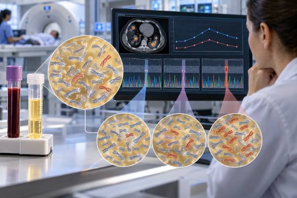
A ctDNA mutation panel is a blood-based genomic test that looks for cancer-related DNA alterations circulating in plasma. It is most often used in people with known advanced cancer when the oncology team needs molecular information for treatment selection, especially if tumor tissue is unavailable, difficult to biopsy, or unlikely to produce results quickly enough. Depending on the assay, a panel may detect single-nucleotide variants, small insertions or deletions, selected gene fusions, copy-number changes, and other biomarkers. Results can identify actionable driver mutations, acquired resistance mechanisms, or variants whose significance is still uncertain. A negative blood panel does not prove that the tumor lacks a targetable alteration because some cancers shed little ctDNA and some alteration types are harder to detect in plasma. For that reason, tissue testing remains important when plasma results are negative, inconclusive, or inconsistent with the cancer’s clinical behavior.
- ctDNA mutation panels profile tumor-derived DNA in blood and can sometimes replace or complement a repeat tissue biopsy for genomic testing.
- Actionable findings may support targeted therapy, but actionability depends on the exact cancer type, stage, alteration, and available treatment.
- Resistance mutations can emerge during therapy and may be detected in plasma at progression or sometimes before clear radiographic progression.
- A negative panel can be non-informative when tumor shedding is low or the assay cannot reliably detect the relevant alteration type.
- Variants from clonal hematopoiesis can mimic tumor mutations, so the source and clinical context of a plasma finding matter.
Table of Contents
- What a ctDNA Mutation Panel Tests
- How Blood-Based Genomic Testing Works
- How to Read Actionable Mutations, VAF, and VUS Results
- Using ctDNA Panels to Select Targeted Treatment
- Resistance Markers and Testing at Cancer Progression
- Why a Negative ctDNA Panel May Need Tissue Confirmation
- Limitations and Practical Next Steps
What a ctDNA Mutation Panel Tests
A ctDNA mutation panel analyzes the tumor-derived portion of cell-free DNA in plasma. It is a form of liquid biopsy genomic profiling. Instead of removing a new piece of tumor with a needle or surgery, the laboratory looks for molecular alterations released from cancer cells into blood.
Panels vary widely in size and design. Some examine a focused group of genes with established treatment relevance. Others perform broad next-generation sequencing across dozens or hundreds of genes. The report may include several classes of genomic alteration:
- Single-nucleotide variants, in which one DNA base is changed.
- Small insertions and deletions, sometimes called indels.
- Gene fusions or rearrangements, when the assay is designed to detect them.
- Copy-number gains or amplifications, which can increase the number of copies of a gene.
- Selected genomic signatures, such as microsatellite instability or other validated biomarkers on certain platforms.
Not every blood panel is equally sensitive for every alteration. Point mutations and short indels are generally easier to detect than some fusions and copy-number changes, especially when little tumor DNA is present. The exact reportable range should therefore be checked rather than assuming that “500 genes tested” means every possible alteration in those genes can be detected with the same sensitivity.
A ctDNA test may be used for several purposes, but a broad mutation panel is primarily designed for genomic profiling. It should not be confused with an ultra-sensitive MRD assay that looks for microscopic residual disease after curative-intent treatment.
Panels can also reveal more than one tumor clone. Because plasma DNA may be released from several metastatic sites, a blood sample can sometimes capture molecular heterogeneity that a single tissue biopsy misses. This is useful when cancer has evolved over time or after several lines of targeted therapy.
How Blood-Based Genomic Testing Works
The process begins with a blood draw. Plasma is separated from blood cells, cell-free DNA is extracted, and the laboratory sequences selected genomic regions. Because ctDNA may represent only a small fraction of all cfDNA, modern assays use deep sequencing and error-suppression methods to distinguish real low-frequency variants from technical noise.
Many panels use unique molecular identifiers or related techniques that label individual DNA molecules before amplification. This helps the laboratory recognize repeated copies of the same original fragment and reduce errors introduced during sequencing.
The amount of tumor-derived DNA strongly affects performance. In a patient with extensive metastatic disease, tumor fraction may be high enough to support broad profiling. In a patient with low-volume disease, the same assay may find no reportable alterations even when the tumor clearly contains mutations.
Preanalytic handling matters. Blood-cell breakdown can release normal genomic DNA into plasma, diluting the tumor signal. Collection tubes, shipping time, processing conditions, plasma volume, and extraction quality all contribute to the reliability of the final result.
The laboratory then compares detected variants with databases and clinical evidence. The report may classify findings as therapeutically relevant, biologically significant, or uncertain. Some reports also identify whether a biomarker is linked to an approved drug, a guideline-supported option, or a clinical trial.
A result can be returned faster than a new tissue-biopsy workflow in some settings, which is one reason plasma testing is useful when treatment decisions are urgent. It also avoids the physical risks of an invasive biopsy and can be repeated during therapy.
However, the blood test cannot show tumor histology or tissue architecture. It does not replace the original pathology diagnosis. If the cancer type is unknown, tissue is usually still needed to determine what the tumor is before molecular findings can be interpreted correctly.
How to Read Actionable Mutations, VAF, and VUS Results
The most important part of a ctDNA panel is not the number of mutations listed. It is the clinical meaning of each finding for that patient’s cancer.
An actionable alteration is one for which evidence supports a specific management option. Actionability is contextual. The same mutation can be a validated drug target in one tumor type, have weaker evidence in another, and have no established treatment implication in a third.
A driver mutation contributes to cancer growth and may or may not be druggable. A resistance mutation helps explain why a tumor is no longer responding to a therapy. A variant of uncertain significance, or VUS, is a change whose clinical effect is not established. A VUS generally should not be used by itself to select an unproven targeted treatment.
Many reports include variant allele fraction, or VAF. If a variant has a VAF of 1%, roughly 1% of analyzable DNA molecules at that position carried the variant under the laboratory’s method. VAF is affected by tumor shedding, total cfDNA, copy number, tumor heterogeneity, treatment, and whether the mutation is present in every cancer cell.
VAF should not be interpreted as the percentage of the body occupied by cancer. A mutation with a 10% VAF does not mean 10% of the patient’s cells are malignant. Likewise, comparing VAF values across different genes or laboratories can be misleading.
Some reports estimate tumor fraction, which is an attempt to measure what proportion of cfDNA appears tumor-derived. A higher tumor fraction can make a negative panel more informative because there was enough tumor signal available for testing. A low tumor fraction makes false-negative results more likely.
The 2026 ASCO ctDNA guideline cautions against using ctDNA fractions, percentages, or concentrations as a stand-alone surrogate measure of disease. These values can support interpretation, but they do not replace imaging or validated response criteria.
One more distinction matters: a variant seen in plasma is not automatically a tumor mutation. Blood-forming cells can acquire age-related mutations through clonal hematopoiesis. Some assays use paired white-blood-cell sequencing or computational methods to help identify this source.
Using ctDNA Panels to Select Targeted Treatment
One of the strongest clinical uses of ctDNA panels is finding tumor alterations that can guide targeted therapy in advanced cancer. Current guidelines support plasma testing when tumor tissue is difficult or unsafe to obtain, when tissue is unavailable or inadequate, when waiting for tissue results could delay an important decision, or when a drug’s approved indication permits or requires ctDNA testing.
Examples of clinically important alterations vary by cancer. Depending on the tumor type and treatment setting, plasma testing may identify changes in genes such as EGFR, KRAS, BRAF, PIK3CA, ESR1, ERBB2, MET, RET, ALK, ROS1, BRCA1, or BRCA2. This list is illustrative, not a universal panel and not a statement that every alteration in these genes is actionable in every cancer.
In advanced non-small cell lung cancer, blood-based genotyping can sometimes identify a targetable driver rapidly. In colorectal cancer, RAS and BRAF alterations can affect treatment selection. In breast cancer, plasma testing may identify PIK3CA or ESR1 alterations relevant to particular hormone receptor-positive disease settings. Other tumor types have their own validated genomic targets.
The KRAS liquid biopsy example shows why context is essential. A KRAS alteration can have different implications in lung, colorectal, and pancreatic cancers, and only certain variants have matched targeted drugs in specific settings.
Guidelines for tumor next-generation sequencing have expanded as more genomic biomarkers gain treatment relevance. ESMO’s 2024 recommendations support tumor NGS in multiple advanced cancers and in metastatic disease when tumor-agnostic alterations can be matched to available therapies.
A blood-based panel can also be valuable when an earlier tissue specimen no longer reflects current disease. A cancer may acquire new alterations after months or years of treatment. Repeat ctDNA profiling can provide a contemporary molecular snapshot without requiring repeated biopsies of difficult metastatic sites.
The result still needs clinical review. A laboratory may list an alteration as potentially actionable based on a drug approved in another tumor type or based on early trial evidence. The oncology team should distinguish a standard-of-care indication from an off-label hypothesis or investigational option.
Resistance Markers and Testing at Cancer Progression
Cancer evolves under treatment pressure. A targeted therapy may eliminate sensitive cells while allowing a resistant clone to expand. ctDNA can sometimes reveal that resistant clone by detecting a new mutation or genomic change in blood.
This is the basis of liquid biopsy resistance testing. The test is often most useful when disease begins to progress and the oncology team needs to understand whether a new molecular mechanism can guide the next therapy.
Well-known examples illustrate the concept. EGFR T790M historically emerged as a resistance mechanism after earlier-generation EGFR inhibitors in lung cancer and became an important plasma-detectable target for later therapy. Other EGFR resistance mechanisms, including C797S in particular treatment contexts, can also occur. The role of a specific alteration depends on which drugs the patient has already received.
In hormone receptor-positive breast cancer, ESR1 mutations can emerge under endocrine treatment and may support selection of therapies with evidence in that molecular setting. An ESR1 liquid biopsy test can therefore answer a treatment-resistance question that was not relevant when the original tumor was first diagnosed.
In metastatic colorectal cancer, resistant subclones involving KRAS, NRAS, BRAF, or EGFR-pathway alterations can emerge during anti-EGFR therapy. Plasma can sometimes capture several resistant clones at once, highlighting the heterogeneity of treatment-resistant disease.
Resistance testing has limits. Finding a resistance mutation does not always identify an effective next drug. Some resistance mechanisms are actionable; others are mainly explanatory. A panel may also miss non-genetic resistance caused by changes in cell state, the tumor microenvironment, drug metabolism, or pathways not represented on the assay.
Serial testing can show a mutation appearing, increasing, decreasing, or disappearing over time. Those patterns can be biologically informative, but treatment should not be changed solely because a low-level variant fluctuates unless evidence supports that action.
The timing of testing matters as well. A sample collected long after treatment has stopped may show a different clonal mixture from one drawn at the moment of progression. The most useful report is tied to a specific clinical question and a clearly documented treatment history.
Why a Negative ctDNA Panel May Need Tissue Confirmation
A negative ctDNA panel is one of the easiest results to misunderstand. “No alterations detected” does not necessarily mean the cancer has no mutations and does not mean a targetable alteration is absent.
The 2026 ASCO guideline recommends seeking tissue-based confirmation when ctDNA results are negative, inconclusive, or inconsistent with the clinical scenario. Earlier ESMO guidance similarly emphasized reflex tissue testing after a non-informative plasma result.
The reason is biological sensitivity. A panel can only detect what is present in the sampled plasma above its analytical limit. Some tumors release abundant ctDNA. Others shed very little. Low-volume disease, certain metastatic locations, treatment response, or naturally low-shedding tumor biology can all reduce detection.
The alteration type matters too. Some plasma assays have lower sensitivity for gene fusions and copy-number alterations than for common point mutations and short indels. A patient whose cancer is driven by a difficult-to-detect rearrangement can therefore have a negative plasma panel despite an informative tissue result.
A negative result becomes more reassuring when the report shows that there was enough tumor-derived DNA for the assay to have a reasonable chance of finding alterations. Even then, the panel’s gene list and technical scope matter.
This is why plasma-first should not be confused with plasma-only. A blood test can be an efficient first step, especially when speed and biopsy risk are important. Tissue remains the backup when plasma does not answer the question.
The reverse can also happen. Tissue testing from an old or small biopsy may miss a resistant clone that ctDNA detects. In that situation, the plasma result can add information not present in the archival sample. The two methods are complementary rather than competitors.
Limitations and Practical Next Steps
Before ordering a ctDNA mutation panel, the clinician should define the decision the test is meant to support. Broad testing is most useful when there is a realistic possibility that a genomic finding will change therapy, clarify resistance, or identify a suitable clinical trial.
Important limitations include:
- Low ctDNA shedding, which raises false-negative risk.
- Uneven sensitivity across alteration types, especially for some fusions and copy-number changes.
- Clonal hematopoiesis, which can create variants that did not come from the solid tumor.
- Variants of uncertain significance, which can generate information without an evidence-based treatment.
- Assay-to-assay differences in genes, depth, limits of detection, bioinformatics, and reporting.
- Tumor evolution, meaning a result can become outdated as treatment selects new clones.
Another common error is using a broad mutation panel as if it were an MRD test. A ctDNA MRD test is optimized for extremely small residual signals after curative-intent therapy. A negative comprehensive genomic profiling panel in that setting does not establish MRD negativity.
After results return, ask several practical questions:
- Which findings are considered clinically actionable in this exact cancer?
- Are they linked to an approved therapy, guideline recommendation, or clinical trial?
- Is any finding a resistance marker that explains the current progression?
- Is the tumor fraction high enough for a negative result to be informative?
- Should tissue testing be performed because plasma was negative or incomplete?
- Could any variant represent clonal hematopoiesis or a possible inherited mutation?
A possible inherited finding requires special care. ctDNA panels are designed mainly to detect somatic tumor alterations, not to diagnose hereditary cancer syndromes. If the pattern suggests a germline variant, confirmation with an appropriate non-tumor sample and genetic counseling may be recommended.
Patients should also know that larger panels are not automatically better. A broad assay can discover rare targets and trial opportunities, but it also produces more variants with uncertain or weak evidence. Clinical value depends on matching a reliable result to an effective treatment, not simply maximizing the number of genes sequenced.
When serial panels are obtained, compare them in the context of therapy dates and imaging. The disappearance of a mutation can accompany response, while a new alteration can signal clonal evolution. Yet a changing VAF should not override a patient’s symptoms, scan findings, or established response criteria without supporting evidence.
The best use of a ctDNA mutation panel is therefore focused precision oncology: obtain molecular information when it can answer a treatment question, understand what the assay may miss, and use tissue confirmation when blood testing cannot provide a reliable answer.
References
- Circulating Tumor DNA Testing in Solid Tumors and Lymphoma: ASCO Guideline 2026 (Guideline)
- Clinical applications of cell-free DNA-based liquid biopsy analysis 2025 (Review)
- Circulating tumor DNA to monitor treatment response in solid tumors and advance precision oncology 2025 (Review)
- Recommendations for the use of next-generation sequencing (NGS) for patients with advanced cancer in 2024: a report from the ESMO Precision Medicine Working Group 2024 (Guideline)
- ESMO recommendations on the use of circulating tumour DNA assays for patients with cancer: a report from the ESMO Precision Medicine Working Group 2022 (Guideline)
Disclaimer
A ctDNA mutation panel can guide cancer treatment only when a detected alteration has validated clinical meaning for the specific tumor and treatment setting. A negative plasma test can miss actionable mutations and may need tissue confirmation. Genomic results, resistance markers, and possible inherited findings should be reviewed with the oncology team and appropriate molecular or genetic specialists.