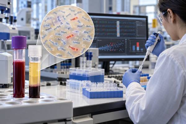
A cell-free DNA cancer test analyzes DNA fragments circulating in blood plasma. Most cell-free DNA, or cfDNA, comes from normal cells, especially blood-forming cells; only a portion may come from a tumor. That cancer-derived portion is called circulating tumor DNA, or ctDNA. Cancer-focused cfDNA testing can look for mutations, copy-number changes, methylation patterns, fragment characteristics, or other genomic signals. The meaning of a result depends on why the test was ordered. In advanced cancer, cfDNA testing can identify treatment-relevant mutations when tumor tissue is limited or difficult to obtain. In early-stage disease, highly sensitive assays may be used to study molecular residual disease or recurrence risk. Screening uses are developing but remain distinct from routine diagnostic testing. A negative result does not prove that no cancer or mutation is present because some tumors release very little DNA into the bloodstream.
- Cell-free DNA is not the same as tumor DNA; ctDNA is the tumor-derived fraction within total cfDNA.
- There is no universal “normal cfDNA cancer level.” Results depend on the assay, target mutations, tumor fraction, and clinical setting.
- A detected cancer-related mutation may guide treatment, but some variants can come from clonal blood cells rather than the tumor.
- A negative blood result can occur when tumor shedding is low, so tissue testing may still be needed.
- Most cfDNA blood draws require no fasting, but correct collection tubes, processing, and timing are important for test accuracy.
Table of Contents
- What Cell-Free DNA Is and Where Tumor DNA Fits
- What a Cell-Free DNA Cancer Test Can Measure
- How Positive, Negative, and Inconclusive Results Are Interpreted
- Clinical Uses: Genotyping, Monitoring, MRD, and Early Detection
- Limitations, False Results, and Clonal Hematopoiesis
- Blood Draw, Preparation, and Sample Quality
- Cell-Free DNA Testing Versus Tissue Biopsy
What Cell-Free DNA Is and Where Tumor DNA Fits
Cell-free DNA is made of short DNA fragments released into body fluids when cells die or actively shed material. In blood, these fragments circulate mainly in plasma. Healthy people have cfDNA too, so finding cfDNA itself is not evidence of cancer.
The cancer-specific portion is circulating tumor DNA. CtDNA can contain the same acquired genomic changes found in tumor cells, such as point mutations, small insertions or deletions, copy-number alterations, rearrangements, and abnormal methylation patterns. A ctDNA test is therefore a cancer-focused use of cfDNA analysis.
The fraction of total cfDNA that comes from the tumor can vary enormously. In advanced metastatic cancer it may be high enough for broad genomic profiling. After surgery for an early-stage tumor, ctDNA can fall to extremely low concentrations—sometimes parts per million—making detection much harder.
This explains why two people with the same cancer type can have very different blood-test results. Tumor size is only one factor. Tumor location, blood supply, metastatic sites, treatment, biological subtype, and how rapidly cells are turning over all affect how much tumor DNA reaches the plasma. Tumors in the brain or certain low-volume sites may be poorly represented in peripheral blood.
The term “cell-free DNA cancer test” can therefore describe several different laboratory products. Some are broad mutation panels for people with known advanced cancer. Others are personalized assays that search for a small set of mutations from a person’s tumor. Still others analyze methylation or fragment patterns for early cancer detection. The intended use is essential to interpreting the result.
What a Cell-Free DNA Cancer Test Can Measure
Cancer-focused cfDNA assays can measure several classes of molecular information. The exact report depends on the platform.
Mutations and small DNA changes
Next-generation sequencing panels can search dozens to hundreds of cancer-associated genes. Examples include EGFR, KRAS, BRAF, PIK3CA, ESR1, BRCA1, BRCA2, ALK, RET, MET, ERBB2, and TP53, although the actual gene list varies. A blood-based genomic mutation panel may identify a driver mutation that supports targeted therapy or a resistance change that emerged during treatment.
Reports often show a variant allele fraction (VAF): the percentage of DNA fragments at that position carrying the variant. VAF is not the same as the percentage of tumor in the body. It is influenced by ctDNA shedding, tumor clonality, copy number, assay depth, and background cfDNA.
Copy-number changes and gene fusions
Some assays can detect gene amplification, deletion, or rearrangement. These changes can be clinically actionable, but they are often harder to identify in plasma when the tumor fraction is low. A negative fusion or copy-number result can therefore be less reassuring than a clearly detected alteration.
Methylation and fragment patterns
Cancer cells can leave distinctive epigenetic and physical signatures in cfDNA. Methylation-based tests assess chemical tags attached to DNA; fragmentomic tests examine fragment length, breakpoints, and genome-wide patterns. These approaches are especially relevant to early detection because they do not depend on finding one specific mutation.
A fragmentomics cancer blood test and a mutation panel can both use cfDNA but answer different questions.
How Positive, Negative, and Inconclusive Results Are Interpreted
A cfDNA cancer report should be interpreted according to the test’s purpose, not as a generic positive-versus-negative cancer screen.
A pathogenic or likely pathogenic alteration detected result means the laboratory found a DNA change with evidence linking it to cancer biology. In someone with a known cancer, the next question is whether that alteration is actionable. Some mutations predict sensitivity to a targeted drug; others indicate resistance, prognosis, or eligibility for a clinical trial.
A no alteration detected result does not mean the tumor lacks all mutations. It may mean that the assay did not detect the targeted alteration in the blood sample. Reasons include low tumor fraction, a mutation outside the tested regions, technical detection limits, or a cancer that does not shed much ctDNA.
An inconclusive or quantity not sufficient result can occur when the sample contains too little DNA, fails quality controls, or has a signal near the assay’s limit of detection. Depending on the clinical situation, the next step may be another blood draw or tissue testing.
Some reports also give a tumor fraction estimate or comment on whether ctDNA shedding appears low. There is no universal cutoff that defines “high” or “low” across all platforms. A numeric percentage from one assay should not be compared directly with a percentage from another unless the methods are validated as equivalent.
The most clinically important rule is that an unexpected blood result should be reconciled with pathology, imaging, treatment history, and prior molecular testing. A molecular result is not interpreted in isolation.
Reports can also use terms that sound more definitive than they are. “Not detected” usually means not detected above the assay’s validated threshold, not biologically absent. “Detected” means the laboratory found enough evidence to call the alteration, but it does not establish that every tumor cell carries it. “Low-level” may indicate a small variant allele fraction, yet a low number can still be clinically important if the alteration has a strong treatment association. Conversely, a relatively high VAF does not automatically mean the cancer is advanced.
When a result changes from one blood draw to the next, compare like with like. Different platforms can have different gene lists, sequencing depths, filters, and limits of detection. A mutation reported at 0.4% by one assay and 0.2% by another cannot be assumed to have fallen by half. Serial monitoring is most interpretable when the same validated method is used at clinically meaningful time points.
Clinical Uses: Genotyping, Monitoring, MRD, and Early Detection
CfDNA testing has different levels of evidence depending on the setting.
Advanced cancer genotyping
The strongest routine use is molecular profiling in advanced solid tumors when tissue is unavailable, insufficient, risky to obtain, or too slow to guide a treatment decision. Plasma can capture DNA from multiple metastatic sites, which may reveal tumor heterogeneity that a single biopsy misses.
Guidelines support validated plasma ctDNA testing for actionable alterations in appropriate advanced cancers. A positive actionable result can be clinically useful. A negative or uninformative result often needs tissue confirmation because plasma sensitivity is imperfect.
Treatment response and resistance
CtDNA levels and mutation patterns can change faster than tumor size on imaging. Serial testing may show that a driver mutation is falling during effective therapy or that a new resistance mutation has emerged. Examples include resistance alterations involving EGFR in lung cancer or ESR1 in hormone receptor-positive breast cancer. A liquid biopsy resistance mutation test uses this principle to look for evolving tumor clones.
Not every change in VAF should trigger a treatment switch. The result needs a validated clinical action and should be interpreted alongside imaging and symptoms.
Molecular residual disease
After surgery or other curative-intent treatment, cfDNA assays can search for tiny amounts of residual ctDNA. A positive ctDNA minimal residual disease test is strongly associated with higher recurrence risk in many solid tumors. However, the value of changing treatment based only on MRD results is still being defined for each cancer type.
Early cancer detection
Multi-cancer detection research uses cfDNA methylation, mutations, fragmentomics, or combinations of biomarkers to screen people without known cancer. This remains a different use case from genomic profiling in a patient with an established diagnosis. Screening tests need exceptionally high specificity because most people tested do not have cancer.
Limitations, False Results, and Clonal Hematopoiesis
The main limitation of cfDNA cancer testing is low or variable tumor shedding. If very little ctDNA is present, even a technically excellent assay can miss a real tumor alteration. Low shedding is particularly common in small-volume, early-stage, or certain anatomically confined cancers.
A second challenge is clonal hematopoiesis. As people age, blood-forming stem cells can acquire mutations and expand into clones without causing leukemia. These blood-cell mutations can release DNA into plasma and appear on a cfDNA test. Commonly involved genes include DNMT3A, TET2, ASXL1, and sometimes TP53 or other cancer-related genes.
That means a mutation detected in plasma is not automatically from a solid tumor. Laboratories use bioinformatic filters, matched white-blood-cell sequencing, variant patterns, and clinical context to reduce this source of false attribution.
Other limitations include:
- Preanalytic contamination: white blood cells can break down after collection and release genomic DNA, diluting the tumor signal.
- Assay-specific detection limits: a variant present below the validated threshold may not be reported.
- Uneven alteration detection: point mutations may be easier to detect than some fusions or copy-number changes.
- Tumor heterogeneity: a plasma sample can contain DNA from several clones, which is useful but can complicate interpretation.
- Germline variants: some alterations may be inherited rather than tumor-specific and may require separate confirmatory genetic testing.
- Biological timing: recent treatment can rapidly change the amount of tumor DNA in circulation.
For these reasons, a cfDNA test is a complement to pathology and clinical assessment, not a universal substitute for tissue.
Blood Draw, Preparation, and Sample Quality
Most plasma cfDNA tests do not require fasting. People can usually take routine medications unless the ordering clinician gives different instructions. The important preparation issues are logistical and laboratory-related rather than dietary.
Blood is collected into tubes designed either for rapid plasma processing or for stabilizing blood cells during transport. If ordinary tubes are left unprocessed too long, white blood cells can rupture and release large amounts of normal genomic DNA. That increases background DNA and can make low-level tumor signals harder to detect.
Plasma is generally preferred over serum for ctDNA analysis because clotting can release additional DNA from blood cells. Laboratories also control centrifugation, storage temperature, freeze-thaw cycles, DNA extraction, sequencing depth, and quality thresholds.
The timing of collection may matter during treatment. A clinician may want a baseline sample before therapy, a defined on-treatment sample, or a postoperative sample after enough time has passed for DNA released by surgery-related tissue injury to clear. The correct timing depends on the specific clinical question and assay.
If a sample fails quality controls, repeating the blood draw is often more useful than overinterpreting a borderline result.
Practical details can differ between laboratories. Some request two tubes to provide enough plasma or allow repeat analysis. Turnaround time may range from several days to a few weeks depending on whether the assay is a focused PCR test, a broad sequencing panel, or a personalized MRD assay that first requires tumor sequencing. Recent transfusion, organ transplantation, pregnancy, or a second malignancy can also complicate interpretation for certain assays, so the ordering team should know relevant medical history.
For serial testing, consistency matters. Samples collected at substantially different points in a treatment cycle can reflect biological fluctuations as well as true disease change. Clinicians therefore often define the collection schedule in advance and interpret trends together with imaging rather than reacting to a single isolated rise or fall.
Cell-Free DNA Testing Versus Tissue Biopsy
CfDNA testing and tissue biopsy are complementary. Blood testing is minimally invasive, can be repeated, and may capture DNA from multiple tumor sites. Tissue provides cellular architecture, histology, protein expression, tumor microenvironment information, and often a stronger signal for comprehensive genomic analysis.
A tissue biopsy is still essential when the diagnosis itself is uncertain. A cfDNA mutation associated with lung cancer, for example, cannot by itself prove that a lung mass is malignant or establish the exact histologic subtype.
Plasma testing is especially useful when a biopsy is unsafe, the tumor is difficult to reach, tissue is exhausted, or rapid genomic information is needed. It can also reveal resistance mutations that were not present in an older tissue sample.
When plasma identifies a well-validated actionable alteration, treatment can sometimes be selected without repeating tissue, depending on the cancer, drug indication, and guideline. When plasma is negative, tissue often remains the better next step if the clinical need for genomic information is high.
There are also situations in which both tests are valuable. A new tissue biopsy can confirm that a lesion is cancer and establish histology, while plasma can simultaneously sample DNA shed from disease elsewhere in the body. If the two results disagree, the difference may reflect tumor heterogeneity, an older tissue specimen, low plasma shedding, a technical limitation, or a variant that originated from blood cells rather than the tumor. The disagreement should be investigated rather than automatically assuming one test is “right” and the other “wrong.”
For hereditary-risk questions, a tumor or plasma finding is not a substitute for a dedicated germline test. If a BRCA1, BRCA2, TP53, or another potentially inherited variant appears at a pattern suggestive of germline origin, the clinician may recommend testing a normal sample—usually blood cells or saliva—through a genetics laboratory and, when appropriate, genetic counseling. That distinction affects not only treatment but also future cancer risk and relatives.
The most useful way to read a cfDNA report is therefore to ask four questions: Why was this test ordered? How much tumor signal was detectable? Which alteration was found or not found? What clinical action is validated for that result? Those questions prevent a common mistake—treating every cfDNA number as a direct measure of how much cancer is present.
References
- Clinical applications of cell-free DNA-based liquid biopsy analysis 2025 (Review)
- Liquid Biopsy in Solid Tumours: An Overview 2025 (Review)
- Clinical Practice Guideline for Blood-based Circulating Tumor DNA Assays 2024 (Guideline)
- Clonal hematopoiesis detection in patients with cancer using cell-free DNA sequencing 2023
- Integrating circulating-free DNA (cfDNA) analysis into clinical practice: opportunities and challenges 2022 (Review)
Disclaimer
Cell-free DNA testing is not a stand-alone method for diagnosing or excluding cancer. Results can be affected by tumor shedding, assay design, clonal hematopoiesis, sample quality, and treatment timing, and negative plasma results may require tissue confirmation. Test interpretation and treatment decisions should be made with the oncology and pathology team familiar with the specific assay.