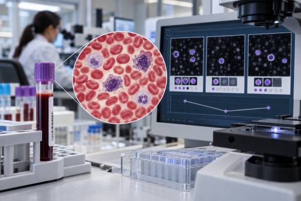
A circulating tumor cell test looks for intact cancer cells that have detached from a tumor and entered the bloodstream. These circulating tumor cells, or CTCs, are rare—often only a few cells among billions of normal blood cells—so testing requires specialized methods to enrich, identify, and count them. In several metastatic cancers, especially breast, prostate, and colorectal cancer, higher CTC counts are associated with worse prognosis and shorter progression-free or overall survival. CTC testing can also be repeated during treatment to study changes over time. However, a CTC result does not by itself diagnose cancer, define tumor stage, or prove that a treatment is working or failing. Different platforms capture different types of cells, and some tumor cells may escape detection. The most established clinical use is prognostic enumeration in selected metastatic cancers, while molecular profiling of CTCs remains an active area of research.
- A CTC test detects intact tumor cells in blood; it is different from ctDNA testing, which analyzes tumor-derived DNA fragments.
- There is no universal normal CTC range across all cancers and platforms; thresholds are disease- and assay-specific.
- Higher CTC counts usually indicate a less favorable prognosis in validated metastatic cancer settings, not a definite amount of tumor burden.
- A low or zero count does not rule out cancer because CTCs are rare and some tumors shed few detectable cells.
- Most CTC blood tests require no fasting, but the sample must be collected and processed according to the platform’s protocol.
Table of Contents
- What Circulating Tumor Cells Are
- How CTC Tests Capture and Identify Tumor Cells
- What CTC Results Mean
- Clinical Uses in Cancer Monitoring and Prognosis
- Limits, False Negatives, and Platform Differences
- CTC Testing Versus ctDNA and Other Liquid Biopsies
- What to Expect Before and After CTC Testing
What Circulating Tumor Cells Are
Circulating tumor cells are whole cells that leave a primary or metastatic tumor and enter the blood. Most do not survive the journey through the circulation. A small fraction may interact with platelets, immune cells, or other CTCs, lodge in distant tissues, and contribute to metastasis.
CTCs are biologically interesting because they provide a moving sample of the cancer. A tissue biopsy captures cells from one place at one time. CTCs may come from several tumor sites and can be collected repeatedly with a blood draw. Researchers can therefore study their number, shape, protein expression, gene mutations, RNA patterns, and other features.
The challenge is rarity. Even in metastatic disease, CTCs can be extremely uncommon compared with red cells and white blood cells. A typical assay may examine 7.5 mL or another defined volume of blood and use enrichment methods to separate likely tumor cells from the background.
A circulating tumor cell count test focuses on enumeration. Other research approaches go further and characterize individual CTCs for markers such as hormone receptors, HER2, androgen receptor variants, epithelial markers, or genomic changes.
CTCs should not be confused with tumor DNA fragments. A CTC contains an intact cell membrane, nucleus, RNA, proteins, and genomic DNA. CtDNA is cell-free material released from tumor cells. Both are forms of liquid biopsy, but they require different technology and answer somewhat different questions.
How CTC Tests Capture and Identify Tumor Cells
A CTC assay generally has two jobs: enrich rare candidate cells and prove that the captured cells meet the assay’s definition of a tumor cell.
Marker-based enrichment
The best-known approach uses antibodies against epithelial cell adhesion molecule, or EpCAM, to pull epithelial tumor cells out of blood with magnetic particles. The CellSearch system then uses staining patterns to distinguish CTCs from white blood cells. A typical CellSearch-defined CTC is nucleated, positive for cytokeratins, and negative for the white-blood-cell marker CD45.
This standardized definition is a major strength because it lets results from different laboratories and studies be compared. It is also a limitation: tumor cells that have reduced epithelial markers during epithelial-to-mesenchymal transition may be missed.
Marker-independent methods
Other platforms enrich cells based on physical properties such as size, deformability, electrical characteristics, or microfluidic behavior. These methods may capture cells with lower EpCAM expression, but they can also recover more non-tumor cells and require different identification rules.
Molecular characterization
After enrichment, some laboratories analyze CTC DNA, RNA, or proteins. This is attractive because an intact cell can reveal information that free DNA cannot, such as protein expression or cellular phenotype. Single-cell sequencing can also show that separate CTCs carry different mutations, illustrating tumor heterogeneity.
Most of these deeper analyses are not yet standardized for routine decision-making. The test report should therefore make clear whether it is providing an established CTC count, an investigational molecular profile, or both.
What CTC Results Mean
CTC results are often reported as cells per defined blood volume, such as cells per 7.5 mL. The number must be interpreted using the specific cancer type, assay, and clinical setting.
There is no single threshold that means “cancer present” or “cancer absent.” In metastatic breast and prostate cancer, classic CellSearch studies used 5 CTCs per 7.5 mL to divide more favorable from less favorable prognostic groups. In metastatic colorectal cancer, a commonly validated threshold is 3 CTCs per 7.5 mL. These are prognostic cutoffs from specific studies, not universal biological boundaries.
A result below a threshold is not “normal” in the same way as a routine blood count. A patient can have substantial cancer with zero CTCs detected in one sample. Likewise, a result above a validated cutoff does not reveal the exact number of metastases or replace imaging.
The trend may provide additional information. In metastatic breast cancer, for example, persistently elevated CTCs during treatment have been associated with shorter survival, while conversion from a higher to a lower count can be associated with a better prognosis. Even so, changing therapy solely because the CTC count changes has not become a universal standard.
A CTC count can also be reported as zero. Zero means no cells meeting that assay’s definition were found in the tested volume; it does not mean that no tumor cells exist anywhere in circulation. The distinction matters when disease is visible on imaging but the blood result is low. The imaging and clinical findings do not become less real because the CTC test is negative.
Likewise, a very high count should not be converted into a tumor size or percentage of the body involved by cancer. CTC number and radiographic tumor burden often correlate only imperfectly. A small but biologically aggressive cancer can shed many cells, while a larger tumor in a low-shedding site can produce fewer detectable CTCs.
If a report gives a count from a non-CellSearch platform, do not apply CellSearch cutoffs automatically. Different technologies sample and define cells differently.
Clinical Uses in Cancer Monitoring and Prognosis
The most established role of CTC enumeration is prognosis in selected metastatic solid tumors. Prognostic means the result is associated with the expected course of disease; it does not necessarily tell which treatment will work best.
Metastatic breast cancer
CTC enumeration has a strong evidence base in metastatic breast cancer. Higher baseline counts and persistently elevated counts after treatment starts are associated with shorter progression-free and overall survival. Large pooled analyses have confirmed that serial counts retain prognostic value across breast cancer subtypes and treatment settings.
This information can complement symptoms, imaging, physical examination, and other tumor markers. It should not replace them.
Metastatic prostate cancer
In metastatic castration-resistant prostate cancer, higher CellSearch CTC counts are also associated with poorer overall survival. CTC changes have been studied as trial endpoints and treatment-response biomarkers. Molecular CTC testing has an additional niche in prostate cancer research, including assays for androgen receptor splice variant 7, or AR-V7, which has been investigated as a treatment-selection marker.
Metastatic colorectal cancer
In metastatic colorectal cancer, counts at or above validated thresholds are associated with shorter progression-free and overall survival. Serial measurements can add prognostic information during therapy, although they are not a substitute for standard radiographic response assessment.
Other solid tumors
CTCs are studied in lung, ovarian, pancreatic, gastric, head and neck, bladder, melanoma, and many other cancers. Evidence for routine use is less mature and varies by platform. International expert consensus published in 2025 supported established prognostic and monitoring roles mainly in metastatic breast and prostate cancer while emphasizing that many other uses remain investigational.
For early-stage cancers, CTCs may be associated with recurrence risk, but sensitivity is challenging because the number of circulating cells can be extremely low. Combining CTCs with ctDNA MRD testing is being explored as a way to capture complementary biological signals.
Limits, False Negatives, and Platform Differences
CTC testing has several practical and biological limitations.
Sampling error is unavoidable. A blood draw contains only a small fraction of the body’s circulating blood volume. When CTCs are extremely rare, a tube can contain no detectable cells even when tumor cells are circulating elsewhere.
Marker selection can bias the result. EpCAM-based systems may miss tumor cells that have changed phenotype and lost epithelial markers. Marker-independent systems may capture a broader range of cells but can have different specificity and reproducibility.
CTCs are heterogeneous. Some travel alone; others form clusters. Some have epithelial features, some mesenchymal features, and many show mixed states. Not every CTC has the same metastatic potential.
Preanalytic handling matters. Collection tubes, transport time, temperature, sample age, and processing steps can affect cell preservation. A damaged CTC cannot be counted or characterized accurately.
Results do not transfer across platforms. A count from one system is not necessarily equivalent to the same numeric count from another. Thresholds need validation for the exact assay.
Clinical utility is narrower than clinical validity. A marker can strongly predict prognosis without proving that changing treatment based on the marker improves outcomes. This distinction explains why CTC enumeration can be informative yet not routinely dictate therapy in every metastatic cancer.
Finally, a CTC test is not a screening test for the general population. Because CTCs are often absent or undetectable in early disease, a zero count cannot exclude an early cancer.
Another source of complexity is cell clustering. CTC clusters can contain several tumor cells and may include platelets, immune cells, or stromal cells. Research suggests that clusters can have different metastatic behavior from single CTCs, but most routine enumeration systems were built around standardized cell counting rather than a complete functional analysis of clusters. Reports that mention clusters therefore need assay-specific interpretation.
The biology can also change during treatment. Therapy may eliminate one tumor clone while allowing another to expand, alter epithelial-marker expression, or change the number of cells entering the bloodstream. This is scientifically useful because CTCs can reveal evolution in real time, but it means that a change in count is not always a simple measure of “more cancer” or “less cancer.”
CTC Testing Versus ctDNA and Other Liquid Biopsies
CTCs and ctDNA provide overlapping but not identical information.
A circulating tumor DNA test analyzes fragments of tumor DNA in plasma. It is generally easier to scale for broad sequencing and is widely used for genomic profiling in advanced cancer. CtDNA can detect actionable mutations and resistance changes without having to capture intact cells.
CTCs preserve the whole cell. That makes it possible to study morphology, proteins, RNA, DNA, and sometimes functional behavior. In theory, CTCs can reveal whether a resistance mechanism is expressed at the cellular level or whether different circulating cells have different phenotypes.
The tradeoff is that CTCs are harder to isolate. CtDNA may be abundant when CTCs are scarce, and the reverse can also occur. Neither biomarker is guaranteed to represent every tumor site.
Other liquid biopsy components include cell-free RNA, exosomes, circulating proteins, tumor-educated platelets, and extracellular vesicles. Multi-analyte approaches may ultimately combine several signal types rather than choosing one biomarker.
Clinically, the test should match the question. For a rapid search for actionable mutations in advanced cancer, ctDNA is often more practical. For validated prognostic enumeration in metastatic breast or prostate cancer, CTCs may add information. For early detection or molecular residual disease, highly sensitive ctDNA and multi-omic methods currently have a larger evidence base than standard CTC counting.
What to Expect Before and After CTC Testing
A CTC test usually requires a routine venous blood draw and no fasting. The laboratory or oncology clinic may specify a dedicated collection tube and a required shipping or processing window. Because intact cells must be preserved, handling instructions are important.
Before the test, ask which platform will be used and why. The answer should identify the cancer type, clinical setting, and decision the result is expected to support. If the purpose is prognosis, it is useful to know whether the result will actually change treatment or simply provide additional risk information.
After testing, review the report in context. A high count may support a less favorable prognosis, but it should be compared with imaging, symptoms, tumor markers, and previous CTC values. A low count should not override clear evidence of progression elsewhere.
For serial monitoring, consistency improves interpretation. Using the same platform, similar collection conditions, and clinically meaningful time points reduces noise. A one-time change can reflect sampling variability; a persistent trend is usually more informative. The interval between tests should be chosen for the treatment and cancer rather than simply repeating the assay as often as possible. Testing too frequently can magnify biological and analytical noise without adding a useful decision point.
When a count rises unexpectedly, clinicians usually look for confirmation in the broader picture. That may include a repeat CTC test, imaging, symptoms, examination findings, or standard laboratory markers. A single discordant value is a reason to investigate, not an automatic instruction to stop an otherwise effective treatment. The purpose is to improve clinical context and risk assessment, not to let one rare-cell measurement overrule stronger evidence.
If a molecular feature is reported from CTCs, ask whether that finding has been clinically validated for treatment selection or remains investigational. Many CTC technologies are scientifically promising but have not yet crossed the threshold from biomarker research to routine therapeutic decision-making.
It is also reasonable to ask whether the laboratory participates in external quality programs and how reproducibility was established. CTC assays operate at the edge of rare-cell detection, so technical consistency matters. A clinically useful report should identify the method, specimen volume, counting definition, and interpretation range rather than present a number without context.
For patients, the most useful takeaway is that CTC testing can add a dynamic layer of information to cancer care, especially in metastatic disease, but it should sit beside—not above—pathology, imaging, symptoms, and standard biomarkers. The strength of the evidence depends on the exact tumor type and platform.
References
- International expert consensus on the clinical integration of circulating tumor cells in solid tumors 2025 (Position Statement)
- Clinical applications of circulating tumor cells in patients with solid tumors 2024 (Review)
- Circulating tumor cells: from new biological insights to clinical practice 2024 (Review)
- Unveiling the impact of circulating tumor cells: Two decades of discovery and clinical advancements in solid tumors 2024 (Review)
- Latest advances in clinical studies of circulating tumor cells in early and metastatic breast cancer 2023 (Review)
Disclaimer
CTC testing provides prognostic or monitoring information only in specific validated cancer settings and does not diagnose cancer by itself. Counts vary by assay, cancer type, disease biology, and sample handling, and results from different platforms are not directly interchangeable. Treatment decisions should be made with the oncology team using the full clinical picture.