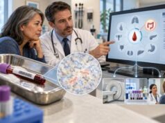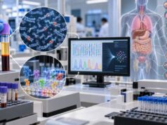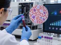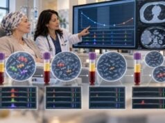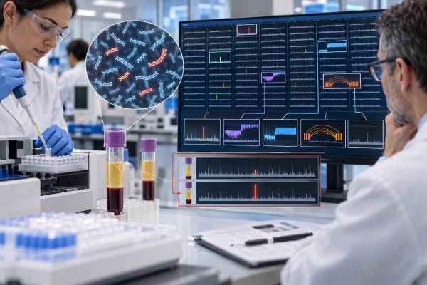
A ctDNA tumor fraction test estimates how much of the cell-free DNA circulating in a blood sample comes from cancer cells. The result can help show whether a liquid biopsy contains enough tumor-derived DNA for reliable genomic analysis and, in some settings, can provide a broad signal of disease activity. Tumor fraction is not a direct measurement of tumor size, however. A large cancer may release relatively little DNA into blood, while a smaller but biologically active or widely metastatic cancer may shed more.
Tumor fraction also depends on the assay, cancer type, treatment timing, metastatic sites, and the total amount of non-tumor cell-free DNA in the sample. For that reason, there is no single percentage that is “normal” or “high” across all laboratories. The most useful interpretation comes from the report’s method, detection limits, accompanying genomic findings, imaging, and changes over time.
- Tumor fraction is the estimated percentage of plasma cell-free DNA that is tumor-derived; it is not the percentage of blood made up of cancer DNA.
- There is no universal normal or abnormal ctDNA tumor fraction range; thresholds are assay- and clinical-context specific.
- A low or undetectable tumor fraction can make a negative liquid-biopsy result less reliable because the tumor may not be shedding enough DNA into blood.
- A falling tumor fraction during treatment can be encouraging, while a rising value may signal increasing molecular disease activity, but neither should be interpreted without clinical context.
- Tumor fraction should not replace imaging, pathology, or tissue testing when those are needed to diagnose, stage, or characterize a cancer.
Table of Contents
- What ctDNA Tumor Fraction Measures
- How the Test Is Performed
- How to Interpret Tumor Fraction Results
- What Affects the Percentage
- How Tumor Fraction Is Used in Cancer Care
- Limitations and Common Mistakes
- Questions to Ask About Your Result
What ctDNA Tumor Fraction Measures
Cell-free DNA, or cfDNA, consists of short DNA fragments released into the bloodstream as cells turn over. Most plasma cfDNA comes from normal blood-forming cells and other non-cancer cells. In a person with cancer, a portion may come from tumor cells. That tumor-derived component is called circulating tumor DNA, or ctDNA.
ctDNA tumor fraction is an estimate of the proportion of total cfDNA that originated from the tumor. If a laboratory reports a tumor fraction of 5%, for example, it is estimating that about 5% of the cell-free DNA molecules represented in that sample are tumor-derived. It does not mean that 5% of the person’s blood, body, or tumor is cancer.
Different assays estimate tumor fraction in different ways. Some infer it from somatic mutation allele frequencies. Others use genome-wide copy-number patterns, methylation signatures, fragment patterns, or combinations of signals. This means two laboratories can produce somewhat different tumor-fraction estimates from the same biological sample.
Tumor fraction is related to, but distinct from, variant allele frequency (VAF). VAF is the percentage of sequencing reads at one genomic position that contain a specific variant. Tumor fraction is an estimate across the sample. A mutation with a 2% VAF does not automatically mean the tumor fraction is 2%, because copy number, tumor heterogeneity, whether a mutation is present in every cancer cell, and normal DNA contamination all influence VAF.
A circulating tumor DNA test may report tumor fraction together with individual mutations, amplifications, fusions, microsatellite instability, or other biomarkers. The fraction helps the clinician judge how much tumor signal was available for those measurements.
How the Test Is Performed
Most tumor-fraction testing starts with a standard peripheral blood draw into specialized collection tubes designed to preserve cell-free DNA. The laboratory separates plasma from blood cells, extracts cfDNA, and analyzes it using a validated molecular method. Many commercial assays use next-generation sequencing, while some newer approaches also analyze methylation or genome-wide fragmentation patterns.
Preparation is usually simple. Fasting is generally not required unless another test ordered at the same time requires it. The more important variables are sample handling, adequate plasma volume, and timing in relation to treatment or major procedures.
A typical workflow is:
- Blood is collected in the laboratory’s required tubes.
- Plasma is separated from blood cells to reduce contamination by genomic DNA.
- cfDNA is extracted and prepared for sequencing or another molecular assay.
- The assay identifies tumor-associated signals and estimates tumor fraction when technically possible.
- The final report combines the tumor-fraction estimate with genomic findings and assay-specific quality information.
Some reports provide a numerical percentage. Others use categories such as low, intermediate, or high, or state that tumor fraction could not be reliably estimated. A result can also be “not detected” or below the assay’s quantifiable range.
Testing may be ordered as part of a broad ctDNA mutation panel. In that setting, tumor fraction is often valuable because the sensitivity of mutation detection depends partly on how much tumor-derived DNA is present. Copy-number changes can require substantially more tumor signal than some point mutations, so a technically successful blood test may still be underpowered for particular alteration types.
How to Interpret Tumor Fraction Results
There is no universal ctDNA tumor fraction reference range. Unlike sodium, hemoglobin, or many routine laboratory tests, tumor fraction does not have a single healthy interval that applies across assays and cancers. Interpretation depends on the analytical method and the reason the test was ordered.
| Report pattern | What it may mean | Important caution |
|---|---|---|
| Undetectable or very low | Little tumor-derived DNA was measurable in plasma | Does not rule out cancer or a tumor mutation |
| Low but measurable | There is a detectable tumor signal, but some genomic findings may be harder to detect | Assay-specific sensitivity matters |
| Higher fraction | More of the sampled cfDNA appears tumor-derived and genomic profiling may be more informative | It is not a direct tumor-volume percentage |
| Falling on serial tests | May reflect molecular response to effective treatment | Timing, therapy type, and assay consistency matter |
| Rising on serial tests | May reflect increasing molecular disease activity or progression | Should be correlated with imaging and clinical findings |
A value such as 1%, 5%, or 10% should not be interpreted using a universal severity scale. Research studies sometimes use cutoffs such as 1%, 5%, or 10% to define groups, but those thresholds answer specific study questions and are not interchangeable diagnostic standards. A 5% threshold used in one lung-cancer study, for example, does not establish that 5% is a general “high” tumor fraction for every cancer.
The most practical question is often not “Is my percentage high?” but “Was there enough ctDNA in this sample for the negative and positive findings to be trusted?” A high-confidence pathogenic alteration can be clinically important even when tumor fraction is modest. Conversely, a report with no actionable alterations and very low tumor fraction may need follow-up tissue testing because the blood sample may simply lack enough tumor signal.
What Affects the Percentage
Tumor fraction reflects both how much DNA the cancer releases and how much non-tumor cfDNA is present. Several factors can change the result without representing a simple change in tumor size.
Cancer type and metastatic site matter. Liver and widespread visceral metastases often shed more ctDNA into plasma than cancers confined to certain sites. Central nervous system tumors can be particularly difficult to assess from plasma because the blood-brain barrier limits release into peripheral circulation. Some indolent, mucinous, or low-volume tumors also shed little ctDNA.
Disease burden matters, but imperfectly. Higher tumor volume often correlates with higher ctDNA levels, yet the relationship varies widely. Tumor vascularity, cell turnover, treatment sensitivity, and biology influence shedding. This is why tumor fraction should be viewed as a molecular biomarker rather than a blood-based measurement of tumor dimensions.
Treatment timing matters. Effective therapy can reduce ctDNA quickly, sometimes before a clear radiographic response is visible. Therapy can also cause short-term changes in DNA release. Serial interpretation is therefore strongest when samples are collected at clinically meaningful, reasonably consistent time points.
Non-tumor cfDNA can dilute the percentage. Infection, inflammation, strenuous physiologic stress, trauma, surgery, and other conditions may increase cfDNA from normal cells. Even if the absolute amount of tumor DNA stays similar, a larger background of normal cfDNA can lower the estimated tumor fraction.
Clonal hematopoiesis can complicate mutation interpretation. Age-related blood-cell clones can carry mutations in genes that also occur in cancer. High-quality assays use bioinformatic methods and, in some settings, paired white-blood-cell analysis to distinguish these signals. A broad liquid biopsy test should therefore be interpreted as a complete report rather than by focusing only on one number.
How Tumor Fraction Is Used in Cancer Care
Tumor fraction has several clinically useful roles, although the strength of evidence differs by application.
Judging how informative a liquid biopsy may be
A low-tumor-fraction sample increases the chance that a truly present alteration will be missed. This is especially important when the result is being used to choose targeted therapy. Major precision-oncology recommendations emphasize that a non-informative or negative plasma result may need reflex tissue testing when feasible.
This principle is especially relevant for tests such as FoundationOne Liquid CDx and other comprehensive plasma genomic assays. The laboratory’s own detection limits, report comments, and companion-diagnostic labeling take priority over any generic tumor-fraction rule.
Following response over time
Serial ctDNA can change faster than imaging in some cancers. Studies have found that substantial decreases in ctDNA or tumor fraction after treatment are associated with better outcomes in several advanced solid tumors, while persistent or rising levels may be associated with poorer outcomes. This is promising for treatment monitoring, but a change should not automatically trigger treatment replacement unless that use is validated for the specific disease, assay, and clinical scenario.
Estimating prognosis and molecular disease activity
Higher tumor fraction has been associated with worse prognosis in multiple advanced cancers. The biological explanation is plausible: more tumor-derived DNA can reflect greater tumor burden, aggressive disease, or both. But prognosis for an individual patient depends on far more than ctDNA, including tumor type, stage, genomic drivers, treatment options, performance status, and response to therapy.
Comparing absolute ctDNA level with tumor fraction
Some reports give tumor fraction, while research assays may also report the concentration of tumor-derived DNA or a molecular response score. These values are related but answer different questions. Tumor fraction is a ratio: the tumor-derived component is divided by the total cfDNA in the sample. The same absolute amount of ctDNA can therefore produce a different fraction if the background amount of normal cfDNA changes.
This distinction can matter after surgery, infection, inflammation, or other physiologic stress that increases non-tumor cfDNA. A lower percentage does not always prove that the absolute number of tumor-derived fragments fell by the same amount. Conversely, an increasing fraction may reflect more ctDNA, less background cfDNA, or both. Laboratories that validate serial molecular-response measurements account for these features in assay-specific ways, which is another reason not to compare percentages from unrelated platforms as though they were interchangeable.
Minimal residual disease is a different use case
After surgery or curative-intent therapy, the relevant question is often whether tiny amounts of tumor DNA remain, not whether the tumor fraction is several percentage points. Specialized ctDNA minimal residual disease testing may use tumor-informed or highly sensitive tumor-naive methods designed for extremely low ctDNA levels. A routine tumor-fraction value from an advanced-cancer profiling assay should not be substituted for an MRD result.
Limitations and Common Mistakes
The largest interpretation error is treating tumor fraction as a direct cancer-burden percentage. It is a proportion within circulating cell-free DNA, not a measurement of how much of the body contains cancer.
Another common mistake is assuming that 0% means cancer-free. Plasma may contain no detectable ctDNA even when cancer is present. Early-stage cancers, small-volume disease, brain-confined disease, and certain tumor types can be poor shedders. A negative blood result therefore cannot replace imaging, pathology, or tissue genomic testing when those are indicated.
Other limitations include:
- Assay differences: tumor fraction may be calculated from mutations, copy-number changes, methylation, fragmentomics, or combined models.
- Analytical floor: values near the assay’s lower limit are less precise and can fluctuate.
- Biological variation: ctDNA release changes over time and across metastatic sites.
- Pre-analytical variation: collection tube, processing delay, plasma quality, and sample volume can affect results.
- Clonal hematopoiesis: blood-cell mutations can resemble tumor mutations unless appropriately filtered.
- Lack of universal action thresholds: a research cutoff is not automatically a treatment threshold.
Serial testing is most interpretable when the same validated assay is used at comparable clinical time points. Switching laboratories can introduce methodological differences that look like biological change.
A second problem is over-reading small changes. A shift from 0.8% to 1.0%, for example, may look like a 25% relative increase, but whether that difference exceeds expected analytical and biological variation depends on the assay. Clinicians generally look for a pattern that is consistent with other evidence rather than treating every decimal-place change as progression. When a test provides a formal molecular-response category, that validated category is usually more useful than inventing a personal cutoff.
Tumor fraction can also be difficult to estimate when no suitable genomic or epigenomic signal is present. A laboratory may still report pathogenic variants while declining to provide a confident global fraction. That is not necessarily a failed test; it means the fraction itself did not meet that platform’s estimation criteria.
For recurrence surveillance, clinicians may choose a tumor-informed ctDNA test when a known tumor profile is available, or a tumor-naive ctDNA test when tissue is not required. Those tests answer a different question from broad tumor-fraction estimation.
Questions to Ask About Your Result
A tumor-fraction report is most useful when you know why it was measured and what the assay can reliably detect at that level. Questions for the oncology team can include:
- What method did this laboratory use to estimate tumor fraction?
- Is the reported percentage above the assay’s validated range for the alteration types being assessed?
- Does a low fraction make any negative findings uncertain?
- Was a specific actionable mutation detected despite a low fraction?
- Should tissue testing be performed because the plasma result was negative or non-informative?
- If this is a repeat test, were the samples analyzed with the same platform and at comparable treatment time points?
- Is the change in tumor fraction large enough to be clinically meaningful, or could it reflect assay variation?
- Will this result change treatment now, or is it mainly prognostic or investigational information?
A single number should rarely be interpreted in isolation. The strongest conclusions come from combining the liquid-biopsy report with pathology, imaging, treatment history, symptoms, and the behavior of the cancer over time.
References
- Role of ctDNA Tumor Fraction in Selecting Immunotherapy Based Regimens in Advanced Non-small Cell Lung Cancer 2026
- Chemotherapy response monitoring with DNA methylation-based ctDNA tumor fraction: Evidence from a real-world cohort of patients with advanced common solid malignancies 2025
- Real-world pan-tumor comprehensive genomic profiling sample adequacy and success rates in tissue and liquid specimens 2024
- ESMO recommendations on the use of circulating tumour DNA assays for patients with cancer: a report from the ESMO Precision Medicine Working Group 2022 (Guideline)
- Clinical and analytical validation of FoundationOne Liquid CDx, a novel 324-Gene cfDNA-based comprehensive genomic profiling assay for cancers of solid tumor origin. 2020 (Validation Study)
Disclaimer
ctDNA tumor fraction is a laboratory-derived biomarker and should be interpreted by a qualified oncology team in the context of the specific assay, cancer, treatment, imaging, and pathology. A low or undetectable tumor fraction does not rule out cancer or exclude a clinically important tumor mutation. Do not change cancer treatment based on a tumor-fraction result without guidance from the treating clinician.