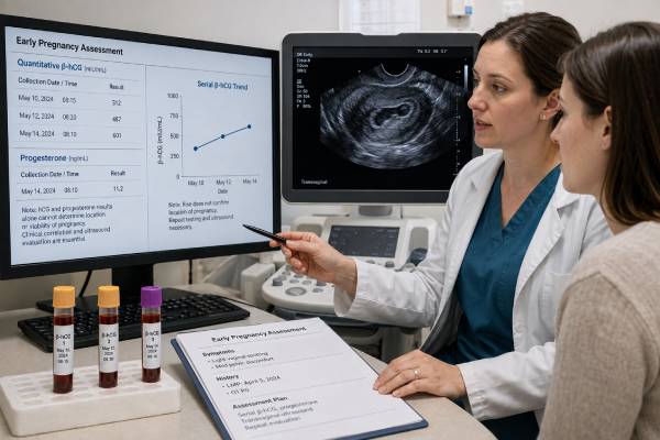
An ectopic pregnancy blood test panel usually centers on serial quantitative human chorionic gonadotropin (hCG), with progesterone sometimes added as a supporting marker. These tests help clinicians manage a positive pregnancy test when pain, bleeding, risk factors, or ultrasound findings leave the pregnancy’s location uncertain. They cannot show where implantation occurred. A transvaginal ultrasound, clinical examination, symptoms, and repeated assessment are required to distinguish an early intrauterine pregnancy from a resolving pregnancy or an ectopic pregnancy. The hCG pattern may rise too slowly, plateau, or fall in an ectopic pregnancy, but some ectopic pregnancies initially rise within ranges seen in viable uterine pregnancies. Progesterone can suggest whether a pregnancy is likely viable, yet it cannot identify location. Because a tubal pregnancy can rupture at a low or declining hCG level, a reassuring number must never override severe symptoms. The panel is most useful as part of a defined follow-up pathway with clear timing and emergency instructions.
- Serial quantitative hCG is more informative than one result, commonly using samples about 48 hours apart.
- A slow rise or plateau increases concern for ectopic pregnancy but does not prove the diagnosis.
- Falling hCG suggests a resolving pregnancy, yet follow-up may be needed until the location is known or hCG is negative.
- Progesterone may help assess viability but cannot distinguish an ectopic pregnancy from a failing intrauterine pregnancy.
- The ultrasound “discriminatory level” is not an automatic treatment threshold and should be applied conservatively.
- Severe one-sided pain, shoulder pain, fainting, weakness, or heavy bleeding requires emergency care regardless of laboratory results.
Table of Contents
- What the Panel Can and Cannot Show
- When Ectopic Testing Is Needed
- How hCG Trends Are Used
- The Role of Progesterone
- Combining Blood Tests With Ultrasound
- Common Result Patterns
- Factors That Complicate Interpretation
- Follow-Up, Treatment, and Emergencies
What the Panel Can and Cannot Show
There is no single blood test that diagnoses ectopic pregnancy. The laboratory component usually includes:
- Quantitative hCG, which measures the amount of pregnancy hormone in serum
- Repeat quantitative hCG, often about 48 hours later, to calculate the direction and percentage of change
- Serum progesterone, used in some services to estimate the likelihood that a pregnancy is viable
- Complete blood count, when bleeding is significant or anemia is possible
- Blood type and Rh status, when bleeding or treatment planning makes this relevant
- Kidney and liver tests, if methotrexate treatment is being considered
Only hCG and progesterone are pregnancy-hormone markers. The other tests assess safety, blood loss, or treatment suitability.
An hCG result confirms that trophoblastic tissue is producing hormone. It cannot tell whether that tissue is in the uterine cavity, a fallopian tube, the cervix, a cesarean scar, the ovary, or another site. Progesterone also cannot identify location. This is why the term pregnancy of unknown location is used when hCG is positive but transvaginal ultrasound has not yet shown either an intrauterine or ectopic pregnancy.
A pregnancy of unknown location is a temporary clinical category. It may later prove to be:
- A normal intrauterine pregnancy that was too early to see
- A nonviable intrauterine pregnancy
- A resolving pregnancy that has already passed
- An ectopic pregnancy
The panel helps guide the next test and its timing. It should never be used to reassure someone with signs of internal bleeding or to justify ending a potentially viable pregnancy from one ambiguous value.
When Ectopic Testing Is Needed
Evaluation is appropriate when a person has a positive pregnancy test plus symptoms or circumstances that raise concern. Symptoms can include pelvic or abdominal pain, vaginal bleeding, spotting, rectal pressure, dizziness, shoulder-tip pain, weakness, or faintness. Some ectopic pregnancies initially cause mild or no symptoms, so risk and ultrasound findings also matter.
Risk factors include a prior ectopic pregnancy, previous tubal surgery, pelvic inflammatory disease, infertility, assisted reproduction, smoking, and pregnancy with an intrauterine device in place. However, many patients diagnosed with ectopic pregnancy have no recognized risk factor.
Testing may also begin after an ultrasound shows no definite intrauterine pregnancy. Whether that is concerning depends on the true gestational age, the quality of the scan, the hCG concentration, and whether the uterus or adnexa contain suspicious findings. A scan performed before about five weeks from the last menstrual period may simply be too early, especially if ovulation occurred late.
The clinician typically records the exact location and severity of pain, vital signs, bleeding amount, abdominal findings, last menstrual period, fertility treatment dates, prior hCG results, and previous ectopic history. This clinical information determines whether outpatient serial testing is safe or immediate intervention is required.
A person who is hemodynamically unstable, has severe pain, shows signs of significant internal bleeding, or has an ultrasound strongly suggesting rupture does not wait for a 48-hour hormone trend. Emergency imaging, gynecologic consultation, and surgery may be necessary.
How hCG Trends Are Used
Quantitative hCG is usually reported in mIU/mL or IU/L. The first measurement establishes a baseline. The second shows whether the level is rising, falling, or changing very little.
In a potentially viable intrauterine pregnancy, the minimum expected 48-hour rise becomes smaller as the starting hCG increases. Approximate lower limits commonly used are around 49% when the initial value is below 1,500 mIU/mL, 40% from 1,500 to 3,000 mIU/mL, and 33% above 3,000 mIU/mL. These are not “normal ranges” and do not require every healthy pregnancy to follow the same curve.
A rise below the expected minimum is abnormal enough to warrant closer assessment. It may reflect ectopic pregnancy, an early intrauterine pregnancy that is not developing normally, inaccurate timing, or occasionally a viable pregnancy with an atypical rise. A doubling-time calculation can summarize the trend, but clinicians often focus on percent change and ultrasound.
A plateau means little change over repeated measurements. Depending on the service, a change of less than roughly 10–15% over 48 hours may be described as plateauing. This pattern is concerning for persistent pregnancy tissue, including ectopic pregnancy, but laboratory variation should be considered when the difference is small.
A falling hCG generally indicates that the pregnancy is not continuing. The rate of decline matters. A decline that is slower than expected can suggest ectopic pregnancy or retained tissue. Even a substantial decline does not locate the pregnancy, and follow-up may continue until hCG is undetectable or a clear diagnosis is established.
An apparently normal rise cannot exclude ectopic pregnancy. A minority of ectopic pregnancies produce increases that overlap with viable intrauterine pregnancies. Symptoms and ultrasound therefore remain essential throughout the process.
The Role of Progesterone
Progesterone supports the endometrium and early pregnancy. A single serum measurement can help estimate whether a pregnancy is likely viable, particularly when the ultrasound is inconclusive. Its main limitation is that low concentrations occur in both ectopic pregnancies and failing intrauterine pregnancies.
Very low progesterone values, often around 5–6 ng/mL or lower in untreated pregnancies, are strongly associated with nonviability. Values above roughly 20–25 ng/mL are more compatible with a viable pregnancy. The broad middle range remains uncertain, and exact thresholds differ among assays and studies.
Progesterone does not answer the most urgent location question. A very low value does not prove an ectopic pregnancy. A high value reduces the likelihood of a failing pregnancy but does not completely rule out ectopic implantation. For this reason, some clinical guidelines do not recommend using progesterone to diagnose ectopic pregnancy in a pregnancy of unknown location.
Supplementation further limits interpretation. Vaginal, oral, intramuscular, or subcutaneous progesterone can raise serum levels to different degrees. A patient who conceived through fertility treatment should provide the exact product, route, dose, and timing of the most recent dose. The measured level may primarily show medication exposure.
Progesterone treatment should not be started merely to “fix” an unexplained low number without confirming the pregnancy situation. It cannot move an ectopic pregnancy to the uterus. Where progesterone is recommended for threatened miscarriage, protocols generally require an ultrasound-confirmed intrauterine pregnancy and a relevant miscarriage history.
Combining Blood Tests With Ultrasound
Transvaginal ultrasound directly assesses pregnancy location. The scan looks for an intrauterine gestational sac with expected structures, an adnexal mass separate from the ovary, a tubal ring, an extrauterine embryo, and free fluid that may represent bleeding.
Clinicians may compare the scan with the hCG concentration. The “discriminatory level” describes a concentration above which an intrauterine gestational sac is usually expected to be visible on high-quality transvaginal ultrasound. In modern practice, a conservative value as high as about 3,500 mIU/mL may be used when avoiding interruption of a desired viable pregnancy is important.
This is not a rigid threshold. Visibility depends on:
- Exact gestational age and ovulation timing
- Ultrasound equipment and operator experience
- Uterine fibroids, adenomyosis, or anatomical variation
- Multiple gestation
- Laboratory assay differences
- Whether the scan is transvaginal or transabdominal
An empty uterus above a chosen threshold increases concern but does not prove ectopic pregnancy. If the patient is stable and no definitive ectopic is seen, repeating ultrasound and hCG may be safer than immediate treatment.
Likewise, a small fluid collection in the uterus is not always a true gestational sac. A definitive intrauterine pregnancy generally requires appropriate structures such as a yolk sac or embryo within the sac. A heterotopic pregnancy, where intrauterine and ectopic pregnancies coexist, is rare in spontaneous conception but more common after assisted reproduction. Seeing an intrauterine pregnancy does not completely exclude an ectopic pregnancy when symptoms or fertility treatment raise concern.
Common Result Patterns
| Laboratory pattern | Possible interpretation | Usual next step |
|---|---|---|
| hCG rises at or above the expected minimum; progesterone relatively high | Potentially viable pregnancy, but ectopic pregnancy not fully excluded | Repeat ultrasound at the appropriate time |
| hCG rises slowly; progesterone low or intermediate | Ectopic or failing intrauterine pregnancy possible | Close serial hCG, ultrasound, symptom monitoring |
| hCG plateaus | Persistent pregnancy tissue or ectopic pregnancy concern | Specialist review and management planning |
| hCG falls quickly; no concerning symptoms | Resolving pregnancy likely | Follow until resolution or location is established |
| hCG falls slowly; pain or bleeding continues | Ectopic pregnancy remains possible | Urgent reassessment, repeat scan and laboratory tests |
| Any hormone pattern with severe pain or instability | Possible rupture or significant bleeding | Emergency care immediately |
These patterns guide care rather than determine it. The clinician may use a formal hCG ratio, prediction model, or local pregnancy-of-unknown-location protocol. Results should not be interpreted with a general internet calculator when the exact sample interval, assay, symptoms, and ultrasound findings are unknown.
Factors That Complicate Interpretation
Several situations make hormone trends less straightforward.
Late ovulation or uncertain dates: A pregnancy may be younger than the last menstrual period suggests. A low hCG and empty uterus may be normal at that stage.
Recent hCG injection: Fertility trigger medication can remain detectable and create an initial positive result. The clinic’s scheduled test date accounts for this better than an early home test.
Multiple pregnancy: hCG may be higher on average, but concentration cannot reliably determine the number or location of embryos.
Vanishing twin: Loss of one implanted embryo can alter hCG patterns and make an otherwise continuing pregnancy look atypical.
Recent miscarriage, abortion, or delivery: Residual hCG can persist for days or weeks. A new rise after falling may indicate a new pregnancy, retained tissue, or another cause.
Assay differences: Results from different laboratories may not be directly comparable. Small changes can reflect analytical variation.
Interfering antibodies: Rare serum assay interference may produce persistent low-positive hCG when urine testing is negative. Repeat testing on another platform can help.
Heterotopic pregnancy: An intrauterine gestation can coexist with an ectopic gestation, especially after embryo transfer. Persistent focal pain still deserves assessment.
A beta-hCG result should therefore be read as one part of a changing clinical picture, not a permanent label.
Follow-Up, Treatment, and Emergencies
Stable patients may be followed as outpatients with written instructions and reliable access to care. A typical plan includes repeat quantitative hCG in about 48 hours, repeat transvaginal ultrasound at a clinically appropriate interval, and immediate reassessment if symptoms worsen.
When ectopic pregnancy is confirmed or strongly suspected, management depends on stability, pain, hCG level and trend, ultrasound findings, pregnancy size, fetal cardiac activity, medical conditions, and ability to return for follow-up. Options include expectant management, methotrexate, or surgery. Hormone results help determine eligibility but do not replace individual assessment.
Expectant management is limited to selected stable patients with low and declining hCG and no concerning findings. Methotrexate requires a reliable diagnosis, exclusion of a viable intrauterine pregnancy, medical screening, and repeated hCG testing afterward. Surgery is required for rupture, instability, substantial bleeding, certain ultrasound findings, or when medical follow-up is unsafe or unsuitable.
After treatment, hCG is followed until it reaches the service’s negative or nonpregnant range. Pain can occur during resolution, but worsening or severe pain still requires urgent review because rupture can occur during follow-up.
Call emergency services or go to an emergency department for sudden severe abdominal or pelvic pain, shoulder-tip pain, fainting, collapse, confusion, marked weakness, rapid breathing, pale or clammy skin, or heavy bleeding. Do not drive yourself if you feel faint or unstable. A low, declining, or previously “reassuring” hCG does not make these symptoms safe to watch at home.
Pregnancy of unknown location deserves particular caution because it is a temporary status, not a final diagnosis. The patient has a positive pregnancy test, but transvaginal ultrasound does not yet show a definite intrauterine or ectopic pregnancy. A very early intrauterine pregnancy, a completed or ongoing miscarriage, and an ectopic pregnancy can all look the same at the first visit. The purpose of serial testing is to move safely from uncertainty to a confirmed location or documented resolution.
Many services use a structured 48-hour pathway. A substantial hCG rise makes a developing intrauterine pregnancy more likely and leads to a repeat scan at an appropriate interval. A large decline suggests a noncontinuing pregnancy, but follow-up continues until the protocol endpoint. A plateau, small rise, or small decline warrants prompt senior review because ectopic pregnancy becomes more likely. Exact thresholds differ, so the local pathway should be followed rather than mixing cutoffs from different guidelines.
Progesterone can estimate the probability of viability but cannot distinguish ectopic from failed intrauterine pregnancy. Very low values occur in both. For that reason, a low progesterone result must never be used to reassure a patient that the pregnancy is “just a miscarriage” when the location has not been established. Conversely, a higher value cannot exclude ectopic implantation.
Management choices after an ectopic diagnosis depend on symptoms, hemodynamic stability, hCG concentration and trend, ultrasound findings, ectopic size and cardiac activity, medical contraindications, and the patient’s ability to return for follow-up. Expectant management may be appropriate for selected stable patients with low and declining hCG. Methotrexate requires eligibility screening and scheduled monitoring. Surgery may be needed for rupture, instability, significant pain, high-risk imaging findings, or when other approaches are unsuitable.
Methotrexate follow-up illustrates why ordinary doubling rules should not be applied to treatment monitoring. hCG may rise initially after the dose. Clinicians commonly compare values on defined treatment days and expect a specified decline before moving to weekly testing. A patient remains at risk for rupture until the ectopic pregnancy has resolved, even when the number is falling.
Blood group and Rh status may be reviewed when bleeding or treatment occurs, according to gestational age and local anti-D guidance. A complete blood count can help assess anemia when bleeding is significant. Kidney and liver tests may be required before methotrexate. These tests support treatment safety but do not diagnose ectopic pregnancy themselves.
The emotional burden of serial assessment is substantial. Patients may spend days between blood draws without knowing whether the pregnancy is viable or where it is located. Good care includes written emergency instructions, a direct contact route, realistic explanations of uncertainty, and a scheduled next step rather than an open-ended request to “repeat the test.”
Immediate evaluation is essential for worsening one-sided pain, shoulder-tip pain, fainting, collapse, marked dizziness, breathlessness, or heavy bleeding. A normal blood pressure at an earlier visit, a low hCG, or a recent decline does not protect against later rupture. Clinical deterioration always overrides the planned laboratory schedule.
Ultrasound terminology can help patients understand why follow-up continues. A “pseudosac” or fluid collection in the uterus is not equivalent to a definite gestational sac with a yolk sac or embryo. An adnexal mass may be suspicious without being conclusive. Free fluid can be physiologic in small amounts or concerning when extensive and accompanied by pain or instability. The radiology report must be interpreted with symptoms and hCG rather than as a list of isolated phrases.
Risk factors such as prior ectopic pregnancy, tubal surgery, pelvic inflammatory disease, smoking, an intrauterine device in place at conception, or assisted reproduction increase suspicion, but many ectopic pregnancies occur without any known risk factor. Absence of risk history therefore cannot justify less follow-up when the location is unknown.
Future fertility questions are best addressed after the acute episode. Treatment choice, condition of the opposite tube, underlying infertility, and prior history influence recurrence risk and time to try again. Patients treated with methotrexate receive specific advice about folate, alcohol, medications, sun exposure, and delaying conception. Those instructions should come from the treating service because dose and local policy matter.
A written record can reduce errors during repeated visits. Keep the hCG values with collection times, ultrasound dates, symptoms, and the name of the reviewing service. If care moves between an emergency department, laboratory, and early-pregnancy clinic, bring the complete sequence rather than the latest value alone. This helps clinicians recognize a plateau, verify that follow-up is complete, and avoid repeating an already answered test.
When discharge from follow-up is appropriate, the service should confirm the endpoint explicitly. A negative test, resolved hCG, confirmed intrauterine pregnancy, or completed treatment each closes a different pathway. Patients should not assume that silence after a blood draw means ectopic risk has ended.
References
- Ectopic pregnancy and miscarriage: diagnosis and initial management 2026 (Guideline)
- β-Human Chorionic Gonadotropin Dynamics in Early Gestation 2024 (Review)
- Exploring Progesterone Deficiency in First-Trimester Miscarriage and the Impact of Hormone Therapy on Foetal Development: A Scoping Review 2024 (Scoping Review)
- Early Pregnancy Diagnosis 2023 (Review)
- Human Chorionic Gonadotropin and Early Embryogenesis: Review 2022 (Review)
Disclaimer
Blood tests cannot rule out or confirm ectopic pregnancy without clinical and ultrasound assessment. Follow the testing schedule and emergency instructions provided by the treating service. Severe pain, fainting, shoulder pain, weakness, or heavy bleeding needs immediate emergency evaluation.