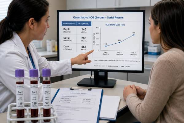
An hCG doubling time test is not a separate laboratory assay. It is an interpretation of two or more quantitative serum human chorionic gonadotropin measurements collected at known intervals, usually about 48 hours apart. The calculation shows how quickly hCG is rising or falling in early pregnancy. Although patients often hear that hCG should double every two days, healthy pregnancies do not all follow that exact rule. The minimum expected rise becomes slower as the starting concentration increases, and an appropriate rise cannot completely exclude ectopic pregnancy. A slow rise, plateau, or fall raises concern for a nonviable or ectopic pregnancy but does not identify the pregnancy’s location. Ultrasound and symptoms remain essential. The trend is most useful before a definitive intrauterine pregnancy is visible, especially in pregnancy of unknown location, bleeding, pain, or fertility-treatment follow-up. Results should be calculated from the actual collection times and preferably the same laboratory rather than judged from home pregnancy-test line darkness.
- Doubling time is calculated from serial quantitative hCG results; there is no separate “doubling” blood assay.
- Exact doubling in 48 hours is not required for every viable pregnancy.
- Minimum expected rises are lower when the initial hCG concentration is higher.
- A reassuring rise does not prove the pregnancy is inside the uterus.
- A falling level does not end ectopic precautions until clinical follow-up is complete.
Table of Contents
- What an hCG Doubling Time Test Is
- Why Clinicians Follow Serial hCG
- How to Calculate Rise and Doubling Time
- Expected Early-Pregnancy Trends
- Slow-Rising, Plateauing, and Falling Results
- hCG Trends and Ultrasound Timing
- Special Situations and Common Mistakes
- Follow-Up Questions and Emergency Symptoms
What an hCG Doubling Time Test Is
Human chorionic gonadotropin is produced mainly by placental trophoblast cells after implantation. A quantitative serum hCG assay reports its concentration, usually in mIU/mL or IU/L. When at least two results are available, clinicians can calculate the percentage change and an estimated doubling or halving time.
The phrase “doubling time test” can be misleading because the laboratory does not directly measure a rate. It measures separate concentrations. The rate is calculated afterward using the values and exact number of hours between blood draws.
Serial hCG is most informative in the early first trimester before ultrasound has established location and viability. Once a viable intrauterine pregnancy with appropriate cardiac activity is confirmed, repeated hCG usually adds little and may create confusion because the rise naturally slows.
A trend provides probability, not proof. Trophoblastic tissue in an ectopic pregnancy can produce a rise that overlaps with viable intrauterine pregnancy. Conversely, a small proportion of viable pregnancies may rise below commonly used minimum thresholds. Clinical decisions should not depend on a single arithmetic rule when ultrasound or follow-up can clarify the diagnosis.
The calculation is different from a qualitative hCG test, which reports positive or negative, and from home-test “line progression.” Consumer test lines are affected by urine concentration, lighting, device design, and saturation. They are not calibrated to measure doubling time.
For reliable comparison, use quantitative serum results from the same laboratory when possible. Assay differences, rounding, and collection intervals can materially alter the calculated rate, especially at low concentrations.
Why Clinicians Follow Serial hCG
Serial hCG is commonly ordered when an early pregnancy cannot yet be fully assessed by ultrasound. Typical reasons include vaginal bleeding, pelvic pain, uncertain dates, prior ectopic pregnancy, an inconclusive scan, or a pregnancy of unknown location.
A pregnancy of unknown location means the pregnancy test is positive but ultrasound does not yet show a definite intrauterine or ectopic pregnancy. It is a temporary classification with three broad possibilities: a very early intrauterine pregnancy, a failing or completed intrauterine pregnancy, or an ectopic pregnancy. The hCG trend helps plan the next ultrasound and level of clinical review.
Fertility clinics may follow hCG after embryo transfer. The scheduled initial test avoids confusion from an hCG trigger injection, and a repeat value can provide early information about progression. The same minimum-rise principles may be considered, but clinic protocols and exact transfer dates matter.
After suspected or confirmed miscarriage, serial testing may be used when ultrasound does not document completion or when the pregnancy location was never established. A falling trend supports resolution, but follow-up must continue to the protocol endpoint because ectopic pregnancy can also decline.
After treatment for ectopic pregnancy, clinicians use condition-specific monitoring schedules. For example, methotrexate follow-up may compare defined days rather than relying on ordinary 48-hour doubling rules. The treating service should provide exact dates and emergency instructions.
Serial hCG is not generally required in an uncomplicated pregnancy solely for reassurance. The ranges are broad, and repeated testing can produce anxiety from minor variations. Once ultrasound can answer the clinical question more directly, imaging becomes the preferred tool.
The test is valuable when it changes management: determining when to repeat ultrasound, identifying a trend that needs urgent review, confirming resolution, or monitoring treatment. It is less useful when numbers are collected without a defined endpoint.
How to Calculate Rise and Doubling Time
The simplest calculation is the percentage rise:
(new hCG − initial hCG) ÷ initial hCG × 100.
If hCG increases from 500 to 800 mIU/mL, the rise is:
(800 − 500) ÷ 500 × 100 = 60%.
The percentage decline uses the same formula, producing a negative result, or can be stated as the amount lost divided by the initial value. A drop from 1,000 to 600 is a 40% decline.
Doubling time uses logarithms because exponential growth is not linear:
Doubling time = elapsed time × ln(2) ÷ ln(new value ÷ initial value).
For example, if hCG rises from 500 to 800 over exactly 48 hours, the estimated doubling time is about 71 hours. A value does not need to double during the collection interval for a doubling time to be calculated.
Actual elapsed time matters. Samples drawn Monday at 8 a.m. and Wednesday at 4 p.m. are 56 hours apart, not 48. Comparing the raw percentage rise with a 48-hour threshold without adjusting for time can misclassify the trend.
Clinicians often use validated minimum percentage rises rather than calculate a formal doubling time. This approach is easier and better aligned with evidence showing that expected growth depends on the initial concentration.
Online calculators can perform the arithmetic, but they cannot assess symptoms, laboratory differences, ultrasound, or pregnancy location. A mathematically reassuring result is not a medical clearance. The ordering clinician should interpret the trend.
When results come from different laboratories, avoid overprecision. A reported doubling time of 68 versus 72 hours may reflect assay variation rather than a biologically meaningful difference. Trends are interpreted as part of a range, not to the nearest minute.
Expected Early-Pregnancy Trends
Early hCG commonly rises rapidly, but the expected minimum rise slows as the starting level increases. Research-based approximate lower boundaries for a viable intrauterine pregnancy over 48 hours are:
| Initial hCG concentration | Approximate minimum 48-hour rise |
|---|---|
| Below 1,500 mIU/mL | About 49% |
| 1,500–3,000 mIU/mL | About 40% |
| Above 3,000 mIU/mL | About 33% |
These figures do not mean that every result above the boundary is normal or every result below it is nonviable. They describe statistical patterns and are used to identify pregnancies that need closer assessment.
An older rule expected a 66% rise or exact doubling in 48 hours. Applying that rule rigidly can incorrectly classify viable pregnancies, especially at higher starting levels. Modern care favors more conservative minimum-rise thresholds and ultrasound confirmation.
NICE uses a practical pregnancy-of-unknown-location pathway in which a rise greater than 63% over 48 hours suggests a developing intrauterine pregnancy, while still acknowledging that ectopic pregnancy cannot be excluded. A decline greater than 50% suggests the pregnancy is unlikely to continue. Intermediate changes receive prompt clinical review. Local protocols may use other evidence-based boundaries.
hCG rise naturally slows near the end of the first trimester. Levels often peak around 8 to 11 weeks, then decline and stabilize. Doubling-time calculations are therefore not meaningful throughout the entire pregnancy.
Embryo-transfer dating can make timing more certain than menstrual dating, but biological variability remains. Twin pregnancies may have higher concentrations on average, yet their doubling patterns overlap with singleton pregnancies. The trend cannot determine fetus number.
A normal trend is encouraging only within context. If severe pain or bleeding occurs, urgent assessment is required even when the increase exceeds a minimum threshold.
Slow-Rising, Plateauing, and Falling Results
A rise below the expected minimum increases concern for a nonviable intrauterine pregnancy or ectopic pregnancy. It does not distinguish between them. Repeat ultrasound, additional hCG measurements, and clinical assessment are usually required before a definitive diagnosis.
A plateau means little change across serial results. Laboratories and protocols define it differently, but a persistently flat pattern is abnormal in most early-pregnancy settings. It is particularly concerning for ectopic pregnancy or retained trophoblastic tissue.
A falling hCG generally indicates that the pregnancy is not continuing. The decline may follow a chemical pregnancy, miscarriage, resolving ectopic pregnancy, or treatment. The speed of decline can help determine whether follow-up is proceeding as expected.
The dangerous misconception is that falling hCG makes ectopic pregnancy harmless. Rupture can occur at low and declining levels. Until the location is known or the clinician confirms resolution, patients must retain emergency precautions.
A trend can also be erratic. Small apparent rises and falls may reflect assay variation, different laboratories, or samples drawn too close together. Repeat testing at an appropriate interval can clarify the direction.
No irreversible treatment should be based only on a borderline trend when a potentially viable intrauterine pregnancy remains possible and the patient is stable. Guidelines emphasize conservative ultrasound criteria and appropriate repeat intervals to avoid misdiagnosis.
When a slow rise is found, ask what the starting level was, how many hours elapsed, whether the same laboratory was used, and what ultrasound showed. A 35% rise may be more concerning at an initial value of 500 than at a value above 3,000, but neither result is interpreted without the complete picture.
hCG Trends and Ultrasound Timing
Transvaginal ultrasound is the only routine test that can directly identify pregnancy location. hCG helps determine when an ultrasound may be informative, but there is no absolute concentration at which every normal intrauterine pregnancy must be visible.
The “discriminatory level” is an approximate threshold above which a gestational sac is usually expected with high-quality transvaginal imaging. It varies by equipment, operator, assay, multiple gestation, and uterine anatomy. A conservative level as high as about 3,500 mIU/mL may be used when avoiding interruption of a potentially viable pregnancy is important.
If hCG is below the local threshold and ultrasound is nondiagnostic, a stable patient may have repeat hCG in about 48 hours and another scan later. If hCG is above the threshold with no intrauterine pregnancy, ectopic pregnancy and pregnancy loss become more concerning, but expert review is still needed.
Ultrasound findings should be interpreted by developmental criteria, not hCG alone. A gestational sac, yolk sac, embryo, and cardiac activity appear in sequence, with normal variation. An incorrect last menstrual period can make a normal early scan appear delayed.
Once a definite intrauterine pregnancy is visible, serial hCG becomes less useful. If viability remains uncertain, a repeat ultrasound after the recommended interval is generally more definitive than continued doubling calculations.
An intrauterine pregnancy usually excludes ectopic pregnancy, but heterotopic pregnancy is possible, particularly after assisted reproduction. Persistent one-sided pain or concerning adnexal findings require evaluation despite an intrauterine sac.
The safest pathway combines symptoms, hemodynamic stability, hCG trend, and ultrasound. The hormone should guide timing and probability, not replace imaging.
Special Situations and Common Mistakes
Fertility medication containing hCG can remain detectable after an ovulation trigger. Testing before the clinic’s scheduled date may measure the injection rather than pregnancy hormone. Later trends are interpreted according to the treatment protocol.
After embryo transfer, patients sometimes compare their initial level with online charts for a particular “days post transfer” date. Laboratory methods, embryo age, implantation timing, and pregnancy number create wide overlap. The trend and ultrasound are more useful than ranking one value.
Multiple gestation may produce higher average hCG, but neither a high starting value nor fast doubling proves twins. Conversely, a normal singleton-range value does not exclude twins.
Vanishing twin can alter early levels and prenatal screening markers. hCG may initially rise strongly and later slow without the remaining pregnancy necessarily being abnormal. Ultrasound provides the relevant diagnosis.
Common interpretation mistakes include:
- using qualitative home-test lines as quantitative data;
- comparing results from different laboratories without caution;
- ignoring the exact time between samples;
- expecting a 100% rise at every starting concentration;
- applying early doubling rules after the first-trimester peak;
- assuming a good rise excludes ectopic pregnancy;
- assuming a decline eliminates rupture risk;
- dating pregnancy from one hCG value.
Rare assay interference can produce persistent low serum hCG. Heterophile antibodies, pituitary hCG near menopause, and other sources are considered when the pattern does not fit pregnancy. A laboratory may compare urine, use another assay platform, or perform dilution and blocking studies.
Extremely high hCG can rarely create a hook effect in some assays, yielding an unexpectedly low result. Dilution testing resolves the issue when clinical findings suggest molar pregnancy or another very high-hCG condition.
Follow-Up Questions and Emergency Symptoms
A useful follow-up plan is specific. It states when the next blood draw will occur, whether repeat ultrasound is scheduled, what trend the clinician expects, and which symptoms require immediate care. Patients should not be asked to monitor portal numbers without access to clinical interpretation.
Questions to ask include:
- What were the exact collection times and percentage change?
- Was the same laboratory used?
- Which minimum-rise or decline framework is being applied?
- Is the pregnancy location known?
- When should ultrasound be repeated?
- What is the endpoint for blood testing?
- Could fertility medication or a recent pregnancy affect the value?
- Which symptoms mean I should go to emergency care now?
Seek urgent evaluation for severe or worsening pelvic or abdominal pain, one-sided pain, shoulder-tip pain, fainting, marked dizziness, weakness, shortness of breath, heavy bleeding, or signs of shock. Do not wait for the next hCG test when these occur.
For stable patients, uncertainty can last several days. This waiting period is emotionally difficult. Clinicians should explain probabilities carefully, avoid promising an outcome from one trend, and provide written safety instructions and a clear contact route.
hCG does not have to double perfectly. Its change should be judged against the initial level, elapsed time, ultrasound, and symptoms. A trend can identify concern and guide follow-up, while definitive conclusions about location and viability generally come from imaging and the complete clinical course.
Mathematical precision can create false certainty. Doubling-time formulas assume exponential change between two measurements, but real hCG production is not observed continuously and may not follow a perfect curve. Rounding by the laboratory can substantially alter a calculated rate when concentrations are low. For clinical decisions, a range and validated minimum rise are more useful than a doubling time reported to one decimal place.
Three or more values can reveal a pattern that two values miss. A rise may initially appear adequate and then plateau, or a small decline may accelerate. Additional testing is ordered only when it will clarify management; collecting many closely spaced samples can amplify analytic noise. The interval and endpoint should be defined by the care team.
The trend after a vanishing twin can be difficult to interpret because hormone production from one gestation stops while another continues. hCG may rise more slowly than expected or change irregularly. Ultrasound, rather than a calculator, determines the status of the remaining pregnancy.
In multiple gestation, higher absolute values do not translate into a special twin doubling rule. A normal twin pregnancy can overlap completely with singleton trends. The number and chorionicity of fetuses are ultrasound diagnoses.
A declining pattern after a known miscarriage should still be connected to symptoms. Retained tissue can cause prolonged bleeding or infection, while ectopic pregnancy remains possible if an intrauterine pregnancy was never confirmed. Fever, foul discharge, worsening pain, faintness, or heavy bleeding requires reassessment regardless of the calculated half-life.
Patients may find repeated testing emotionally consuming. It can help to avoid checking unreviewed portal results overnight, identify one clinician who will explain the sequence, and ask for probabilities rather than promises. A trend supports clinical interpretation; it does not measure effort, stress, or anything the patient did to cause the outcome.
Once ultrasound establishes appropriate intrauterine development, stopping serial hCG is usually a sign that a better diagnostic tool has taken over, not that monitoring has been abandoned. Later prenatal assessment follows anatomy, growth, maternal health, and fetal well-being rather than continued doubling.
The calculation should also account for the laboratory’s reporting limits. A result shown as “less than 5” is not an exact value of zero and cannot be used in a doubling formula. Likewise, values rounded to whole numbers create proportionally more uncertainty near the detection threshold. When the first result is borderline, clinicians may describe the change qualitatively until a clearly measurable concentration is available. If the next result becomes negative, recent loss or absence of an ongoing pregnancy is considered in context; if it rises, the serial pathway continues. Arithmetic is most reliable once both measurements are within the assay’s quantitative range.
If only one quantitative value is available, no doubling time exists yet. Estimating a rate from a qualitative test, home-test line, or an assumed starting value is mathematically and clinically invalid.
Clinicians may report a rise as “appropriate,” “suboptimal,” or “indeterminate” rather than giving a formal doubling time. These terms should be linked to a next action. An indeterminate trend is not the same as a confirmed loss; it means that additional observation or imaging is required before the outcome can be classified.
The final interpretation should name the pregnancy status only when evidence supports it: developing intrauterine pregnancy, pregnancy of unknown location, failing pregnancy, resolved pregnancy, or confirmed ectopic pregnancy. A doubling-time value by itself is not one of those diagnoses.
References
- β-Human Chorionic Gonadotropin Dynamics in Early Gestation 2024 (Review)
- Human Chorionic Gonadotropin 2025 (Review)
- Ectopic pregnancy and miscarriage: diagnosis and initial management 2026 (Guideline)
- Early Pregnancy Diagnosis 2023 (Review)
- Human Chorionic Gonadotropin and Early Embryogenesis: Review 2022 (Review)
Disclaimer
This article provides general education and cannot diagnose pregnancy viability, miscarriage, or ectopic pregnancy. Serial hCG trends must be interpreted by a clinician with exact timing, symptoms, ultrasound, and treatment history. Seek emergency care for severe pain, shoulder-tip pain, fainting, weakness, breathing difficulty, or heavy bleeding.