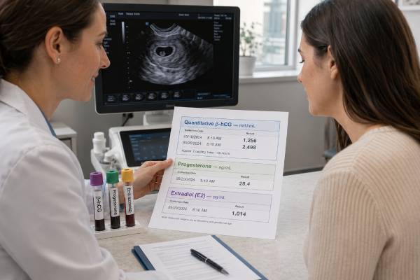
An early pregnancy blood test panel may measure human chorionic gonadotropin (hCG), progesterone, and estradiol to provide a broader view of implantation and early hormonal support. It is most often used when pregnancy dates are uncertain, symptoms such as bleeding or pain occur, or a fertility clinic is monitoring treatment. The three hormones do not carry equal diagnostic weight. Serial quantitative hCG and transvaginal ultrasound are usually the main tools for assessing early pregnancy development and location. Progesterone can add information about the likelihood of viability, while estradiol is more useful in assisted-reproduction protocols than in routine spontaneous pregnancy care. No single cutoff proves that a pregnancy is normal, ectopic, or destined to miscarry. Results overlap widely, change quickly, and can be altered by supplements or fertility medications. A panel is therefore best treated as a set of clues interpreted with gestational timing, symptoms, treatment history, and ultrasound rather than as a pass-or-fail test.
- Quantitative hCG is the panel’s main trend marker; two measurements about 48 hours apart are often more useful than one value.
- Progesterone supports the uterine lining, but one low result cannot determine where the pregnancy is located.
- Estradiol normally rises in pregnancy, yet routine estradiol monitoring has limited value outside fertility treatment.
- Hormone supplements can make progesterone and estradiol results look reassuring without proving embryo or placental development.
- Wide laboratory and gestational ranges mean that results should be compared with the same lab and the clinical timeline.
- Pain, fainting, shoulder pain, or heavy bleeding needs urgent assessment regardless of panel results.
Table of Contents
- What the Panel Includes
- Why Clinicians Order It
- Timing and Preparation
- Understanding Each Hormone
- Reading the Results Together
- Fertility Treatment Considerations
- Limitations and Common Mistakes
- Next Steps After Testing
What the Panel Includes
An early pregnancy hormone panel is not a universally standardized package. One clinic may order only quantitative hCG and progesterone, while another adds estradiol because the patient conceived through in vitro fertilization or is using hormonal support. The requisition should list the exact tests.
The typical components are:
- Quantitative hCG: Reports a numerical concentration in mIU/mL or IU/L. hCG is produced mainly by developing placental tissue after implantation.
- Progesterone: Reports a serum concentration, commonly in ng/mL or nmol/L. Early progesterone comes largely from the corpus luteum before placental production takes over.
- Estradiol: Reports a serum concentration, commonly in pg/mL or pmol/L. It is produced by the ovary early and increasingly through the fetoplacental unit as pregnancy progresses.
These tests answer different questions. hCG is useful because its rate of change can be followed over a short interval. Progesterone fluctuates in pulses and is often interpreted as a broad viability marker rather than a precise trend. Estradiol varies with ovarian activity, the number of stimulated follicles, placental development, and medication exposure.
The panel should not be confused with routine first-prenatal-visit laboratory testing, which usually includes blood type, Rh status, complete blood count, infection screening, urinalysis, and other tests. It is also different from first-trimester genetic screening, which may measure PAPP-A and free beta-hCG to calculate chromosome-condition risk.
A person who only needs pregnancy confirmation may not benefit from all three measurements. A standard hCG blood test may be enough. Broader testing becomes more reasonable when the clinical situation is uncertain or treatment decisions depend on hormone support.
Why Clinicians Order It
The panel is usually ordered to clarify a specific concern rather than simply to collect more data. Common reasons include:
- A positive pregnancy test with uncertain dates
- Vaginal bleeding or pelvic pain in the first trimester
- Previous ectopic pregnancy or repeated early pregnancy loss
- Pregnancy after ovulation induction, intrauterine insemination, or embryo transfer
- Monitoring after a fertility clinic changes progesterone or estradiol support
- A pregnancy of unknown location on transvaginal ultrasound
- Concern that hCG is rising more slowly than expected
- Follow-up after a very early loss or treatment for ectopic pregnancy
When ectopic pregnancy is possible, the panel cannot replace ultrasound. hCG helps determine when and how to repeat imaging, but it does not reveal location. Progesterone may be low in both ectopic and failing intrauterine pregnancies. Estradiol adds little location information.
For threatened miscarriage, clinicians may use hCG trends and ultrasound to determine whether development is continuing. Progesterone can sometimes help stratify risk, especially when ultrasound is inconclusive. The progesterone test in pregnancy should still be interpreted as supportive evidence, not a definitive miscarriage test.
In assisted reproduction, the reasons are different. The clinic may be checking whether a medication regimen produces adequate circulating hormone levels, whether a trigger injection has cleared, or whether an embryo-transfer pregnancy is producing an appropriate hCG signal. Protocol-specific targets can differ substantially from ranges used in spontaneous conception.
Routine serial hormone panels after a normally developing intrauterine pregnancy is visible are rarely useful. Ultrasound becomes a more direct measure of growth, heartbeat, and location, while hCG’s early rapid rise naturally slows.
Timing and Preparation
No fasting is generally required. A standard venous blood sample is collected, and the laboratory analyzes serum or plasma. Time of day is not critical for hCG, although progesterone can vary because secretion is pulsatile. If a clinic is comparing results closely, collecting at a similar time and using the same laboratory may reduce avoidable variation.
Accurate timing information is essential. Before the draw, record:
- The first day of the last menstrual period
- Typical cycle length
- Estimated ovulation date or positive ovulation-predictor result
- Insemination, egg-retrieval, and embryo-transfer dates
- Whether the embryo was transferred on day 3, day 5, or another day
- Date and dose of any hCG trigger injection
- Names, doses, routes, and timing of progesterone and estradiol medication
- Prior hormone results and ultrasound findings
Testing extremely early can create uncertainty. hCG may still be below the detection threshold before or just after implantation. A low first value can be completely appropriate if conception occurred later than assumed. Repeating hCG after about 48 hours often clarifies whether the pregnancy signal is developing.
Progesterone results can change with route of treatment. Vaginal progesterone may create high concentrations in uterine tissue without producing a proportionally high serum level. Intramuscular progesterone tends to produce higher and steadier blood concentrations. Oral formulations have different absorption and metabolism. A laboratory result should therefore be judged against the exact treatment protocol rather than a generic target found online.
Estradiol medication can also dominate the measured value. In a programmed frozen embryo transfer, the blood result may largely reflect the prescribed estradiol dose. It cannot independently show that the placenta is functioning normally.
Understanding Each Hormone
hCG: the main dynamic marker
A quantitative hCG result is most useful as part of a series. In early viable pregnancies, the level generally rises substantially every 48 hours, but exact doubling is not required. The minimum expected rise slows as the starting concentration increases. Rough guideposts often used in clinical assessment are about a 49% rise when the initial level is below 1,500 mIU/mL, 40% at 1,500–3,000 mIU/mL, and 33% above 3,000 mIU/mL over roughly 48 hours.
These thresholds identify pregnancies that deserve closer evaluation; they do not diagnose outcome. Some healthy pregnancies rise below a simple doubling rule, and some ectopic pregnancies rise at an apparently normal rate. The hCG doubling-time calculation is an aid, not a substitute for ultrasound.
A falling hCG level usually means trophoblastic tissue is no longer growing. Continued follow-up may still be required until the value becomes negative or the pregnancy location is established.
Progesterone: a broad viability clue
Progesterone transforms and stabilizes the endometrium, reduces uterine contractility, and supports immune tolerance. Serum levels overlap between healthy, ectopic, and failing pregnancies. Very low values are associated with nonviability, while higher values are more reassuring, but the middle range is large and inconclusive.
Clinicians sometimes use values below about 5–6 ng/mL as strongly concerning and values above about 20–25 ng/mL as more compatible with viability in untreated pregnancies. These are not universal cutoffs. Assay differences, gestational age, pulsatile secretion, and supplementation can shift interpretation.
Most importantly, progesterone cannot identify location. A low value does not prove ectopic pregnancy, and a high value does not rule it out.
Estradiol: context-dependent information
Estradiol helps regulate implantation, uterine blood flow, placental vascular development, and breast changes. It rises across pregnancy, but spontaneous pregnancies show broad ranges. A single low or high result has no widely accepted role in diagnosing miscarriage or ectopic pregnancy.
In fertility treatment, estradiol may help evaluate ovarian response before conception and medication exposure after transfer. Some clinics follow trends, but evidence does not support using a universal estradiol target to declare an early pregnancy viable. An estradiol test in pregnancy is best interpreted by the clinician who knows the stimulation or transfer protocol.
Reading the Results Together
The three results should be integrated rather than scored independently. The following patterns illustrate common interpretations, but they are not diagnostic rules.
| Pattern | Possible meaning | Typical follow-up |
|---|---|---|
| hCG rises appropriately; progesterone adequate; estradiol stable or rising | More reassuring, especially with known dates | Ultrasound at the appropriate gestational time |
| hCG rises slowly; progesterone low | Abnormal intrauterine pregnancy or ectopic pregnancy possible | Repeat hCG, transvaginal ultrasound, symptom review |
| hCG falls; progesterone falls | Pregnancy is likely resolving | Confirm location or follow to negative as advised |
| hCG plateaus; progesterone intermediate | Pregnancy of unknown location or ectopic pregnancy concern | Close serial testing and ultrasound |
| hCG rises; progesterone and estradiol high on supplements | Medication may explain steroid levels | Judge hCG and ultrasound separately; follow clinic protocol |
A single abnormal component should trigger questions, not an automatic conclusion. For example, a low progesterone result may be a consequence of a failing pregnancy rather than its cause. Giving progesterone can raise the measured level but cannot correct an ectopic location or a major chromosomal problem.
Likewise, a high estradiol result may reflect multiple stimulated follicles or prescribed tablets. It does not prove that placental development is stronger. A high initial hCG may reflect earlier dates or multiple gestation, but ultrasound is needed to count gestational sacs and embryos.
Symptoms can override an otherwise reassuring panel. An ectopic pregnancy may rupture with low or falling hCG, so stability cannot be judged from laboratory values alone.
Fertility Treatment Considerations
Assisted-reproduction pregnancies require protocol-specific interpretation. The date of fertilization is more certain, but medication exposure and embryo factors add complexity.
After an hCG trigger injection, residual medication can produce an early positive test. Clinics usually schedule the first beta-hCG far enough after retrieval or transfer to reduce this effect. Testing at home sooner may create a false impression of implantation.
Progesterone support is common after fresh and frozen embryo transfer. Clinics may use vaginal capsules or gel, intramuscular injections, subcutaneous formulations, or combinations. A serum threshold appropriate for one route may not fit another. Missed doses, incorrect administration, or poor absorption should be discussed promptly rather than corrected without guidance.
Estradiol is frequently prescribed in programmed frozen transfer cycles because there may be no functioning corpus luteum. In that setting, abruptly stopping medication can be harmful. Patients should continue the prescribed schedule until the fertility team provides a taper or stop date, even if a single hormone result appears high.
Early hCG levels after embryo transfer can help predict outcome at a population level, but there is substantial overlap. Embryo age, transfer day, fresh versus frozen cycle, preimplantation testing, singleton versus multiple implantation, and exact blood-draw timing all influence values. Comparing one result with another patient’s online result is rarely meaningful.
A fertility clinic may repeat hCG every 48–72 hours until the trend is clear and then perform ultrasound around the time a gestational sac and fetal structures should be visible. Once ultrasound provides direct evidence, hormone testing usually becomes secondary.
Limitations and Common Mistakes
The panel can produce false certainty when its limitations are ignored.
- Treating one value as a diagnosis: Wide normal ranges make isolated numbers difficult to interpret.
- Demanding exact hCG doubling: Viable pregnancies do not all follow a 48-hour doubling schedule.
- Using progesterone to locate pregnancy: Progesterone reflects viability imperfectly and cannot distinguish uterine from ectopic implantation.
- Ignoring medication: Supplemented progesterone and estradiol may mainly measure the prescription.
- Switching laboratories: Different assays may not be directly comparable, especially when changes are small.
- Repeating too soon: Short intervals magnify laboratory noise and increase anxiety without adding useful information.
- Using gestational ranges as strict boundaries: Published ranges are extremely broad and depend on dating method and population.
- Assuming a reassuring panel excludes emergency: Rupture risk depends on the ectopic pregnancy and symptoms, not on a safe hCG number.
Another common mistake is ordering all three tests without a plan for how each result will be used. More information can create more ambiguous findings. Before testing, it is reasonable to ask which result would change treatment, when it will be repeated, and when ultrasound will become the deciding test.
An abnormal panel does not identify the cause of pregnancy loss. Most early miscarriages involve chromosomal abnormalities that hormone testing cannot prevent or diagnose. Endocrine, anatomical, immune, and clotting evaluations are separate questions and are not automatically indicated after one loss.
Next Steps After Testing
The next step depends on symptoms and the combined pattern.
For a stable patient with an early positive hCG and no concerning symptoms, the clinician may repeat quantitative hCG in about 48 hours and schedule transvaginal ultrasound based on dates. If the trend is clearly falling, follow-up may continue until the result is negative or a known intrauterine loss is complete.
If the rise is slower than expected or no pregnancy is visible when it might reasonably be expected, care shifts to a pregnancy-of-unknown-location pathway. This usually includes serial hCG, repeat ultrasound, and clear emergency instructions. Treatment should not be based on a single hormone threshold when a viable intrauterine pregnancy remains possible.
Contact the treating clinic promptly for missed hormone doses, vomiting that prevents medication use, new bleeding, increasing pain, or uncertainty about whether to continue supplements. Do not start or stop progesterone or estradiol solely because of an online reference range.
Seek emergency care for severe or worsening one-sided pelvic pain, shoulder pain, fainting, collapse, marked dizziness, weakness, shortness of breath, pale or clammy skin, or heavy bleeding. These symptoms can indicate internal bleeding from ectopic pregnancy and require immediate evaluation even when recent hormone results seemed reassuring.
Panel interpretation becomes most useful when results are organized by the biological process they reflect. hCG is primarily a trophoblast signal and is best evaluated as a timed trend. Progesterone is a broad viability marker with substantial overlap and no ability to locate the pregnancy. Estradiol reflects ovarian and, later, placental steroid production, but its interpretation is highly dependent on fertility treatment and gestational timing. None of the three independently confirms an embryo or heartbeat.
Discordant patterns are common. A strong hCG rise with an intermediate progesterone value may still occur in a viable pregnancy, particularly when the sample was drawn at a different time of day. A high progesterone result after supplementation cannot prove that endogenous hormone production is adequate. Estradiol may look low after a programmed frozen embryo-transfer cycle if the sample timing or medication route differs from the clinic’s protocol. The correct response is usually to verify treatment details and prioritize the marker that answers the clinical question.
A useful timeline is more informative than a list of isolated values. Record the date and time of each draw, the laboratory, hCG concentration, progesterone and estradiol units, medication dose and route, bleeding or pain, and ultrasound findings. For assisted reproduction, include the trigger date, retrieval date, embryo age, and transfer date. This prevents a result from being compared with the wrong gestational day.
The panel should also have a stopping rule. Once ultrasound confirms a normally developing intrauterine pregnancy, repeated hCG usually adds little. Progesterone and estradiol monitoring may continue in some fertility protocols, but the clinic should state the target, frequency, and planned discontinuation of support. In spontaneous pregnancies, serial steroid testing without a defined action can increase anxiety and lead to unnecessary dose changes.
When results are concerning, clinicians should avoid treating the panel as a score in which one “good” hormone cancels one “bad” hormone. A low progesterone cannot be rescued mathematically by a high hCG, and a rising hCG cannot exclude ectopic pregnancy. The combined assessment is anatomical and clinical: symptoms determine urgency, ultrasound determines location and developmental findings, and blood tests show biochemical trends.
Patients can ask for the interpretation in plain terms: Is the pregnancy location known? Is the pattern reassuring, indeterminate, or concerning? Which result is driving that conclusion? What is the next test, and when will uncertainty be resolved? Clear answers are more helpful than a broad statement that the hormones are “normal.”
Medication changes are a common source of error. Patients may see a lower steroid value and increase progesterone or estrogen on their own, or skip a dose because the result seems high. Serum concentration does not directly measure endometrial exposure, especially with vaginal medication, and fertility protocols are built around specific routes and schedules. Dose changes should come from the prescribing clinic.
The panel also has limited value for predicting later pregnancy outcomes. A reassuring early profile does not screen for chromosome conditions, anatomy problems, preeclampsia, gestational diabetes, or preterm birth. Those questions are addressed through separate prenatal screening, ultrasound, maternal health assessment, and later surveillance. Early hormone results should be treated as stage-specific information rather than a certificate for the rest of pregnancy.
When a panel result is released in stages, wait for the clinician’s integrated review. One analyte may post before the others, and an automatically flagged value may be expected for the medication or gestational day. Partial portal data should not trigger self-treatment or cancellation of a planned scan.
References
- β-Human Chorionic Gonadotropin Dynamics in Early Gestation 2024 (Review)
- Exploring Progesterone Deficiency in First-Trimester Miscarriage and the Impact of Hormone Therapy on Foetal Development: A Scoping Review 2024 (Scoping Review)
- The pathophysiological role of estrogens in the initial stages of pregnancy: molecular mechanisms and clinical implications for pregnancy outcome from the periconceptional period to end of the first trimester 2023 (Review)
- Ectopic pregnancy and miscarriage: diagnosis and initial management 2026 (Guideline)
- Human Chorionic Gonadotropin and Early Embryogenesis: Review 2022 (Review)
Disclaimer
Early pregnancy hormone panels cannot independently confirm pregnancy location or viability. Results should be interpreted by a clinician who knows the timing, symptoms, ultrasound findings, and any fertility medications. Severe pain, fainting, shoulder pain, weakness, or heavy bleeding requires urgent medical care.