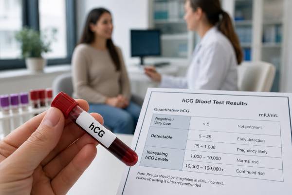
A human chorionic gonadotropin blood test detects hCG, a hormone made mainly by placental tissue after an embryo implants. Serum testing can identify very small amounts of hCG and may become positive before a urine pregnancy test. A qualitative blood test reports positive or negative, while a quantitative test gives a concentration in milli-international units per milliliter (mIU/mL). Most laboratories consider less than 5 mIU/mL negative and 25 mIU/mL or higher positive, with an intermediate zone that may require repeat testing, but each laboratory sets its own limits. One number cannot confirm that a pregnancy is healthy or located inside the uterus. When symptoms, dates, or ultrasound findings are uncertain, clinicians interpret hCG over time and pair it with transvaginal ultrasound. Fertility medicines containing hCG, a very recent pregnancy, rare antibody interference, and several medical conditions can also affect results. Severe pain, heavy bleeding, fainting, or shoulder pain requires urgent assessment regardless of the hCG value.
- Serum hCG may become detectable about 8–11 days after ovulation, although timing varies with implantation.
- Less than 5 mIU/mL is often negative; 5–24 mIU/mL is commonly indeterminate; 25 mIU/mL or more is often positive.
- A positive result confirms hCG is present but does not by itself prove a viable intrauterine pregnancy.
- Serial quantitative results are commonly compared about 48 hours apart when early pregnancy progress is uncertain.
- Fertility injections containing hCG and a recent pregnancy can cause a positive result without a new ongoing pregnancy.
Table of Contents
- What the hCG Blood Test Measures
- Qualitative and Quantitative hCG Tests
- When to Test and How to Prepare
- Positive, Negative, and Borderline Results
- hCG Levels and Early Pregnancy Trends
- Using hCG With Ultrasound
- False Results and Nonpregnancy Causes
- Follow-Up and Urgent Symptoms
What the hCG Blood Test Measures
Human chorionic gonadotropin is a glycoprotein hormone produced after implantation by trophoblast cells that later form much of the placenta. Its best-known early role is to stimulate the corpus luteum in the ovary so progesterone production continues. Progesterone supports the uterine lining until placental hormone production becomes established.
The hCG molecule has alpha and beta subunits. The alpha portion resembles subunits found in luteinizing hormone, follicle-stimulating hormone, and thyroid-stimulating hormone. Pregnancy assays use antibodies directed toward the beta portion or other hCG-specific sites to reduce cross-reaction with those hormones. Different platforms recognize different combinations of intact hCG and hCG variants, which is one reason results from separate laboratories may not be perfectly interchangeable.
hCG appears in maternal blood after implantation, not immediately after fertilization. Implantation timing varies. A blood test may detect hCG roughly 8 to 11 days after ovulation in some pregnancies, but a negative result that early can simply mean implantation occurred later or the concentration has not crossed the assay threshold.
Serum testing has several uses:
- confirming a suspected pregnancy;
- evaluating an early pregnancy with pain, bleeding, uncertain dates, or an inconclusive ultrasound;
- following hCG after miscarriage, ectopic pregnancy, abortion, or treatment of gestational trophoblastic disease;
- confirming pregnancy after assisted reproduction;
- occasionally evaluating hCG production from a tumor or other nonpregnancy source.
A standard serum hCG test is different from the free beta-hCG marker used in first-trimester screening. Prenatal chromosome screening converts free beta-hCG to MoM and combines it with other markers. A pregnancy-confirmation assay reports whether hCG is present or how much is present.
The test does not measure fetal heartbeat, chromosome number, placental location, or future pregnancy outcome. Those questions require ultrasound, other screening or diagnostic tests, and clinical follow-up.
Qualitative and Quantitative hCG Tests
A qualitative hCG blood test provides a binary or near-binary answer: positive or negative. It is useful when the immediate question is whether hCG is detectable. Some laboratories include an equivocal category. Because it does not provide a concentration, it cannot show the amount of change between samples.
A quantitative hCG blood test, sometimes called quantitative beta-hCG, reports a number in mIU/mL or IU/L. These units are numerically equivalent: 100 mIU/mL equals 100 IU/L. Quantitative testing is more useful when the clinician needs to compare serial values, correlate the result with ultrasound, or document that hCG has returned to a nonpregnant level.
| Test type | Typical report | Best use | Main limitation |
|---|---|---|---|
| Qualitative serum hCG | Positive, negative, or equivocal | Rapid pregnancy confirmation | Cannot calculate a rise or fall |
| Quantitative serum hCG | Numeric value in mIU/mL | Serial trends and ultrasound correlation | One value is rarely diagnostic |
| Urine hCG | Positive or negative | Convenient home or clinic testing | More affected by urine dilution and timing |
Blood testing is often more analytically sensitive than urine testing, but sensitivity alone does not guarantee a clinically useful answer. An extremely early positive can identify a biochemical pregnancy that later stops developing before ultrasound can show it. That information can be valuable in fertility treatment, yet it may also create uncertainty that requires repeat testing.
When serial comparison is planned, use the same laboratory whenever possible. Different assays may produce modestly different values from the same sample because they detect hCG forms differently. A small apparent change across two unrelated laboratories can reflect method variation rather than pregnancy biology.
Clinicians should order the test that answers the actual question. A qualitative test may be sufficient before an imaging study or medication. A quantitative hCG blood test is preferable for an early pregnancy of unknown location or when a numeric decline must be followed to resolution.
When to Test and How to Prepare
A blood test can become positive before the expected menstrual period, but testing later reduces the chance of a false-negative result. For spontaneous conception, a practical time is the day the period is expected or afterward. For fertility treatment, follow the clinic’s scheduled test date; testing early can detect injected hCG rather than pregnancy-produced hormone.
No fasting is needed. Food, water, prenatal vitamins, and ordinary activity do not change the result in a clinically meaningful way. The sample is drawn from a vein, usually in the arm. Minor bruising, soreness, and brief lightheadedness are the most common effects.
Tell the ordering clinician about:
- the first day of the last menstrual period and usual cycle length;
- the date of ovulation, insemination, egg retrieval, or embryo transfer when known;
- any hCG trigger injection or other fertility medicine;
- recent pregnancy, delivery, miscarriage, abortion, or ectopic pregnancy;
- current pain, bleeding, dizziness, or fainting;
- prior ectopic pregnancy or tubal surgery;
- a history of gestational trophoblastic disease;
- an unexpected positive hCG result when pregnancy is not possible.
An hCG trigger shot can remain detectable for days. The exact clearance depends on the dose, route, body size, kidney function, and assay. Fertility clinics therefore select a testing date that limits confusion and interpret the result in relation to the injection.
After a pregnancy ends, hCG does not become negative immediately. The time to clear depends on the starting concentration and whether pregnancy tissue remains. A positive test days or weeks later may represent expected decline, retained tissue, ectopic pregnancy, persistent trophoblastic disease, or a new conception. Serial measurement and clinical context distinguish these possibilities.
Results may be available within hours or by the next day. An emergency department may process hCG rapidly because it can influence imaging and treatment choices, but urgent symptoms should be assessed before waiting for a laboratory portal to update.
Positive, Negative, and Borderline Results
Laboratory cutoffs vary, so the reference interval printed on the report controls interpretation. A common framework is:
| Serum hCG result | Common laboratory interpretation | Usual next step when pregnancy status is uncertain |
|---|---|---|
| Less than 5 mIU/mL | Negative | Repeat only if testing was early or symptoms remain concerning |
| 5–24 mIU/mL | Borderline or indeterminate | Repeat quantitative hCG in about 48 hours |
| 25 mIU/mL or higher | Positive | Interpret with dates, symptoms, and ultrasound as appropriate |
A positive result means the assay detected hCG above its decision threshold. In a person who could be pregnant, pregnancy is the most likely explanation. It does not establish gestational age precisely, prove normal development, or show where implantation occurred.
A negative result means hCG was below the assay’s negative threshold at that moment. It usually excludes an established pregnancy, but it may be too early. Late ovulation, late implantation, an incorrect expected-period date, or a very dilute urine sample can explain an early negative urine test; blood is less affected by dilution but still cannot detect hCG before production begins.
A borderline result is not a weak positive that can be permanently classified from one sample. Repeat measurement shows whether the concentration is rising, falling, or remaining low. A rising value may indicate a very early pregnancy. A falling value may represent a biochemical loss or resolving recent pregnancy. A persistently low value may require investigation for assay interference, pituitary hCG, retained trophoblastic tissue, or another source.
Very high hCG values can occur in normal pregnancy, particularly with multiple gestation, but also with incorrect dating and gestational trophoblastic disease. The degree of nausea does not reliably predict the number. Ultrasound is the proper test for confirming twins or evaluating a molar pregnancy.
Never compare a result directly with a broad internet “week-by-week normal” chart to date a pregnancy. Normal ranges overlap by thousands of units. Ultrasound dating is more accurate once appropriate structures are visible.
hCG Levels and Early Pregnancy Trends
In early viable intrauterine pregnancies, hCG usually rises rapidly, but it does not have to double every 48 hours. The expected minimum rise becomes slower as the starting concentration increases. Commonly used lower-bound estimates over 48 hours are approximately:
- 49% when the initial hCG is below 1,500 mIU/mL;
- 40% when the initial value is 1,500–3,000 mIU/mL;
- 33% when the initial value is above 3,000 mIU/mL.
These estimates describe most viable pregnancies, not every viable pregnancy. Timing intervals also matter. Samples drawn 36 or 60 hours apart should not be judged as though exactly 48 hours passed. Clinicians may calculate the percentage change or use a validated ratio rather than relying on mental “doubling.”
A rise slower than expected suggests an abnormal pregnancy but cannot distinguish ectopic pregnancy from an intrauterine pregnancy that is failing. Some ectopic pregnancies show an apparently reassuring rise. Conversely, an initially slow rise can rarely be followed by a viable outcome. Treatment should not be based on a marginal trend without considering symptoms and ultrasound.
A falling hCG usually indicates that pregnancy tissue is no longer developing. The size of the decline helps guide follow-up, but decline alone does not prove the pregnancy was inside the uterus. An ectopic pregnancy can decline, plateau, or fluctuate and can rupture at low values. If no intrauterine pregnancy was ever confirmed, monitoring generally continues until the location is established or hCG becomes nonpregnant.
hCG typically peaks around 8 to 11 weeks and then falls to a lower plateau. Serial doubling rules are therefore inappropriate later in the first trimester. Once ultrasound can evaluate embryo development and cardiac activity, repeated hCG often adds little and can create anxiety.
A doubling-time calculation can summarize a trend, but the percentage rise and clinical setting are more useful than a single calculated number. Two well-timed measurements are a starting point; complicated cases may require more samples.
Using hCG With Ultrasound
hCG and transvaginal ultrasound answer complementary questions. hCG describes trophoblastic hormone production. Ultrasound can show whether a gestational sac is in the uterus, whether a yolk sac or embryo is present, and whether cardiac activity is visible when expected.
The discriminatory level is an hCG concentration above which an intrauterine gestational sac is usually expected to be seen with high-quality transvaginal ultrasound. Historical cutoffs around 1,500–2,000 mIU/mL were sometimes applied too rigidly. To reduce the chance of interrupting a viable pregnancy, clinicians may use a conservative level as high as about 3,500 mIU/mL when the patient is stable and the pregnancy is desired.
The concept has limits. Visibility depends on ultrasound equipment, operator experience, uterine anatomy, multiple gestation, exact gestational age, and whether the structure being seen is a true gestational sac. A value below the threshold does not exclude ectopic pregnancy. A value above it with no intrauterine sac increases concern but is not always enough for immediate treatment in a stable patient.
A pregnancy of unknown location means the pregnancy test is positive but ultrasound has not identified an intrauterine or ectopic pregnancy. It is a temporary classification, not a final diagnosis. Management often includes repeat hCG in about 48 hours, repeat ultrasound, and clear emergency precautions.
Once an intrauterine pregnancy with appropriate structures is confirmed, ectopic pregnancy is largely excluded, except for rare heterotopic pregnancy in which intrauterine and ectopic pregnancies coexist. Heterotopic risk is higher after assisted reproduction. Worsening one-sided pain or internal bleeding signs still require assessment even after an intrauterine sac has been seen.
Ultrasound criteria for miscarriage are designed to avoid false diagnosis. Dates, hCG, and symptoms can support the evaluation, but definitive ultrasound findings or appropriate follow-up are used before treatment when the patient is stable.
False Results and Nonpregnancy Causes
Most hCG blood results are accurate, but unexpected findings deserve confirmation rather than assumptions.
Fertility medication: Injectable hCG used to trigger ovulation or support treatment can cause a true analytic positive that does not represent pregnancy. The effect fades as the medication clears.
Recent pregnancy: hCG may persist after delivery, miscarriage, abortion, ectopic pregnancy, or treatment of a molar pregnancy. A new positive result must be compared with prior values and events.
Heterophile antibody interference: Rare antibodies in the patient’s serum can bridge assay antibodies and produce a false-positive blood result. Clues include a persistently low serum value, negative urine hCG, no change over time, and a clinical picture inconsistent with pregnancy. Laboratories can test serial dilutions, use blocking reagents, or repeat the sample on another platform.
Pituitary hCG: The pituitary can produce small amounts of hCG, especially during perimenopause and after menopause when gonadotropins rise. This can cause a stable low-level serum result. Follicle-stimulating hormone, age, and suppression testing may help distinguish it.
Tumor-related hCG: Gestational trophoblastic disease and some germ-cell or other tumors can produce intact hCG or hCG subunits. This is uncommon, but persistent or rising hCG when pregnancy has been excluded requires medical evaluation.
Hook effect: Extremely high hCG can rarely overwhelm a sandwich immunoassay and produce an inappropriately low or negative result. Diluting the sample can reveal the true concentration. This phenomenon is more often discussed with urine tests and very high-hCG conditions.
Biotin and platform-specific interference: Very high-dose biotin supplements can interfere with some immunoassays, depending on design. The laboratory can advise whether biotin affects its hCG method and how long to stop it before repeat testing. Do not stop prescribed treatment without medical advice.
When a result conflicts with the clinical situation, compare serum and urine, repeat the test, contact the laboratory, and use an alternate assay. Starting chemotherapy, surgery, or other consequential treatment on an unverified low-level result can cause harm.
Follow-Up and Urgent Symptoms
Follow-up depends on why the test was ordered. After a routine positive result without concerning symptoms, prenatal care and dating ultrasound may be arranged according to local practice. After an equivocal result, repeat quantitative testing in about 48 hours is common. After a pregnancy loss or ectopic treatment, follow the clinician’s schedule until hCG reaches the target or becomes undetectable.
Useful questions for the clinician include:
- Was this qualitative or quantitative testing?
- What are this laboratory’s negative and positive thresholds?
- Does the result fit the estimated time since ovulation or embryo transfer?
- Should the next step be repeat hCG, ultrasound, or both?
- Was an intrauterine pregnancy ever confirmed?
- Could medication, a recent pregnancy, or assay interference explain the value?
- Which symptoms should trigger emergency care?
Seek urgent evaluation for severe or increasing abdominal or pelvic pain, pain focused on one side, shoulder-tip pain, heavy vaginal bleeding, fainting, marked weakness, shortness of breath, confusion, or signs of shock. Ectopic pregnancy can rupture even when hCG is low or falling, so symptoms take priority over a reassuring-looking number.
After miscarriage, contact the care team for fever, foul discharge, persistent heavy bleeding, or worsening pain. After methotrexate for ectopic pregnancy, keep every scheduled hCG appointment and follow medication, alcohol, folate, exercise, and travel instructions supplied by the treating team.
Emotional follow-up matters as well. Repeated hCG testing can turn each number into a source of hope or fear, yet early results often remain uncertain. Ask the clinician to explain what the next measurement can and cannot establish and when ultrasound will become more informative. A clear plan reduces unnecessary testing and prevents one value from carrying more meaning than it can support.
Unexpected low-level positives deserve a measured approach. When serum hCG remains just above the laboratory cutoff without the rise expected in pregnancy, the clinician reviews recent pregnancy, fertility medication, menopausal status, and assay interference. Comparing serum with urine, repeating the sample on another platform, or asking the laboratory to perform dilution and antibody-blocking studies may clarify a persistent signal. These investigations are appropriate when the pattern conflicts with dates and imaging, not as the first explanation for every early low result.
A positive hCG result should also be separated from prenatal screening uses of hCG. The concentration measured to confirm pregnancy or follow early progression is not interpreted like free beta-hCG in first-trimester aneuploidy screening or total hCG in a quad screen. Those programs convert a gestational-age-specific marker to MoM and combine it with other information. A routine pregnancy concentration cannot be substituted.
After a result is received, the next step should answer the clinical question. A stable patient with a straightforward positive test may need only prenatal follow-up. A borderline result may need one repeat. Pain, bleeding, uncertain location, or a prior ectopic pregnancy often requires quantitative testing and ultrasound. Persistent hCG after a pregnancy outcome requires a defined endpoint. The value is useful when it leads to a clear plan, not when it is repeatedly measured without deciding what information is still missing.
References
- Human Chorionic Gonadotropin 2025 (Review)
- β-Human Chorionic Gonadotropin Dynamics in Early Gestation 2024 (Review)
- Human Chorionic Gonadotropin and Early Embryogenesis: Review 2022 (Review)
- Early Pregnancy Diagnosis 2023 (Review)
- Ectopic pregnancy and miscarriage: diagnosis and initial management 2026 (Guideline)
Disclaimer
This article is for general education and cannot diagnose pregnancy location, viability, miscarriage, or ectopic pregnancy from an individual hCG result. Interpretation requires the laboratory method, timing, symptoms, prior results, and ultrasound findings. Seek emergency care for severe pain, heavy bleeding, shoulder pain, fainting, or marked weakness.