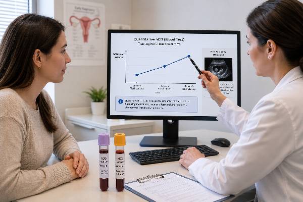
A quantitative hCG blood test measures the concentration of human chorionic gonadotropin in serum and reports a number, usually in milli-international units per milliliter. It can confirm very early pregnancy, establish a baseline, and show how hCG changes when the test is repeated. Clinicians commonly use it for bleeding or pain, uncertain dates, pregnancy of unknown location, suspected ectopic pregnancy, miscarriage follow-up, fertility treatment, and gestational trophoblastic disease. One value cannot determine the exact gestational age, prove viability, or show where a pregnancy implanted because normal ranges overlap widely. The trend is often more useful, but even an appropriate rise does not completely exclude ectopic pregnancy, and a fall does not guarantee that ectopic tissue has resolved. Ultrasound, symptoms, and clinical examination remain essential. Interpretation should use the same laboratory when possible, the exact interval between samples, the starting value, and the patient’s symptoms rather than relying on a rigid rule that hCG must double every 48 hours.
- Quantitative serum hCG gives a numerical concentration rather than a positive or negative label.
- A single level confirms detectable hormone but cannot accurately date or locate a pregnancy.
- Expected rises slow as the starting hCG concentration increases.
- Serial results are most informative when drawn about 48 hours apart in the same laboratory.
- Urgent symptoms require evaluation regardless of whether the number is low, high, rising, or falling.
Table of Contents
- What Quantitative hCG Measures
- Why the Test Is Ordered
- Sample Timing, Units, and Preparation
- Interpreting a Single hCG Level
- Understanding Rise, Doubling Time, and Decline
- hCG and Ultrasound in Early Pregnancy
- Unexpected High, Low, or Persistent Results
- Follow-Up, Safety, and Practical Questions
What Quantitative hCG Measures
Human chorionic gonadotropin is produced mainly by syncytiotrophoblast cells after implantation. Its early function is to maintain the ovarian corpus luteum so progesterone production continues until the placenta can support steroid-hormone production. hCG becomes detectable in maternal serum before it is reliably detectable in urine.
A quantitative assay reports the measured concentration rather than simply positive or negative. Common units are mIU/mL or IU/L; these are numerically equivalent because 1 mIU/mL equals 1 IU/L. Laboratories may call the test beta-hCG, serum hCG, total hCG, or quantitative pregnancy test. Most pregnancy assays detect intact hCG and one or more related forms.
The number reflects trophoblastic hormone production at one moment. It does not directly measure embryo size, cardiac activity, placental blood flow, or chromosome status. Normal pregnancies at the same gestational age can differ by many-fold, making broad online week-by-week charts unsuitable for determining whether one result is normal.
The assay is highly sensitive. Many laboratories consider values below about 5 mIU/mL negative, values around 5 to 24 or 25 indeterminate, and values at or above roughly 25 positive. These are common conventions, not universal rules. The report’s reference interval and clinical context take priority.
After an early peak around the end of the first trimester, hCG generally declines and then plateaus at a lower concentration for the remainder of pregnancy. Therefore, first-trimester trend expectations should not be applied later in gestation.
Quantitative hCG is also used outside routine pregnancy confirmation. It can monitor treatment of ectopic pregnancy or gestational trophoblastic disease and may serve as a tumor marker for certain germ-cell and other hCG-producing tumors. In those settings, targets and interpretation differ substantially from early-pregnancy care.
Why the Test Is Ordered
A quantitative test is useful whenever the actual concentration or its change over time could influence management. Common indications include early vaginal bleeding, pelvic or abdominal pain, uncertain menstrual dates, an inconclusive ultrasound, a pregnancy of unknown location, prior ectopic pregnancy, recurrent loss, or monitoring after assisted reproduction.
In a straightforward pregnancy without symptoms, serial hCG is not routinely needed. A home or qualitative test may confirm pregnancy, followed by prenatal care and ultrasound at an appropriate time. Repeated numbers can create anxiety without improving outcomes when there is no clinical question.
For a pregnancy of unknown location, hCG is commonly measured at presentation and again about 48 hours later. The trend helps determine urgency and timing of repeat ultrasound, but it cannot by itself classify the pregnancy as intrauterine or ectopic. Symptoms always outweigh a reassuring-looking trend.
After miscarriage, clinicians may follow hCG when ultrasound does not fully document completion, when the pregnancy location was never confirmed, or after medical or surgical treatment. The endpoint may be a negative urine test, a very low serum value, or a specified decline, depending on the case and guideline.
After methotrexate treatment for ectopic pregnancy, a defined quantitative monitoring protocol is essential. The schedule is not interchangeable with ordinary 48-hour viability testing. Patients should follow the treating service’s dates because rupture can occur while hCG remains detectable or even while it is falling.
Fertility clinics use quantitative hCG on a scheduled day after embryo transfer or insemination. A second value may assess early progression. Trigger injections containing hCG can cause a temporary positive result if testing occurs too soon, so the clinic’s timing is important.
Persistent or unexpectedly rising hCG after pregnancy can prompt evaluation for retained trophoblastic tissue or gestational trophoblastic disease. In nonpregnant patients, unexplained hCG requires a different diagnostic approach, including review for assay interference and medical causes.
Sample Timing, Units, and Preparation
The test uses blood drawn from a vein. Fasting is usually unnecessary, and ordinary food, water, and prenatal vitamins do not meaningfully change the concentration. Patients should report fertility medicines containing hCG, recent pregnancy outcomes, and any high-dose supplements that the laboratory asks about.
When serial testing is planned, the interval between samples must be recorded accurately. “Two days later” may mean 39 hours or 60 hours, which changes the calculated percentage rise. Many protocols use approximately 48 hours because it balances biological change with practical follow-up.
Using the same laboratory is preferable. Different platforms may have calibration differences large enough to mimic a small rise or fall. When an emergency requires another laboratory, clinicians interpret the change cautiously and may repeat on the original platform.
The result may appear as:
- Below 5 mIU/mL: often reported negative;
- 5–24 or 25 mIU/mL: often indeterminate or borderline;
- ≥25 mIU/mL: often reported positive.
Exact cutoffs vary. A result of 4.8 and one of 5.2 should not be treated as biologically opposite without considering analytic uncertainty and timing.
A very early borderline value is commonly repeated after 48 hours. It may represent a new pregnancy, declining hormone after a chemical pregnancy, residual hCG after a recent pregnancy, exogenous hCG, or a persistent low-level signal.
Unlike urine testing, first-morning collection is not necessary. Hydration does not dilute serum hCG in the same direct way it dilutes urine. Time of day has little practical importance compared with the interval between serial samples.
For assisted reproduction, provide the embryo-transfer date, embryo age at transfer, trigger-shot date, and medication details. For spontaneous conception, record the last menstrual period and possible ovulation date, while recognizing that cycle estimates may be wrong.
Interpreting a Single hCG Level
A single quantitative value can confirm that hCG is present and give a baseline. It cannot reliably answer whether the pregnancy is viable, whether it is ectopic, or exactly how many weeks have passed. The ranges for each gestational week overlap so widely that one value may fit several weeks.
Low numbers are not inherently abnormal when testing occurs very early. A value of 30 mIU/mL could be appropriate shortly after implantation and concerning much later if dates are certain. Late ovulation is a common reason a result seems lower than expected.
Likewise, a high result does not prove twins. Multiple gestations have higher average hCG, but singleton and twin distributions overlap. Ultrasound determines the number of gestational sacs and embryos.
A single value becomes more useful when paired with ultrasound. If a definite intrauterine pregnancy with appropriate development is visible, the ultrasound generally provides more direct viability information than the hCG concentration. If no pregnancy is visible, the level can help plan follow-up but should not trigger an irreversible decision by itself.
The idea of a discriminatory level describes the hCG concentration above which an intrauterine gestational sac is often expected to be visible with high-quality transvaginal ultrasound. The threshold is not absolute. It varies with equipment, operator, multiple gestation, uterine anatomy, and assay. To avoid interrupting a potentially viable pregnancy, some experts use a conservative level near 3,500 mIU/mL when making management decisions.
A value above a local threshold with no intrauterine sac increases concern for ectopic pregnancy or pregnancy loss, but it is not proof. Repeat expert ultrasound, serial hCG, symptoms, and patient preferences guide management.
A low or falling value also does not exclude ectopic pregnancy. Ectopic rupture can occur at low concentrations. The clinical condition remains the priority.
Understanding Rise, Doubling Time, and Decline
The phrase “hCG should double every 48 hours” is an oversimplification. Many viable pregnancies rise more slowly, especially at higher starting concentrations. Modern interpretation often asks whether the increase meets a minimum expected rise rather than whether it exactly doubles.
Approximate minimum 48-hour rises observed in viable intrauterine pregnancies are:
| Initial hCG concentration | Approximate minimum expected rise over 48 hours |
|---|---|
| Below 1,500 mIU/mL | About 49% |
| 1,500–3,000 mIU/mL | About 40% |
| Above 3,000 mIU/mL | About 33% |
These values are statistical boundaries, not guarantees. A rise below them increases concern for ectopic or failing pregnancy, but a small number of viable pregnancies may not follow the expected pattern. A rise above them is reassuring but does not exclude ectopic pregnancy.
Percentage rise is calculated as:
(new hCG − initial hCG) ÷ initial hCG × 100.
For example, an increase from 800 to 1,240 is 55%. The doubling time can be calculated mathematically, but clinicians usually compare the percentage change over the actual interval.
A plateau—little change over time—is abnormal in most early-pregnancy contexts and raises concern for ectopic pregnancy or nonviability. A decline generally indicates a noncontinuing pregnancy. The speed of decline helps determine follow-up, but there is no single fall that proves location.
A rapidly falling value after a known intrauterine miscarriage may support resolution. When the pregnancy location was never confirmed, follow-up must continue because a resolving ectopic pregnancy can also decline. New or worsening pain requires assessment regardless of the trend.
NICE uses a different triage framework for pregnancy of unknown location: an increase greater than 63% over 48 hours suggests a developing intrauterine pregnancy but does not exclude ectopic pregnancy; a decrease greater than 50% suggests the pregnancy is unlikely to continue; changes between those patterns warrant prompt clinical review. Local protocols may differ.
The hCG doubling time test is therefore best understood as serial quantitative testing interpreted with validated thresholds, not a separate assay or a pass-fail doubling requirement.
hCG and Ultrasound in Early Pregnancy
hCG and ultrasound provide complementary information. hCG reflects trophoblastic activity; ultrasound shows anatomy. Neither should be interpreted in isolation when pregnancy location or viability is uncertain.
Transvaginal ultrasound may identify a gestational sac around 4.5 to 5 weeks, a yolk sac around 5 to 5.5 weeks, an embryo around 6 weeks, and cardiac activity thereafter, but normal timing varies. Dating errors of only several days can change what should be visible.
When ultrasound shows neither a definite intrauterine pregnancy nor an ectopic pregnancy, the condition is called pregnancy of unknown location. This is a temporary classification, not a diagnosis. Possibilities include a very early intrauterine pregnancy, a completed or ongoing miscarriage, and an ectopic pregnancy.
Management commonly includes:
- symptom assessment and emergency precautions;
- baseline quantitative hCG;
- repeat hCG after approximately 48 hours;
- repeat transvaginal ultrasound at a clinically appropriate interval;
- continued follow-up until location or resolution is established.
Progesterone may sometimes be measured as a viability marker, but it cannot locate the pregnancy. Some guidelines advise against using it as an adjunct to diagnose ectopic versus viable intrauterine pregnancy in a pregnancy of unknown location.
Once ultrasound confirms a viable intrauterine pregnancy, routine serial hCG usually adds little. hCG may naturally slow, plateau, and later decline even while the pregnancy progresses normally. Ultrasound milestones become more informative.
Heterotopic pregnancy—simultaneous intrauterine and ectopic pregnancy—is rare in spontaneous conception but more likely after assisted reproduction. Seeing an intrauterine pregnancy greatly reduces ectopic concern but does not completely exclude heterotopic pregnancy in a high-risk symptomatic patient.
Unexpected High, Low, or Persistent Results
An hCG result can appear unexpected for many reasons. Incorrect dates are common. Late ovulation makes the level look low; an underestimated gestational age can make it look high. A data or unit error should be excluded before complex conclusions are drawn.
A lower-than-expected result may occur with a normally developing but earlier pregnancy, impending miscarriage, ectopic pregnancy, or laboratory variation. The next measurement and ultrasound are more informative than comparison with a population chart.
A higher-than-expected result may occur with multiple gestation, incorrect dates, or gestational trophoblastic disease. Some chromosome conditions alter hCG patterns in prenatal screening, but a routine quantitative hCG level is not a chromosome test.
Extremely high hCG can be associated with severe nausea and vomiting, thyroid stimulation, or molar pregnancy, but symptoms and ultrasound are required. Rare assay “hook effect” can paradoxically cause an inaccurately low result when concentrations are extraordinarily high; laboratories can test a diluted sample when suspected.
Persistent low-level hCG after pregnancy can reflect retained products, resolving ectopic tissue, quiescent gestational trophoblastic disease, a new pregnancy, pituitary hCG, or assay interference. Heterophile antibodies can produce a false serum signal. The laboratory may compare urine testing, repeat on another platform, perform dilutions, or use blocking reagents.
After delivery or pregnancy loss, hCG does not disappear immediately. Clearance depends on the starting value and whether trophoblastic tissue remains. Follow-up duration varies from days to weeks or longer in trophoblastic disease.
Unexpected results should not be managed by repeatedly ordering isolated tests without a plan. Define the clinical question, confirm dates and medications, use serial values from the same laboratory, and connect the trend with imaging.
Follow-Up, Safety, and Practical Questions
A good follow-up plan specifies when the next blood draw or ultrasound will occur, which change would prompt review, and which symptoms require emergency care. Patients should not be left to interpret portal numbers alone.
Seek urgent care for severe or worsening one-sided pelvic pain, shoulder-tip pain, fainting, marked dizziness, weakness, shortness of breath, heavy bleeding, or signs of shock. Ectopic rupture is a clinical emergency and can occur with low or declining hCG.
Common practical questions include:
Does a doubling value guarantee a healthy pregnancy? No. It improves the probability of viability but cannot exclude ectopic pregnancy or later complications.
Does a slow rise prove ectopic pregnancy? No. It raises concern for ectopic or nonviable pregnancy and requires follow-up, but location comes from ultrasound or definitive clinical evidence.
Does a falling value mean follow-up can stop? Not automatically. The pregnancy location, symptoms, treatment, and protocol determine the endpoint.
Can hCG date pregnancy? Only roughly. Menstrual or embryo-transfer dates and ultrasound are better.
Should levels be checked every day? Usually not. Daily biological and analytic variation can confuse interpretation; approximately 48-hour intervals are more useful in many early-pregnancy protocols.
Can two laboratories be compared? Large changes may still be meaningful, but small differences are harder to interpret because assays vary.
Review the complete report, not only the number. Confirm the unit, sample dates and times, initial concentration, percentage change, symptoms, ultrasound findings, and medications. The safest interpretation is a coordinated one: quantitative hCG describes the hormone trend, ultrasound determines anatomy, and clinical evaluation identifies urgency.
Interpreting a quantitative series requires more than two numbers. The clinician should document the percentage rise or fall, actual elapsed hours, starting concentration, assay location, and whether the patient’s symptoms changed. A trend that looks reassuring on paper may be clinically unsafe if pain or bleeding is worsening. Conversely, a borderline rise in a stable patient may justify another measurement and repeat scan rather than immediate treatment.
The hCG concentration is not a direct measure of miscarriage tissue burden. After uterine aspiration or medication management, the value may decline at different rates depending on gestational age and completeness. Ultrasound, bleeding pattern, pathology, and symptoms can be more useful than expecting one universal percentage fall.
Gestational trophoblastic disease uses specialized surveillance. After molar pregnancy, hCG is followed according to a defined protocol because plateauing or rising values can indicate persistent disease. Patients may be advised to avoid pregnancy during surveillance so that a new conception does not obscure interpretation. This use of quantitative hCG is different from early viability monitoring and should be managed by the responsible specialist team.
For tumors that produce hCG, laboratories may measure intact hCG, beta subunits, or related markers. A standard pregnancy assay may not capture every clinically relevant form. Persistent unexplained values in a nonpregnant patient require coordinated laboratory and medical evaluation rather than repeated obstetric interpretation.
Biotin, heterophile antibodies, and other assay effects are considered when the pattern is inconsistent. Laboratory specialists can often investigate by dilution, alternate platforms, urine comparison, or blocking reagents. Patients should not assume interference merely because a result is unwanted; it is a diagnosis of discordance supported by testing.
Online gestational charts commonly list enormous ranges and may use weeks since conception rather than weeks from the last menstrual period. Comparing a result with the wrong dating convention can create a two-week error. Even with correct dating, the overlap is too wide for one value to confirm normal development.
The endpoint of serial testing should be explicit. It may be visualization of a definitive intrauterine pregnancy, an hCG decline to a specified value, a negative test, or completion of a treatment protocol. Knowing the endpoint prevents both premature discharge and unnecessary repeated blood draws.
Communication is especially important when values are visible before a clinician reviews them. A percentage increase that falls just below a published minimum is not a self-diagnosis, and an increase above it is not permission to ignore symptoms. The team may consider confidence intervals, assay variation, exact dating, and the next ultrasound window. Patients can ask for the calculation and the plan in writing, including who will review results after hours and whether another value is needed if the scan remains nondiagnostic.
For telehealth review, patients should have the full laboratory reports and ultrasound wording available. A screenshot that omits the unit, collection time, or reference comment can lead to an incorrect comparison.
A patient should also know whether the result was ordered as a stat test or routine test. Turnaround time affects when the next decision can be made, but it does not change the biology of the trend. Drawing an extra sample before the planned interval solely because the first result posted quickly may reduce interpretability rather than provide earlier certainty.
References
- Human Chorionic Gonadotropin 2025 (Review)
- β-Human Chorionic Gonadotropin Dynamics in Early Gestation 2024 (Review)
- Early Pregnancy Diagnosis 2023 (Review)
- Ectopic pregnancy and miscarriage: diagnosis and initial management 2026 (Guideline)
- Human Chorionic Gonadotropin and Early Embryogenesis: Review 2022 (Review)
Disclaimer
This article provides general education and cannot diagnose pregnancy viability, miscarriage, ectopic pregnancy, or trophoblastic disease. Quantitative hCG must be interpreted by a clinician with symptoms, exact timing, ultrasound, and treatment history. Seek emergency care for severe pain, shoulder-tip pain, fainting, marked weakness, or heavy bleeding.