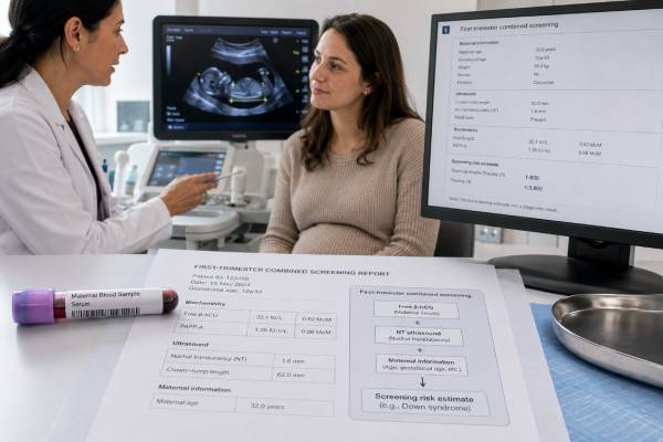
The free beta-hCG test measures one portion of human chorionic gonadotropin in maternal blood and is mainly used as part of first-trimester chromosome screening. It is not the same as a standard pregnancy test or a quantitative hCG test used to follow an early pregnancy. Laboratories combine free beta-hCG with pregnancy-associated plasma protein A (PAPP-A), ultrasound measurement of nuchal translucency, gestational age, maternal age, and other details to estimate the chance of trisomy 21, trisomy 18, or trisomy 13. The result is usually converted to a multiple of the median, or MoM, rather than interpreted from the raw concentration alone. A high or low value does not diagnose a chromosome condition, and many pregnancies with an unusual marker result have a healthy outcome. The report should be read as a complete risk calculation, followed by counseling about cell-free DNA screening, detailed ultrasound, or diagnostic testing when appropriate.
- Free beta-hCG is a first-trimester screening marker, not a stand-alone diagnostic test.
- Testing is commonly performed from 10 weeks through 13 weeks 6 days, often with PAPP-A and nuchal translucency.
- Higher free beta-hCG is associated with trisomy 21 screening patterns; lower levels can occur with trisomy 18 or 13.
- Results are adjusted and reported in MoM, so raw values from different pregnancies should not be compared.
- A screen-positive result means increased probability, not confirmation that the fetus has a chromosome condition.
Table of Contents
- What Free Beta-hCG Measures
- Role in First-Trimester Screening
- Timing, Preparation, and Sample Collection
- MoM and the Risk Calculation
- High Free Beta-hCG Results
- Low Free Beta-hCG Results
- Screen-Positive Results and Follow-Up Options
- Limitations and Special Situations
What Free Beta-hCG Measures
Human chorionic gonadotropin is a hormone produced mainly by placental trophoblast cells after implantation. The intact hormone has two protein parts, an alpha subunit and a beta subunit. The beta subunit gives hCG much of its biological specificity. Some beta subunits circulate unattached to the alpha subunit; laboratories call this fraction free beta-hCG.
A free beta-hCG assay is designed for prenatal risk assessment. A routine hCG blood test for pregnancy usually measures total hCG or the beta-hCG signal needed to report a positive, negative, or numeric pregnancy result. Those tests answer questions such as whether hCG is present and how its concentration changes. Free beta-hCG screening asks a different question: is the marker higher or lower than expected for a pregnancy at the same gestational age, after relevant adjustments?
The placenta releases large amounts of hCG early in pregnancy. Concentrations rise rapidly, peak near the end of the first trimester, and then decline. Because the marker changes from week to week, a raw number has little meaning without an accurate gestational age. A value that is ordinary at 10 weeks could be unusual at 13 weeks. The laboratory therefore compares the patient’s result with a reference median for the exact stage of pregnancy.
Free beta-hCG does not directly count fetal chromosomes. It reflects placental biology that, in population studies, differs on average among pregnancies affected by certain chromosome conditions. The overlap between affected and unaffected pregnancies is substantial. That is why the marker is combined with other independent information instead of being treated as a yes-or-no result.
Free beta-hCG also should not be used by itself to judge miscarriage, ectopic pregnancy, fetal heartbeat, or placental function. In a patient with pain, bleeding, or an uncertain early pregnancy location, clinicians use symptoms, ultrasound, and serial quantitative hCG rather than a prenatal free beta-hCG screening result.
Role in First-Trimester Screening
The combined first-trimester screen typically includes two blood markers—free beta-hCG and PAPP-A—plus an ultrasound measurement of the fluid-filled space at the back of the fetal neck, called nuchal translucency. A computer algorithm combines these findings with maternal age and pregnancy details to estimate an individual probability for trisomy 21, trisomy 18, and sometimes trisomy 13.
Characteristic patterns help the algorithm separate higher-risk from lower-risk pregnancies:
| Screening condition | Free beta-hCG pattern | PAPP-A pattern | Other common contribution |
|---|---|---|---|
| Trisomy 21 | Often higher than the gestational median | Often lower | Nuchal translucency may be increased |
| Trisomy 18 | Often lower | Often lower | Ultrasound findings may increase risk |
| Trisomy 13 | Often lower or not clearly elevated | Often lower | Ultrasound findings may increase risk |
| Unaffected pregnancy | Wide normal overlap | Wide normal overlap | Risk is calculated from all inputs |
These are statistical tendencies, not rules. A pregnancy with trisomy 21 can have a free beta-hCG result near 1.0 MoM, and an unaffected pregnancy can have a high result. The combined calculation performs better than either serum marker alone because each input contributes partly independent information.
Programs use different reporting cutoffs. One service may call a risk of 1 in 250 screen positive, while another uses 1 in 300 or a locally defined threshold. The same numeric risk can therefore receive different labels in different systems. The actual probability is usually more informative than the color or category printed on the report.
First-trimester serum screening remains useful in settings where cell-free DNA is not available, is not covered, or is chosen as a second step after an intermediate result. It can also provide placental-marker information that cell-free DNA does not provide in the same way. However, cell-free DNA generally has higher screening performance for the common trisomies. Patients should receive counseling about all available screening and diagnostic choices rather than being directed automatically to one pathway.
Screening is optional. A person may choose screening for early information, choose diagnostic testing for a definitive chromosome answer, or decline testing after discussing benefits, limitations, timing, cost, and personal values.
Timing, Preparation, and Sample Collection
Free beta-hCG for combined first-trimester screening is generally collected from about 10 weeks through 13 weeks 6 days. The exact window depends on the laboratory and the screening program. Nuchal translucency ultrasound is usually performed when the fetal crown-rump length is within the validated range, commonly 45 to 84 mm, which corresponds approximately to 11 weeks through 13 weeks 6 days.
The blood draw and ultrasound do not always have to occur on the same day. Some programs collect blood earlier so the biochemical results are available at the ultrasound appointment. What is essential is that the laboratory receives the correct gestational age and required clinical details.
No fasting is usually required. Prenatal vitamins, ordinary meals, and normal activity do not meaningfully prepare the marker. Unless the ordering clinician gives different instructions, patients can eat, drink, and take routine medicines as usual. The blood sample is obtained from a vein in the arm. Brief discomfort, bruising, or lightheadedness can occur, as with any blood draw.
Before collection, verify the following information:
- gestational age and how it was established;
- date of the ultrasound or expected crown-rump length;
- singleton, twin, or higher-order pregnancy;
- maternal weight, because blood-volume dilution affects serum concentrations;
- smoking status and relevant demographic adjustment factors used by the laboratory;
- conception by in vitro fertilization, donor egg, or other assisted reproduction;
- insulin-treated diabetes, when requested by the screening program;
- a recent fetal loss in a multiple pregnancy, sometimes called a vanishing twin.
A dating error is one of the most important correctable causes of an unexpected result. If an early ultrasound changes the due date, the laboratory may be able to recalculate the screen without repeating the blood draw. Recalculation is only valid when the program’s rules allow it and the new dating information falls within the proper testing window.
Results may take several days. The laboratory often sends the complete screen to the prenatal clinician rather than releasing a free beta-hCG concentration in isolation. A report that lists only an analyte value may be incomplete until the ultrasound and risk calculation are added.
MoM and the Risk Calculation
Prenatal serum markers are usually expressed as multiples of the median. A result of 1.0 MoM means the concentration equals the median among pregnancies of the same gestational age in the laboratory’s reference population. A result of 2.0 MoM is twice that median; 0.5 MoM is half of it.
The laboratory first converts the measured concentration to an unadjusted MoM. It then applies validated correction factors. These may account for maternal weight, smoking, diabetes, ethnicity or ancestry variables used by the program, assisted conception, and number of fetuses. Modern laboratories periodically review these factors because population characteristics and assay methods change.
There is no universal “normal range” that works like a standard chemistry panel. Values near 1.0 MoM are common, but a marker at 1.8 MoM is not automatically abnormal, and a marker at 0.6 MoM is not automatically dangerous. The distribution is continuous. The software uses the exact value to modify the age-related starting probability.
For example, suppose two patients both have free beta-hCG of 2.2 MoM. One has PAPP-A near the median and a small nuchal translucency; the other has low PAPP-A and an increased nuchal translucency. Their final trisomy 21 probabilities may be very different. Maternal age also changes the starting probability, so identical marker values do not produce identical final risks at different ages.
Reports commonly show results such as “1 in 2,000” or “1 in 80.” A risk of 1 in 80 means that among 80 pregnancies with the same calculated pattern, approximately one would be expected to have the condition and 79 would not. It does not mean the fetus is “80% likely” to be affected. Converting the fraction to a percentage can help: 1 in 80 is 1.25%, while 1 in 2,000 is 0.05%.
The positive predictive value of a screen depends strongly on the underlying prevalence. A high-risk label is therefore not equivalent to a diagnosis. Conversely, a low-risk result reduces risk but cannot exclude every chromosome condition, structural anomaly, single-gene disorder, or pregnancy complication.
High Free Beta-hCG Results
A high free beta-hCG MoM contributes to the classic first-trimester screening pattern for trisomy 21. The degree of elevation matters, but only within the full model. Laboratories may regard values above approximately 2.0 MoM as elevated for descriptive purposes, yet there is no single threshold that diagnoses Down syndrome or mandates one specific next step.
Common explanations for a high result include:
- normal biological variation in an unaffected pregnancy;
- underestimated gestational age or incorrect dating information;
- a multiple pregnancy that was not entered correctly;
- trisomy 21 screening pattern when accompanied by other risk factors;
- assay or data-entry problems, which are uncommon but worth checking when the value is extreme;
- placental variation that may be associated with later pregnancy outcomes in some studies.
Very high values deserve careful review because routine screening algorithms may cap or truncate extreme MoMs. The clinician should confirm dating, fetus number, weight, and conception details before interpreting the risk. If the value is implausibly high, a repeat sample may be considered according to laboratory policy, but repeating an analyte simply to obtain a more reassuring number is not always appropriate.
A high free beta-hCG result is not the same as a high quantitative total hCG result in early pregnancy. The assays measure different forms and serve different clinical purposes. It also does not prove twins, molar pregnancy, or placental disease. Those diagnoses require the appropriate total hCG test, ultrasound, examination, and clinical history.
Some studies link extreme first-trimester free beta-hCG results with higher rates of outcomes such as hypertensive disorders, growth differences, or preterm birth, but associations vary and are not sufficiently specific to predict an individual outcome. An isolated high marker with normal chromosome screening does not automatically make a pregnancy high risk. The obstetric clinician may consider the complete history, ultrasound findings, and local protocols when deciding whether additional growth or placental surveillance is useful.
A patient who receives a high marker result should ask for the final calculated risk, not rely on the word “high” beside the analyte. The calculated risk determines whether genetic counseling, cell-free DNA, or diagnostic testing should be discussed.
Low Free Beta-hCG Results
Low free beta-hCG can contribute to screening patterns associated with trisomy 18 and trisomy 13, especially when PAPP-A is also low or ultrasound findings are present. Low values also occur in unaffected pregnancies, and many isolated low results do not lead to a chromosome diagnosis.
The first step is to check data quality. Overestimated gestational age can make a measured concentration appear lower than expected. Maternal weight, diabetes adjustments, assisted conception, and multiple gestation can also change the corrected MoM. The laboratory—not a hand calculation from an internet chart—should perform any recalculation.
Low free beta-hCG should not be used to diagnose a failing pregnancy. A first-trimester screening sample is usually taken after pregnancy viability and dating have already been assessed by ultrasound. If there is bleeding, pelvic pain, loss of symptoms, or concern about development, the clinician evaluates the pregnancy directly rather than interpreting the screening MoM as a viability test.
The significance of a low value depends on the rest of the screen:
- Low free beta-hCG with low PAPP-A and increased nuchal translucency: the combined risk may be substantially increased and prompt diagnostic discussion.
- Low free beta-hCG with normal PAPP-A and normal ultrasound: the final risk may remain low.
- Low marker with a dating discrepancy: updated ultrasound dating may materially change the calculation.
- Low marker in twins: interpretation is less precise because the blood result reflects both placentas.
Low serum markers have also been studied as signs of placental dysfunction, fetal growth restriction, preeclampsia, or pregnancy loss. These associations are strongest for certain marker patterns and extreme values, and they do not function as stand-alone predictions. A normal anatomy scan and routine prenatal follow-up remain important regardless of screening category.
Patients sometimes search for a way to “raise” free beta-hCG through food, supplements, or rest. There is no evidence-based treatment aimed at changing a prenatal screening marker. Management focuses on clarifying fetal chromosome risk and monitoring the pregnancy when the overall clinical picture supports it.
Screen-Positive Results and Follow-Up Options
A screen-positive result means the calculated probability crossed the program’s cutoff. It does not confirm trisomy. The next conversation should cover the exact condition involved, the numeric risk, ultrasound findings, gestational age, and the difference between another screening test and a diagnostic test.
Common follow-up options include:
- Genetic counseling. A genetics professional can explain the report, family history, residual risk, and possible outcomes without directing the patient toward one decision.
- Cell-free DNA screening. This maternal blood test analyzes placental DNA fragments and is more accurate for trisomy 21, 18, and 13 than traditional serum screening. It remains a screening test. Using it after a positive combined screen can reduce invasive procedures, but it delays a definitive answer and may miss chromosome findings outside its scope.
- Chorionic villus sampling. CVS samples placental tissue, usually during the late first trimester. Chromosome analysis can provide a diagnostic answer. Placental mosaicism can occasionally require further clarification.
- Amniocentesis. Usually performed from about 15 weeks, amniocentesis samples amniotic fluid and fetal cells for diagnostic testing.
- Detailed ultrasound. Ultrasound may identify structural findings that change counseling, but a normal scan cannot exclude trisomy.
No irreversible pregnancy decision should be based on a free beta-hCG result or screening calculation alone. A diagnostic result is the appropriate basis when certainty about a chromosome condition is needed.
A low-risk result also requires accurate interpretation. It substantially reduces the chance of the screened trisomies but does not provide a guarantee. All pregnancies should still receive a second-trimester anatomy ultrasound, commonly at 18 to 22 weeks. Screening for open neural tube defects may require second-trimester maternal serum AFP or ultrasound because first-trimester free beta-hCG does not screen for those conditions.
Avoid undergoing multiple unrelated screening pathways without a clear plan. Repeated screening can create discordant results and confusing false positives. A clinician or genetic counselor can help choose one coherent strategy based on what has already been done.
Limitations and Special Situations
Free beta-hCG screening performs best when the laboratory has correct clinical data and the pregnancy fits the population for which the algorithm was validated. Accuracy can be lower or interpretation can be more complex in several situations.
Twins and higher-order multiples: Serum analytes reflect contributions from more than one placenta. Twin-specific algorithms may be available, but screening is less precise than in singleton pregnancy, and a blood result cannot identify which twin is affected. Higher-order pregnancies have fewer validated options.
Vanishing twin: Placental tissue from a demised co-twin can affect serum markers and cell-free DNA for weeks. The timing of the loss and remaining fetal findings influence which tests are reliable.
Donor eggs and assisted reproduction: Risk calculation should use the details requested by the laboratory, which may include egg-donor age, conception method, and embryo-transfer dating. Entering the pregnant patient’s age where donor age is required can distort the prior risk.
Abnormal ultrasound: A markedly increased nuchal translucency or structural anomaly can indicate chromosome, genetic, or cardiac conditions even when serum risk is low. Diagnostic testing and specialized ultrasound or fetal echocardiography may be offered based on the finding itself.
Previous cell-free DNA: A patient with a low-risk cell-free DNA result generally should not add an independent serum aneuploidy screen solely for reassurance. Serum AFP and the anatomy scan may still be used for structural-defect screening. Clinical programs differ, so coordinate the plan rather than ordering overlapping tests.
Laboratory differences: Assays, medians, correction factors, and cutoffs vary. Do not compare a raw concentration or MoM with a friend’s result, a prior pregnancy, or a chart from another laboratory. The final report and the laboratory’s validated interpretation take priority.
Contact the prenatal team promptly if the report is incomplete, the gestational age is wrong, a twin was not recorded, or the result is labeled screen positive. Seek urgent medical care for heavy bleeding, severe one-sided pelvic or abdominal pain, shoulder pain, fainting, or marked weakness; these symptoms require direct pregnancy assessment and are unrelated to waiting for a screening calculation.
References
- Prenatal Genetic Screening 2025 (Review)
- Biochemical Screening for Fetal Trisomy 21: Pathophysiology of Maternal Serum Markers and Involvement of the Placenta 2023 (Review)
- Extreme βHCG levels in first trimester screening are risk factors for adverse maternal and fetal outcomes 2023
- Clinical consequences of maternal serum PAPP-A and free beta hCG levels above 2.0 multiple of median in the first trimester screening 2023
- The role of the first trimester screen in the face of normal cell-free DNA 2022
Disclaimer
This article provides general education about free beta-hCG prenatal screening and does not interpret an individual pregnancy result. Screening results should be reviewed with an obstetric clinician or genetic counselor using the complete report, ultrasound findings, and medical history. Seek urgent care for severe pain, heavy bleeding, fainting, or other acute pregnancy symptoms.