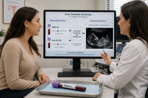
Pregnancy-associated plasma protein A, or PAPP-A, is a protein produced largely by the placenta and measured in maternal blood during first-trimester screening. Laboratories combine it with free beta-hCG, nuchal translucency ultrasound, maternal age, gestational age, and other details to estimate the chance of trisomy 21, trisomy 18, and trisomy 13. PAPP-A is usually reported as a multiple of the median, or MoM, rather than interpreted from the raw concentration. Low PAPP-A contributes to the screening patterns for all three common trisomies and has also been associated with placental complications such as fetal growth restriction, preeclampsia, preterm birth, and stillbirth. However, most pregnancies with an isolated low result do not develop a severe complication, and PAPP-A cannot diagnose placental insufficiency. A high result is usually less clinically important. Accurate pregnancy dating and correct adjustment factors are essential. The final risk calculation—not the isolated PAPP-A number—determines whether genetic counseling, cell-free DNA screening, diagnostic testing, or additional pregnancy surveillance should be discussed.
- PAPP-A is a placental protein used mainly in combined first-trimester chromosome screening.
- Testing is commonly performed from 10 weeks through 13 weeks 6 days.
- Low PAPP-A increases calculated risk for trisomy 21, 18, or 13 when combined with other markers.
- Values are reported in MoM and adjusted for gestational age, weight, smoking, diabetes, and pregnancy details.
- Very low PAPP-A may lead to later fetal-growth or placental surveillance, depending on the complete risk profile.
Table of Contents
- What PAPP-A Is
- PAPP-A in First-Trimester Screening
- Timing, Preparation, and Sample Details
- Understanding MoM and Screening Risk
- Low PAPP-A Results
- High PAPP-A Results
- Follow-Up After an Abnormal Result
- Placental Risk and Test Limitations
What PAPP-A Is
PAPP-A is a large zinc-binding metalloproteinase produced by placental trophoblast cells and other tissues. During pregnancy, maternal serum concentrations rise markedly as the placenta develops. The protein influences insulin-like growth factor signaling by cleaving binding proteins that normally limit growth-factor availability. Through this pathway, it is involved in placental implantation, vascular development, and nutrient-related growth signaling.
PAPP-A is not a pregnancy hormone in the same sense as hCG or progesterone, but it is a pregnancy-associated placental marker. Outside pregnancy, small amounts can be produced in other tissues and have been studied in cardiovascular disease and cancer research. Prenatal laboratories use a pregnancy-specific assay and gestational reference model.
The test does not measure fetal chromosomes directly. Instead, population studies show that first-trimester PAPP-A tends to be lower in pregnancies affected by trisomy 21, 18, or 13. The distributions overlap, so no single concentration can confirm or exclude a chromosome condition.
The same marker has attracted attention because very low values are associated with smaller placental mass or impaired placentation in some pregnancies. That association is biologically plausible: reduced PAPP-A activity may limit local insulin-like growth factor availability and placental growth. Yet the pathway is not simple enough for a blood result to predict one patient’s outcome.
PAPP-A should not be confused with pregnancy-associated plasma protein A2, a related protein studied in preeclampsia research. It is also different from maternal serum AFP, which is used later to screen for open neural tube defects.
A patient may see a raw concentration, percentile, or MoM on the report. The MoM and final risk calculation are the clinically useful values. Raw concentrations cannot be compared between laboratories or gestational weeks.
PAPP-A in First-Trimester Screening
The combined first-trimester screen typically uses PAPP-A, free beta-hCG, and nuchal translucency ultrasound. The algorithm also incorporates maternal age and details such as gestational age, weight, smoking status, diabetes, fetus number, and assisted conception.
Common patterns include:
| Condition | PAPP-A pattern | Free beta-hCG pattern | Ultrasound contribution |
|---|---|---|---|
| Trisomy 21 | Often low | Often high | Nuchal translucency may be increased |
| Trisomy 18 | Often very low | Often low | Structural findings or increased NT may occur |
| Trisomy 13 | Often low | Often low | Structural findings or increased NT may occur |
| Unaffected pregnancy | Wide overlap | Wide overlap | Usually no marker pattern |
The markers are combined because each contributes partly independent information. Low PAPP-A alone has limited detection performance. A low value plus high free beta-hCG and increased nuchal translucency produces a very different risk from the same PAPP-A result with normal values elsewhere.
Screening programs report a probability, such as 1 in 100 or 1 in 5,000. A predefined cutoff determines whether the result is labeled screen positive. Cutoffs differ among countries and laboratories. The actual probability is more informative than the label.
Cell-free DNA screening generally performs better for the common trisomies, but combined screening remains available and can be part of sequential or contingent strategies. PAPP-A may also provide placental-marker information that a low-risk cell-free DNA result does not fully address. Even so, clinicians should avoid running multiple independent aneuploidy screens without a coordinated plan because discordant results can create confusion.
A screen-positive result is not a diagnosis. Chorionic villus sampling or amniocentesis is required for definitive fetal chromosome testing. A low-risk screen reduces probability but cannot guarantee an unaffected fetus.
Timing, Preparation, and Sample Details
PAPP-A is generally collected between about 10 weeks and 13 weeks 6 days. Some programs draw the blood earlier than the nuchal translucency scan so the final risk can be available at the ultrasound appointment. The laboratory’s validated window controls acceptance.
No fasting is usually required. Patients can normally eat, drink, take prenatal vitamins, and continue ordinary activity. The sample is drawn from a vein. Minor bruising or lightheadedness can occur.
Accurate data are crucial because PAPP-A changes rapidly with gestational age. Verify:
- estimated due date and whether it is based on ultrasound;
- crown-rump length when available;
- singleton or multiple pregnancy;
- maternal weight;
- smoking status;
- insulin-treated diabetes, if the program uses that factor;
- conception by IVF, donor egg, or other assisted reproduction;
- a vanishing twin or recent multifetal reduction;
- demographic adjustment factors requested by the laboratory.
A dating error can make PAPP-A appear falsely low or high. If ultrasound revises the gestational age, the clinician may ask the laboratory to recalculate the screen. Recalculation is preferable to comparing the original MoM with an internet chart.
Maternal weight changes measured concentration through blood-volume dilution. Smoking and assisted conception may shift marker distributions. Twin pregnancies require separate algorithms because both placentas contribute to maternal serum and the blood result cannot assign risk to one twin.
Results usually take several days. The portal may release PAPP-A before the nuchal translucency or free beta-hCG is complete. An isolated preliminary value is not the final screen. Wait for the integrated report or contact the ordering team if the clinical data are wrong.
Understanding MoM and Screening Risk
MoM means multiple of the median. A corrected PAPP-A of 1.0 MoM equals the median for pregnancies at the same gestational age after the laboratory’s adjustments. A result of 0.5 MoM is half the median; 0.2 MoM is one-fifth.
There is no universal sharp boundary between normal and abnormal. Values below about 0.4–0.5 MoM are often described as low, and values at or below the fifth percentile—around 0.4 MoM in some systems—receive particular attention. Extremely low values, such as below 0.2 MoM, carry stronger associations. These are descriptive thresholds, not diagnostic cutoffs.
The final chromosome risk begins with a prior probability, influenced strongly by maternal or egg age. The marker likelihoods then raise or lower that probability. For example, identical PAPP-A values can produce different final risks at ages 25 and 42, or when free beta-hCG and nuchal translucency differ.
A report of 1 in 200 means an estimated 0.5% probability; roughly 199 of 200 pregnancies with that calculated profile would not have the condition. A report of 1 in 10,000 means 0.01% residual risk, not zero.
Do not interpret the analyte flag as the screening category. A laboratory may flag PAPP-A below its percentile while the overall trisomy risk remains low. Conversely, a PAPP-A near 1.0 MoM cannot override a markedly abnormal ultrasound or cell-free DNA result.
When reviewing the report, identify:
- corrected PAPP-A MoM;
- free beta-hCG MoM;
- nuchal translucency measurement and quality;
- gestational age used;
- condition-specific probabilities;
- the program’s positive cutoff;
- any placental follow-up recommendation.
These details prevent a single low number from being given more certainty than it carries.
Low PAPP-A Results
Low PAPP-A has two distinct implications: it can alter the first-trimester chromosome risk, and it may identify a pregnancy with increased placental risk even when chromosome screening is low risk.
For aneuploidy, low PAPP-A contributes to trisomy 21, 18, and 13 patterns. The significance depends on free beta-hCG, nuchal translucency, maternal age, and ultrasound. A screen-positive calculation should lead to counseling about cell-free DNA or diagnostic testing, not a diagnosis from PAPP-A alone.
For placental outcomes, low PAPP-A has been associated with:
- fetal growth restriction or a small-for-gestational-age infant;
- preeclampsia and gestational hypertension;
- preterm birth;
- placental abruption;
- stillbirth;
- pregnancy loss, particularly at extreme values.
Most patients with isolated low PAPP-A still have a healthy pregnancy. Its positive predictive value for any one complication is limited. The marker is better understood as one risk factor that may justify closer attention to growth or placental symptoms.
Before accepting the result, verify dating, weight, fetus number, smoking, and conception method. A vanishing twin can distort serum markers and may reduce reliability.
Management varies. A clinician may review aspirin eligibility based on the patient’s complete preeclampsia risk, but low PAPP-A alone does not automatically mean everyone should start aspirin. Low-dose aspirin is time sensitive and has contraindications, so it should be discussed promptly rather than self-started.
Later follow-up may include an anatomy scan, one or more third-trimester growth ultrasounds, Doppler studies if growth is low, or antenatal fetal surveillance depending on the percentile and other conditions. The exact schedule differs by guideline and local practice.
There is no treatment that directly raises PAPP-A. Nutrition, rest, progesterone, and supplements do not normalize the marker. Care focuses on established risk reduction and surveillance.
High PAPP-A Results
High PAPP-A is usually less clinically concerning than low PAPP-A. It may reflect normal variation, later gestational dating, larger placental mass, multiple pregnancy, or assay-specific distribution. It does not diagnose twins or a large fetus.
In first-trimester aneuploidy screening, higher PAPP-A generally shifts risk away from the low-PAPP-A trisomy patterns, but it cannot guarantee normal chromosomes. The rest of the screening inputs still control the calculation.
A value above the laboratory median or even several MoMs high is not treated with medication. When extreme, the laboratory or clinician may verify dating, fetus number, units, and data entry. If the final screen is low risk and ultrasound is normal, no PAPP-A-specific intervention is commonly needed.
Research has explored high PAPP-A in relation to fetal size, placental growth, and metabolic outcomes. Findings are inconsistent and do not support a universal high-value cutoff for surveillance. Group-level association should not be converted into an individual diagnosis.
High PAPP-A also should not be confused with high PAPP-A outside pregnancy, where different assays and cardiovascular research questions apply. Prenatal MoM is tied to gestational reference data.
If a portal labels the result high, ask whether the flag is from a nonpregnant reference interval. Some laboratory systems automatically display inappropriate ranges even though the ordering prenatal algorithm handles the value correctly. The final report is more reliable than a generic flag.
Follow-Up After an Abnormal Result
Follow-up depends on whether the concern is chromosome risk, placental risk, or both.
For a screen-positive trisomy result, options include:
- Genetic counseling to review the exact probability, ultrasound, family history, and values.
- Cell-free DNA screening, which is a more accurate secondary screen for common trisomies but remains non-diagnostic.
- Chorionic villus sampling, usually in the late first trimester, for diagnostic placental chromosome testing.
- Amniocentesis, generally from 15 weeks, for diagnostic testing of fetal cells.
- Detailed ultrasound, which may identify structural findings but cannot exclude a chromosome condition by itself.
No irreversible pregnancy decision should be based on PAPP-A or a screening calculation alone.
When chromosome risk is low but PAPP-A is very low, the obstetric team may create a growth and placental surveillance plan. Ask when growth ultrasound will occur, whether aspirin is indicated for other risk factors, and which symptoms should prompt urgent assessment.
A second-trimester anatomy scan remains important for all pregnancies. Screening for open neural tube defects may require maternal serum AFP or ultrasound because PAPP-A does not address them.
Avoid ordering a repeat PAPP-A simply to see whether it rises. The first-trimester MoM is tied to a validated screening window, and later raw changes are not a standard monitoring tool. Clinical follow-up should use fetal growth, blood pressure, symptoms, and established surveillance methods.
Placental Risk and Test Limitations
PAPP-A is useful because it provides early placental information, but its limitations are substantial. It is influenced by dating, weight, smoking, diabetes, assisted reproduction, multiple pregnancy, and assay calibration. It predicts probabilities across populations rather than outcomes for a single fetus.
A low value cannot show whether placental blood flow is currently adequate. Ultrasound growth, amniotic fluid, uterine and umbilical artery Doppler, maternal blood pressure, and fetal surveillance are more direct when complications are suspected.
Special situations include:
- Twins: Screening accuracy is lower and one serum result cannot identify an affected twin.
- Vanishing twin: Residual placental tissue can affect PAPP-A and free beta-hCG.
- Donor egg: The algorithm may require donor age for chromosome prior risk.
- Abnormal ultrasound: Increased nuchal translucency or a structural anomaly deserves diagnostic counseling even if serum risk is low.
- Low-risk cell-free DNA: This greatly reduces common-trisomy risk but does not erase a very low PAPP-A association with placental outcomes.
Contact the prenatal team promptly for severe headache, visual changes, right-upper-abdominal pain, sudden swelling, vaginal bleeding, fluid leakage, contractions, or reduced fetal movement. These symptoms require direct assessment and should not wait for a scheduled growth scan.
PAPP-A is most useful when it leads to an appropriately scaled plan: accurate chromosome counseling when the combined screen is positive, and proportionate placental surveillance when the value is markedly low. It is least useful when treated as a diagnosis or a target to be “fixed.”
The degree of reduction matters when considering placental associations. A PAPP-A just below a laboratory percentile carries a different population risk from a value that is extremely low. Even at very low levels, however, prediction for an individual pregnancy remains limited. Many patients deliver healthy infants, while some placental complications occur despite normal PAPP-A.
Surveillance plans vary because evidence does not support one universal schedule for every isolated low result. The obstetric team may consider a third-trimester growth scan, serial growth assessment, uterine- or umbilical-artery Doppler in selected settings, and antenatal testing later in pregnancy when additional risk factors are present. The plan should account for blood pressure, prior preeclampsia, fetal growth history, smoking, chronic disease, and findings on the anatomy scan.
Low-dose aspirin is recommended for patients who meet established preeclampsia-risk criteria, but an isolated low PAPP-A is handled differently across guidelines and health systems. Patients should not start aspirin based only on an internet threshold. Benefit depends on indication, dose, and starting time, while bleeding risk and contraindications require review.
A low PAPP-A value is not expected to “improve” through diet, rest, or supplements, and repeating the analyte later is not a standard way to monitor the placenta. The useful follow-up measures outcomes directly: fetal size and growth velocity, amniotic fluid, Doppler blood flow, maternal blood pressure, symptoms, and fetal movement.
When both chromosome screening and placental risk are being discussed, keep the pathways separate. A diagnostic chorionic villus sampling or amniocentesis result can resolve the tested chromosome question but does not erase a placental association. Conversely, normal growth later in pregnancy does not retrospectively change the first-trimester chromosome calculation. Each question has its own test and endpoint.
Patients should receive concrete advice about symptoms. Severe headache, visual disturbance, right-upper-abdominal pain, sudden swelling, vaginal bleeding, fluid leakage, contractions, or reduced fetal movement requires prompt assessment. Waiting for a planned growth scan is not appropriate when new warning signs develop.
PAPP-A is an early risk marker that can justify proportionate attention, but it does not define the pregnancy’s outcome. Accurate screening counseling and a tailored surveillance plan are appropriate responses.
PAPP-A can be profoundly low in some pregnancies with abnormal placental development, but the marker should not be used to diagnose preeclampsia before clinical criteria are present. Angiogenic-marker tests, where available and indicated, answer a different question in symptomatic patients later in pregnancy. Blood pressure, urine protein or organ-function tests, symptoms, and fetal assessment remain central.
Multiple pregnancy requires a different counseling framework. Serum markers reflect more than one placenta, nuchal translucency is measured for each fetus, and diagnostic options depend on chorionicity and which fetus appears affected. A single PAPP-A MoM cannot identify the source of risk.
After a reassuring diagnostic chromosome result, patients may still ask why PAPP-A was low. The honest answer is often that the exact reason cannot be identified. The value becomes a risk flag rather than a disease label. Regular prenatal care and appropriately timed growth assessment are the practical response.
PAPP-A results should also be documented for the clinician performing the anatomy scan and later growth assessment. The sonographer does not diagnose placental disease from the blood value, but awareness of the screening history helps the obstetric team integrate fetal size, placental appearance, Doppler findings, and maternal risk factors. Normal interval growth is reassuring, yet one normal scan does not always replace a planned series when the value was extremely low or additional risks are present.
Fetal movement awareness later in pregnancy remains important regardless of PAPP-A. A patient who notices a meaningful reduction should contact the maternity service promptly rather than waiting for the next scheduled surveillance appointment.
Delivery timing is not determined from PAPP-A alone. It is based on fetal growth, surveillance, maternal condition, gestational age, and any complications that actually develop.
References
- Prenatal Genetic Screening 2025 (Review)
- Biochemical Screening for Fetal Trisomy 21: Pathophysiology of Maternal Serum Markers and Involvement of the Placenta 2023 (Review)
- Abnormal maternal serum markers and adverse pregnancy outcomes 2025 (Guideline)
- Antenatal Fetal Surveillance 2024 (Review)
- The effect of a vanishing twin on first- and second-trimester maternal serum markers and nuchal translucency 2023 (Review)
Disclaimer
This article provides general education about PAPP-A screening and cannot predict an individual fetal chromosome result or placental outcome. The complete screening report should be interpreted by an obstetric clinician or genetic counselor using accurate dating, ultrasound, and medical history. Seek prompt care for reduced fetal movement, bleeding, severe headache, visual changes, or significant abdominal pain.