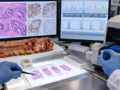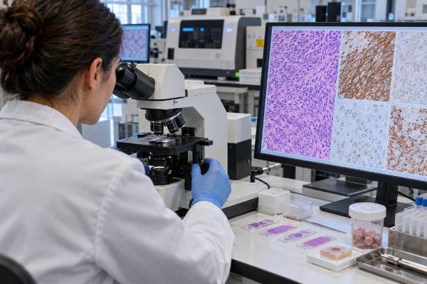
GIST diagnosis combines tumor appearance under the microscope with immunohistochemistry and mutation testing. KIT (CD117) and DOG1 are the most useful immunostains for confirming the GIST lineage, while KIT and PDGFRA mutation testing helps predict sensitivity or resistance to specific tyrosine-kinase inhibitors. These are not interchangeable markers. A tumor can stain strongly for KIT without carrying a KIT mutation, and a small minority of genuine GISTs can be negative for KIT or DOG1 staining. Molecular testing becomes especially important before systemic therapy because mutation subtype can change drug choice and dose. PDGFRA D842V, for example, is resistant to imatinib but sensitive to avapritinib. When KIT and PDGFRA are both wild type, additional workup for SDH deficiency, NF1-related disease, BRAF/RAS alterations, or rare fusions may be needed. The panel therefore serves both diagnostic and treatment-selection purposes.
- KIT/CD117 and DOG1 immunostaining support GIST diagnosis, but neither stain alone is perfectly sensitive or specific.
- KIT and PDGFRA mutation testing predicts response to tyrosine-kinase inhibitors and should be available before neoadjuvant or advanced-disease treatment.
- PDGFRA exon 18 D842V is a key imatinib-resistant mutation with a dedicated targeted option.
- KIT/PDGFRA-wild-type GIST may require SDHB immunostaining and broader genomic testing.
- Mitotic rate, tumor size, site, and rupture status remain essential for recurrence-risk assessment; biomarkers do not replace pathology staging.
Table of Contents
- How KIT, DOG1, and morphology establish GIST diagnosis
- What KIT and PDGFRA mutation testing looks for
- What KIT/PDGFRA-wild-type GIST means
- How biomarkers guide GIST targeted therapy
- What the biomarker panel does not replace
- What to check on a GIST pathology and molecular report
- Practical next steps after GIST biomarker testing
How KIT, DOG1, and morphology establish GIST diagnosis
Most GISTs have spindle-cell, epithelioid, or mixed morphology and arise in the stomach or small intestine. Immunohistochemistry then tests lineage. KIT/CD117 is positive in most GISTs, and DOG1 is similarly sensitive, including in some KIT-negative tumors. CD34 may support the diagnosis but is less specific.
A positive KIT stain does not mean a KIT gene mutation is present. Immunostaining measures protein expression; sequencing identifies a DNA variant. Conversely, a tumor with a KIT mutation can occasionally have weak or unusual staining. Experienced pathology review is important because melanoma, mast-cell lesions, some sarcomas, and other tumors can express KIT. The diagnosis is strongest when morphology, site, immunophenotype, and molecular findings agree.
What KIT and PDGFRA mutation testing looks for
KIT mutations most often involve exon 11, followed by exon 9, with less common primary alterations in exons 13 or 17. PDGFRA mutations occur mainly in gastric GIST and often involve exon 18, including D842V. Sequencing should be sensitive enough to detect variants in samples with limited tumor content.
Mutation subtype matters clinically. KIT exon 11 tumors are usually sensitive to standard-dose imatinib. KIT exon 9 disease can show greater benefit from higher-dose imatinib in advanced disease in some settings. PDGFRA D842V is characteristically resistant to imatinib. Molecular results also help confirm difficult KIT/DOG1-negative tumors and distinguish true wild-type GIST from a false-negative limited assay.
What KIT/PDGFRA-wild-type GIST means
Wild type means no pathogenic KIT or PDGFRA alteration was detected by the assay; it does not mean the tumor lacks a molecular driver. A major subgroup is succinate-dehydrogenase-deficient GIST, often identified by loss of SDHB staining. These tumors are enriched in younger patients, gastric primaries, multifocal disease, and certain hereditary syndromes.
Other wild-type GISTs can be associated with NF1, BRAF or RAS pathway alterations, and very rare NTRK fusions. The appropriate next test depends on age, site, morphology, family history, and SDHB staining. Broad NGS can be particularly useful after high-quality KIT/PDGFRA testing is negative. The label “wild type” should therefore be treated as the start of a second diagnostic branch, not the end of the workup.
How biomarkers guide GIST targeted therapy
Imatinib transformed treatment of KIT- or PDGFRA-sensitive GIST, but the benefit depends on genotype. Primary resistant variants such as PDGFRA D842V require a different strategy, and secondary KIT mutations can emerge during therapy and drive acquired resistance. Later-line TKIs differ in the spectrum of resistant kinase conformations they inhibit.
In localized disease, mutation testing is especially important before neoadjuvant imatinib and before adjuvant therapy is committed, because an insensitive genotype should not be exposed to ineffective treatment. In advanced disease, a new biopsy is not always required at every progression, but molecular information from tissue or research-grade liquid biopsy can sometimes clarify resistance. Drug selection should follow the current guideline and label rather than mutation name alone.
What the biomarker panel does not replace
No biomarker works in isolation. A technically accurate result can still be clinically misleading if it is applied to the wrong cancer type, disease stage, specimen, or treatment question. Cutoffs may also differ by assay, drug label, guideline, and country. For that reason, the laboratory’s own interpretive criteria and the treating team’s current guideline should take priority over a generic internet threshold.
Tumors are heterogeneous, meaning different areas can carry different alterations or levels of protein expression. A small biopsy may miss a positive clone, while a blood-based assay can miss disease that sheds little DNA into the circulation. Conversely, a detectable alteration may be real but not be the main driver of the current disease. Pre-analytic issues such as delayed fixation, decalcification, low tumor content, recent transfusion, or poor plasma handling can also affect some assays.
The safest interpretation separates three questions: analytical validity—did the assay measure what it claims to measure; clinical validity—does the result correlate with the cancer feature of interest; and clinical utility—does acting on the result improve a meaningful decision for this patient. A result can be strong in one category and limited in another.
For GIST specifically, recurrence risk still depends heavily on tumor size, mitotic rate measured over a standardized area, anatomic site, and tumor rupture. A small gastric GIST with a low mitotic rate can have a very different natural history from a large ruptured small-bowel GIST even if both carry a KIT exon 11 mutation.
What to check on a GIST pathology and molecular report
Look for tumor site, size, histologic type, mitotic rate, necrosis, margins when resected, and rupture status. For immunohistochemistry, confirm KIT/CD117 and DOG1 results and whether SDHB was performed when appropriate. For molecular testing, record the gene, exon, exact amino-acid change, variant allele fraction when reported, and assay coverage.
If the report says KIT/PDGFRA wild type, ask whether the method had adequate sensitivity and whether SDH, NF1, BRAF/RAS, or fusion testing is indicated. If systemic therapy is planned, ask explicitly whether the detected genotype is expected to be sensitive to that drug. That single question catches one of the most consequential GIST testing errors.
Practical next steps after GIST biomarker testing
A biomarker result should be read beside the pathology report, stage, imaging, treatment history, and the exact specimen tested. A useful question for the oncology team is not simply “is this positive?” but “what decision does this result change now?” That keeps the result tied to a concrete action such as confirming a diagnosis, choosing a drug, deciding whether hereditary evaluation is needed, or setting a surveillance plan.
If a result seems inconsistent with the clinical picture, ask whether the sample had enough viable tumor, whether the method covered the relevant alteration, and whether a newer metastatic or recurrent specimen would be more representative. Repeating a test is most useful when there is a specific reason to think the original specimen was inadequate, old, or biologically different from the disease being treated today.
Patients should also keep a copy of the complete molecular or pathology report, not only a portal summary. The full report usually lists the method, specimen, tumor percentage, assay limitations, exact variant or staining score, and interpretive comments. Those details matter when seeking a second opinion, transferring care, or checking eligibility for a targeted therapy or clinical trial.
One practical way to avoid overreading Gastrointestinal Stromal Tumor (GIST) Biomarker Panel is to separate the laboratory finding from the clinical decision. The report may be analytically clear while the next step remains conditional on stage, prior therapy, other biomarkers, and patient goals. For example, a result that is highly relevant in metastatic disease may have no established treatment role after curative surgery. Likewise, a biomarker that predicts drug resistance is not necessarily a marker of worse overall prognosis. Keeping those categories separate makes the report easier to use and prevents a single word such as “positive” from carrying more meaning than the evidence supports.
The specimen date deserves attention. Cancer evolves under treatment, and the sample used for Gastrointestinal Stromal Tumor (GIST) Biomarker Panel may have been collected months or years before the current decision. Early driver alterations often remain stable, but protein expression, copy number, and acquired resistance mechanisms can change. Retesting is most valuable when there is a plausible biological reason for change and when a new result could alter management. Repeating testing merely because a value is available is less useful than choosing the specimen that best represents the disease being treated now.
Laboratory reports also vary in how much interpretation they provide. Some give only a final category; others show raw staining percentages, copy-number estimates, variant allele fractions, quality metrics, and assay limitations. For Gastrointestinal Stromal Tumor (GIST) Biomarker Panel, the detailed version is preferable because treatment criteria can evolve. A result that was not actionable when the tissue was first tested may become relevant later, and the original numerical or molecular detail may allow the oncology team to reassess eligibility without immediately repeating a biopsy.
Finally, biomarker testing works best as part of multidisciplinary care. Pathologists judge specimen quality and assay interpretation; medical oncologists connect the finding to treatment; surgeons and gastroenterologists provide disease context; genetic counselors address possible inherited risk when appropriate. Patients do not need to master every technical detail, but they benefit from knowing the purpose of the test, the exact result, what uncertainty remains, and what concrete decision follows. Those four questions turn a complex biomarker report into a usable plan.
One practical way to avoid overreading Gastrointestinal Stromal Tumor (GIST) Biomarker Panel is to separate the laboratory finding from the clinical decision. The report may be analytically clear while the next step remains conditional on stage, prior therapy, other biomarkers, and patient goals. For example, a result that is highly relevant in metastatic disease may have no established treatment role after curative surgery. Likewise, a biomarker that predicts drug resistance is not necessarily a marker of worse overall prognosis. Keeping those categories separate makes the report easier to use and prevents a single word such as “positive” from carrying more meaning than the evidence supports.
The specimen date deserves attention. Cancer evolves under treatment, and the sample used for Gastrointestinal Stromal Tumor (GIST) Biomarker Panel may have been collected months or years before the current decision. Early driver alterations often remain stable, but protein expression, copy number, and acquired resistance mechanisms can change. Retesting is most valuable when there is a plausible biological reason for change and when a new result could alter management. Repeating testing merely because a value is available is less useful than choosing the specimen that best represents the disease being treated now.
Laboratory reports also vary in how much interpretation they provide. Some give only a final category; others show raw staining percentages, copy-number estimates, variant allele fractions, quality metrics, and assay limitations. For Gastrointestinal Stromal Tumor (GIST) Biomarker Panel, the detailed version is preferable because treatment criteria can evolve. A result that was not actionable when the tissue was first tested may become relevant later, and the original numerical or molecular detail may allow the oncology team to reassess eligibility without immediately repeating a biopsy.
Finally, biomarker testing works best as part of multidisciplinary care. Pathologists judge specimen quality and assay interpretation; medical oncologists connect the finding to treatment; surgeons and gastroenterologists provide disease context; genetic counselors address possible inherited risk when appropriate. Patients do not need to master every technical detail, but they benefit from knowing the purpose of the test, the exact result, what uncertainty remains, and what concrete decision follows. Those four questions turn a complex biomarker report into a usable plan.
One practical way to avoid overreading Gastrointestinal Stromal Tumor (GIST) Biomarker Panel is to separate the laboratory finding from the clinical decision. The report may be analytically clear while the next step remains conditional on stage, prior therapy, other biomarkers, and patient goals. For example, a result that is highly relevant in metastatic disease may have no established treatment role after curative surgery. Likewise, a biomarker that predicts drug resistance is not necessarily a marker of worse overall prognosis. Keeping those categories separate makes the report easier to use and prevents a single word such as “positive” from carrying more meaning than the evidence supports.
The specimen date deserves attention. Cancer evolves under treatment, and the sample used for Gastrointestinal Stromal Tumor (GIST) Biomarker Panel may have been collected months or years before the current decision. Early driver alterations often remain stable, but protein expression, copy number, and acquired resistance mechanisms can change. Retesting is most valuable when there is a plausible biological reason for change and when a new result could alter management. Repeating testing merely because a value is available is less useful than choosing the specimen that best represents the disease being treated now.
Laboratory reports also vary in how much interpretation they provide. Some give only a final category; others show raw staining percentages, copy-number estimates, variant allele fractions, quality metrics, and assay limitations. For Gastrointestinal Stromal Tumor (GIST) Biomarker Panel, the detailed version is preferable because treatment criteria can evolve. A result that was not actionable when the tissue was first tested may become relevant later, and the original numerical or molecular detail may allow the oncology team to reassess eligibility without immediately repeating a biopsy.
Finally, biomarker testing works best as part of multidisciplinary care. Pathologists judge specimen quality and assay interpretation; medical oncologists connect the finding to treatment; surgeons and gastroenterologists provide disease context; genetic counselors address possible inherited risk when appropriate. Patients do not need to master every technical detail, but they benefit from knowing the purpose of the test, the exact result, what uncertainty remains, and what concrete decision follows. Those four questions turn a complex biomarker report into a usable plan.
One practical way to avoid overreading Gastrointestinal Stromal Tumor (GIST) Biomarker Panel is to separate the laboratory finding from the clinical decision. The report may be analytically clear while the next step remains conditional on stage, prior therapy, other biomarkers, and patient goals. For example, a result that is highly relevant in metastatic disease may have no established treatment role after curative surgery. Likewise, a biomarker that predicts drug resistance is not necessarily a marker of worse overall prognosis. Keeping those categories separate makes the report easier to use and prevents a single word such as “positive” from carrying more meaning than the evidence supports.
The specimen date deserves attention. Cancer evolves under treatment, and the sample used for Gastrointestinal Stromal Tumor (GIST) Biomarker Panel may have been collected months or years before the current decision. Early driver alterations often remain stable, but protein expression, copy number, and acquired resistance mechanisms can change. Retesting is most valuable when there is a plausible biological reason for change and when a new result could alter management. Repeating testing merely because a value is available is less useful than choosing the specimen that best represents the disease being treated now.
References
- Gastrointestinal stromal tumour (GIST): British Sarcoma Group clinical practice guidelines 2024 (Guideline)
- 2023 GEIS Guidelines for gastrointestinal stromal tumors 2023 (Guideline)
- FDA approves avapritinib for gastrointestinal stromal tumor with a rare mutation 2020 (Official)
- FDA approves ripretinib for advanced gastrointestinal stromal tumor 2020 (Official)
Disclaimer
This article explains biomarker testing for educational purposes and is not a diagnosis or treatment plan. Cancer testing and treatment should be interpreted by the oncology and pathology teams using the complete medical record, current guidelines, and the specific laboratory method. Seek prompt medical care for new or rapidly worsening symptoms rather than relying on a biomarker result alone.