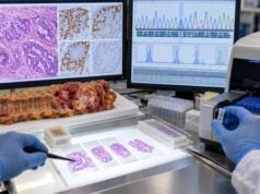
The AFP-L3 test measures a specific form of alpha-fetoprotein (AFP) that is more closely associated with hepatocellular carcinoma (HCC), the most common primary liver cancer, than total AFP alone. It is usually reported as AFP-L3%, meaning the percentage of total AFP made up by the AFP-L3 fraction. A higher percentage can increase concern for HCC, especially in a person who already has cirrhosis, chronic hepatitis B, or another major liver-cancer risk factor. However, AFP-L3 is not a stand-alone cancer diagnosis and should not replace liver imaging. Its value is greatest when clinicians interpret it alongside total AFP, imaging findings, liver disease history, and sometimes des-gamma-carboxy prothrombin (DCP/PIVKA-II). The exact meaning also depends on the assay used and whether a prior AFP-L3 value is available for comparison. A trend over time often provides more useful information than one isolated result.
- AFP-L3% measures the proportion of total AFP that belongs to the Lens culinaris agglutinin-reactive AFP fraction.
- An AFP-L3% around 10% or higher is commonly considered elevated, but the laboratory’s own reference range takes priority.
- A high AFP-L3 result raises HCC concern but does not confirm liver cancer; contrast-enhanced CT or MRI may be needed.
- AFP-L3 is more useful for risk stratification and selected monitoring than for routine HCC surveillance by itself.
- Serial results are most informative when interpreted with total AFP, DCP/PIVKA-II, ultrasound, CT, or MRI.
Table of Contents
- What the AFP-L3 Test Measures
- How to Interpret AFP-L3 Results
- When the AFP-L3 Test Is Used
- AFP-L3 With AFP and DCP/PIVKA-II
- Limitations and False Results
- What Happens After an Abnormal Result
- Practical Questions About AFP-L3 Testing
What the AFP-L3 Test Measures
AFP-L3 is not a completely separate blood protein from AFP. It is a glycosylated subtype of AFP that binds to the plant lectin Lens culinaris agglutinin. Laboratories separate AFP into fractions according to this binding behavior. The result most often reported is AFP-L3%, calculated as the AFP-L3 fraction divided by total AFP and multiplied by 100.
Total AFP can rise for several reasons, including HCC, active hepatitis, liver regeneration, pregnancy, and some germ-cell tumors. AFP-L3 is generally more specific for malignant liver-cell behavior than total AFP, which is why clinicians may use it when total AFP is difficult to interpret or when additional risk information would help.
A typical report may show:
- total AFP in ng/mL;
- AFP-L3% as a percentage;
- an interpretation such as normal, elevated, or high risk; and
- the assay-specific reference interval.
The percentage matters because two people can have similar total AFP values but very different AFP-L3 fractions. For example, a person with total AFP of 20 ng/mL and AFP-L3 of 2% has a different biomarker pattern from someone with total AFP of 20 ng/mL and AFP-L3 of 25%.
AFP-L3 is most relevant in people who already have a reason to be evaluated for HCC. It is not designed as a general population screening test. Routine HCC surveillance is usually based on the person’s underlying liver-cancer risk and imaging schedule, not on AFP-L3 alone. For broader context, a liver cancer biomarker panel may combine AFP-L3 with AFP and DCP/PIVKA-II.
How to Interpret AFP-L3 Results
The most important point is that an elevated AFP-L3% increases suspicion but does not prove HCC. The result must be interpreted with imaging, total AFP, liver function, and the person’s baseline cancer risk.
A cutoff near 10% is commonly used to define an elevated AFP-L3 fraction, although some studies and laboratories use other thresholds. The report from the testing laboratory should therefore be the primary reference. Modern high-sensitivity assays can also quantify AFP-L3 in people with relatively low total AFP, whereas older methods were less reliable when total AFP was very low.
| AFP-L3 pattern | General interpretation | Typical next consideration |
|---|---|---|
| Below the laboratory cutoff | Does not show an increased AFP-L3 fraction | Continue risk-appropriate surveillance or follow-up |
| At or above the cutoff | Raises concern for HCC, especially in a high-risk patient | Review imaging, total AFP, DCP, and clinical context |
| Rising on serial tests | May be more concerning than one stable borderline value | Clinician may shorten the interval to diagnostic imaging |
| Falling after treatment | Can support a treatment response if AFP-L3 was elevated beforehand | Interpret alongside imaging and other tumor markers |
A normal AFP-L3 does not rule out HCC. Some liver cancers produce little AFP, and some produce AFP without a large AFP-L3 fraction. Likewise, a small early tumor may not release enough marker to cause an abnormal blood result. This is why liver imaging remains central.
The meaning of an elevated result also changes with pretest probability. An AFP-L3 of 15% in a person with cirrhosis and a new liver lesion is more concerning than the same value in a person with no known liver disease and normal imaging. Biomarkers work best as part of a clinical probability assessment rather than as yes-or-no tests.
When the AFP-L3 Test Is Used
Clinicians may order AFP-L3 in several situations. One is HCC risk stratification in a person with chronic liver disease. Another is evaluation of a confusing AFP pattern, such as a modestly elevated total AFP that could reflect either inflammation or tumor. AFP-L3 can also be followed after HCC treatment if it was abnormal at baseline.
In the United States, AFP-L3 is not recommended as a replacement for standard HCC surveillance. Current liver-cancer guidance supports surveillance of appropriate high-risk groups with ultrasound and AFP at regular intervals, commonly every six months. If surveillance finds a suspicious lesion or markedly abnormal marker pattern, diagnostic multiphasic CT or contrast-enhanced MRI is generally used.
AFP-L3 may be useful when the clinician wants a more detailed biomarker picture, but it should not delay imaging when imaging is already indicated. A person with cirrhosis, a growing nodule, unexplained weight loss, worsening liver function, or a clearly rising AFP may need direct diagnostic evaluation whether AFP-L3 is normal or not.
After treatment for HCC, a marker that was elevated before therapy can sometimes act as a personal disease-tracking signal. The best comparison is usually the patient’s own previous value measured with the same assay. A falling AFP-L3 after resection, ablation, embolization, radiation, or systemic therapy can be reassuring, but only when imaging supports the same conclusion. A rising level may prompt earlier imaging rather than automatically proving recurrence.
People often encounter AFP-L3 alongside DCP testing for liver cancer, because the two markers reflect different tumor biology and can complement one another.
AFP-L3 With AFP and DCP/PIVKA-II
AFP-L3 becomes more clinically meaningful when considered with total AFP and DCP, also called PIVKA-II. These markers are not interchangeable.
AFP is the broadest and most widely used serum marker in HCC surveillance. It is sensitive to changes in liver biology but is not highly specific because hepatitis and liver regeneration can raise it.
AFP-L3% focuses on the proportion of AFP with a glycosylation pattern associated more strongly with HCC. It can add specificity and may correlate with biologically aggressive tumor features in some studies.
DCP/PIVKA-II is an abnormal prothrombin produced when malignant hepatocytes have impaired vitamin K-dependent carboxylation. It can be high even when AFP is not.
These complementary properties led to multiparameter models such as GALAD, which incorporates gender, age, AFP, AFP-L3, and DCP. Research studies have shown promising performance for detecting HCC, including earlier-stage disease, but practice guidelines still distinguish promising biomarker panels from established surveillance standards.
A practical interpretation is therefore:
- Use the patient’s HCC risk to decide whether surveillance is needed.
- Use ultrasound and AFP according to the surveillance plan.
- Use AFP-L3 or DCP selectively when they add useful risk information.
- Use CT, MRI, or pathology when diagnostic confirmation is needed.
A person whose DCP is the main abnormal marker may benefit from reviewing the dedicated PIVKA-II test interpretation rather than assuming AFP-L3 and PIVKA-II mean the same thing.
Limitations and False Results
AFP-L3 has several important limitations. First, not every HCC produces AFP-L3. Sensitivity for early-stage tumors is not high enough for AFP-L3 to be used alone as a screening or surveillance test. A normal result cannot reassure a high-risk patient who has suspicious imaging.
Second, the percentage depends on how reliably the assay can measure both the numerator and denominator. When total AFP is extremely low, an AFP-L3 percentage may be unavailable or may require a high-sensitivity method. Laboratories differ in analytic platforms, so trends are easier to interpret when testing is performed consistently.
Third, liver disease itself can complicate tumor-marker interpretation. Total AFP can fluctuate with hepatitis activity, and severe hepatic injury can create unusual biomarker patterns. AFP-L3 is more specific than total AFP but is not immune to biological and analytic variability.
Fourth, biomarkers do not establish the location or size of a tumor. A high AFP-L3 cannot tell whether a lesion is 1 cm or 5 cm, whether it invades a vessel, or whether there are metastases. Those questions require imaging and sometimes biopsy.
Finally, a percentage can look dramatic when the total AFP concentration is modest. Clinicians therefore review the absolute AFP, AFP-L3%, previous results, and imaging together. A one-time borderline value often leads to confirmation or trend monitoring rather than an immediate conclusion.
Common interpretation mistakes include assuming that any AFP-L3 elevation equals cancer, ignoring the laboratory cutoff, comparing percentages from different methods without caution, or using a normal AFP-L3 to dismiss a suspicious liver lesion.
What Happens After an Abnormal Result
An abnormal AFP-L3 result usually triggers a structured review rather than an automatic cancer diagnosis. The next step depends on why the test was ordered and whether other abnormalities are present.
A clinician may first confirm:
- the total AFP level and its trend;
- DCP/PIVKA-II if available;
- the latest liver ultrasound, CT, or MRI;
- whether the person has cirrhosis or chronic hepatitis B;
- recent hepatitis activity, biliary problems, or liver decompensation; and
- whether a prior HCC has been treated.
If a high-risk patient has a suspicious lesion, multiphasic CT or contrast-enhanced MRI is commonly the decisive next test. HCC can often be diagnosed noninvasively in at-risk patients when imaging shows the characteristic arterial enhancement and washout pattern. Biomarkers support the assessment but do not substitute for those criteria.
If imaging is negative but AFP-L3 is repeatedly rising, the clinician may repeat the biomarker, review image quality, or shorten the imaging interval based on the overall risk picture. The exact response should be individualized; there is no universal rule that one AFP-L3 value automatically requires biopsy.
People with an already diagnosed HCC should ask whether AFP-L3 was elevated before therapy. If it was, serial testing may provide additional monitoring information. If it was never elevated, it may be less useful as a personal follow-up marker.
Practical Questions About AFP-L3 Testing
Do you need to fast?
Fasting is usually not required for AFP-L3 testing. The sample is typically a standard venous blood draw. Follow the laboratory’s instructions if AFP-L3 is being collected with other tests that do require fasting.
How quickly do results come back?
Turnaround varies by laboratory. AFP-L3 may be a send-out test in some health systems, so results can take longer than routine AFP.
Is AFP-L3 the same as AFP?
No. Total AFP measures all AFP in the sample, while AFP-L3% measures the fraction with a specific lectin-binding pattern. A person can have a modest total AFP but a relatively high AFP-L3 percentage.
Is AFP-L3 used to screen healthy people?
No. The test is most relevant in people with known liver-cancer risk or an existing HCC. HCC surveillance itself is targeted to defined high-risk groups, not the general population.
Can AFP-L3 replace an MRI or CT?
No. A blood marker cannot characterize a liver mass with the detail needed for diagnosis and staging. A high or rising AFP-L3 can justify closer evaluation, but imaging determines whether a suspicious lesion is present.
The most useful question to ask after receiving a result is not simply, “Is this positive?” but, “How does this value change my HCC probability compared with my previous AFP, imaging, and liver disease risk?” That framing keeps the test in its proper role and helps prevent both overreaction to a single abnormal number and false reassurance from a normal one.
How AFP-L3 changes the meaning of a total AFP result
AFP-L3 is often most helpful when total AFP is neither clearly normal nor dramatically high. Consider two common clinical situations. In the first, a patient with cirrhosis has total AFP that fluctuates between 15 and 30 ng/mL during episodes of active hepatitis. If AFP-L3 remains low and imaging is stable, the pattern may fit inflammatory liver activity better than an HCC-specific signal. In the second, total AFP remains modest but AFP-L3 rises repeatedly above the laboratory cutoff. That does not establish cancer, but it can increase concern enough to justify a closer imaging review or a shorter interval to repeat diagnostic imaging.
The reverse situation also occurs. A patient may have a clearly rising total AFP while AFP-L3 remains low. This pattern cannot be dismissed, because some HCCs do not produce a large AFP-L3 fraction. The clinician still evaluates the AFP trend, liver inflammation, pregnancy status when relevant, germ-cell tumor possibilities, and imaging.
Modern high-sensitivity AFP-L3 assays have improved measurement at lower total AFP concentrations, but laboratories do not all use identical methods. This is why comparing a percentage from one laboratory with a percentage from another can be misleading. If a trend is clinically important, repeating the test on the same platform is preferable.
What AFP-L3 can and cannot tell you about tumor aggressiveness
Research has linked elevated AFP-L3 with features such as vascular invasion, poor differentiation, larger tumors, and higher recurrence risk. These associations are useful at the population level, but they should not be converted into a personal staging system. An AFP-L3 of 35% does not prove that a tumor has invaded a blood vessel, and a value of 5% does not prove that a tumor is indolent.
Imaging and pathology answer those questions more directly. Multiphasic CT or MRI can show the number and size of lesions, vascular invasion, and extrahepatic spread. Pathology can describe grade and microscopic features when tissue is available. AFP-L3 adds a biologic signal, but it does not replace those measures.
This distinction matters when patients see a percentage and assume it represents the percentage of liver affected by cancer. It does not. AFP-L3% is only the fraction of circulating AFP that has the L3 lectin-binding pattern.
How surveillance and diagnostic evaluation differ
Surveillance is scheduled testing in people at elevated risk who do not currently have a known suspicious lesion. Diagnostic evaluation begins when surveillance, symptoms, or another test raises concern. AFP-L3 may contribute to both settings, but the next step changes once a lesion is present.
For a high-risk patient with no lesion, clinicians generally continue a guideline-based surveillance program. For a patient with a new or enlarging liver observation, the priority shifts to high-quality contrast imaging and, when needed, multidisciplinary review. Repeating AFP-L3 alone is not an adequate response to a suspicious imaging finding.
A useful way to prepare for a follow-up visit is to bring the full laboratory report rather than only the percentage. Ask whether total AFP was measured at the same time, whether the assay was a high-sensitivity method, whether DCP was also obtained, and how the result changes the imaging plan. Those questions turn a difficult biomarker into a practical decision tool.
References
- AASLD Practice Guidance on prevention, diagnosis, and treatment of hepatocellular carcinoma 2023 (Guideline)
- Existing and emerging biomarkers in hepatocellular carcinoma: relevance in staging, determination of minimal residual disease, and monitoring treatment response: a narrative review 2023 (Review)
- Biomarkers for diagnosis and therapeutic options in hepatocellular carcinoma 2024 (Review)
- Hepatocellular Carcinoma: The Search for an Optimal Screening Test 2025 (Review)
Disclaimer
AFP-L3 results cannot diagnose or exclude hepatocellular carcinoma by themselves. Interpretation should be individualized by a clinician using the laboratory method, total AFP, imaging, liver disease history, and other relevant findings. Seek prompt medical evaluation for new jaundice, rapidly worsening abdominal swelling, severe right-upper-abdominal pain, vomiting blood, confusion, or other signs of serious liver disease.