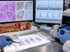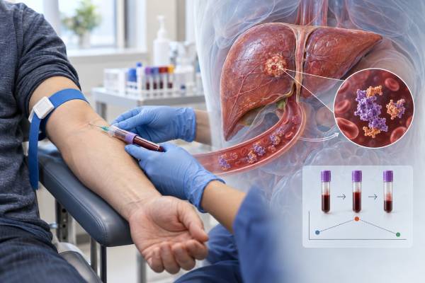
PIVKA-II is a blood biomarker that can rise in hepatocellular carcinoma (HCC), the most common primary liver cancer. It is also called des-gamma-carboxy prothrombin (DCP). The marker is an abnormal form of prothrombin produced when vitamin K-dependent carboxylation is incomplete. HCC cells can produce PIVKA-II, so the test is used in several countries as an adjunct for HCC detection, risk assessment, prognosis, and monitoring. However, a high result is not specific for liver cancer. Vitamin K deficiency, warfarin or other vitamin K antagonists, cholestasis, malabsorption, severe liver dysfunction, and some medications can also increase PIVKA-II. The numerical cutoff also depends on the assay and units, so a value cannot be interpreted without the laboratory reference range. In the United States, major HCC surveillance guidance continues to center on ultrasound with or without AFP rather than PIVKA-II alone. PIVKA-II is most useful when combined with imaging, AFP, clinical risk factors, and repeated trends rather than treated as a stand-alone cancer diagnosis.
- High PIVKA-II can occur in HCC, but it can also rise from vitamin K deficiency, warfarin therapy, cholestasis, or malabsorption.
- PIVKA-II and DCP refer to the same biomarker, although assays may report different units and cutoffs.
- A single high result cannot diagnose HCC; multiphase CT or MRI and the patient’s liver-disease risk determine follow-up.
- PIVKA-II can complement AFP because the two markers are biologically different and may identify partly different HCC cases.
- Falling levels after treatment can support response, while a sustained rise can raise concern for residual or recurrent disease when interpreted with imaging.
Table of Contents
- What PIVKA-II Is
- Why PIVKA-II Rises in HCC
- How to Interpret High and Normal Results
- Noncancer Causes of High PIVKA-II
- PIVKA-II for HCC Surveillance and Diagnosis
- Using PIVKA-II After Liver Cancer Treatment
- Limitations and Practical Next Steps
What PIVKA-II Is
PIVKA-II stands for protein induced by vitamin K absence or antagonist-II. It is an abnormal prothrombin molecule that has not undergone normal vitamin K-dependent gamma-carboxylation. The same analyte is widely called des-gamma-carboxy prothrombin, or DCP.
Normal hepatocytes produce prothrombin as part of the blood-clotting system. Vitamin K is required for proper post-translational modification of several clotting factors. When this process is disrupted, undercarboxylated prothrombin forms can appear in blood.
Hepatocellular carcinoma cells can produce PIVKA-II through abnormal cellular processing even when systemic vitamin K status is adequate. This creates its value as a tumor biomarker.
Unlike AFP, which is an oncofetal protein, PIVKA-II reflects abnormal prothrombin processing. The two markers therefore provide partly independent information. Some HCCs produce AFP but little PIVKA-II; others show the opposite pattern. Combining markers can improve sensitivity compared with either one alone in some populations.
PIVKA-II is not a liver enzyme. It should not be confused with ALT, AST, alkaline phosphatase, or bilirubin. Those tests reflect hepatocyte injury, cholestasis, or liver function rather than a tumor-associated abnormal prothrombin molecule.
It is also not the same as prothrombin time or INR. Warfarin can increase INR and can also elevate PIVKA-II through vitamin K antagonism, but the tests measure different things.
Why PIVKA-II Rises in HCC
HCC cells can have abnormal activity of enzymes involved in vitamin K metabolism and prothrombin carboxylation. The result is secretion of incompletely carboxylated prothrombin into the bloodstream.
Higher PIVKA-II values have been associated in many studies with larger tumors, vascular invasion, more aggressive tumor biology, and worse prognosis. This does not mean every high value reflects advanced disease, but the association has made PIVKA-II useful for risk stratification in addition to detection.
PIVKA-II can also contribute to composite HCC biomarker models. The GALAD score combines gender, age, AFP, AFP-L3, and DCP/PIVKA-II. Other models such as GAAD and ASAP use overlapping clinical and biomarker variables. Recent research and consensus work suggest these panels may improve early HCC detection compared with individual markers, especially when ultrasound quality is limited.
However, promising biomarker-panel performance is not the same as a universal surveillance recommendation. AASLD guidance still emphasizes semiannual ultrasound and AFP for many at-risk adults and does not recommend replacing imaging with PIVKA-II or GALAD alone outside appropriate validated use.
The reason is that a surveillance test must do more than distinguish known cancer from noncancer in a study. It must improve early detection and outcomes in the real population being screened without causing excessive false positives, unnecessary imaging, biopsies, cost, or anxiety.
How to Interpret High and Normal Results
There is no single universal PIVKA-II cutoff because assays use different antibodies, calibrators, and units. Results may be reported in mAU/mL, AU/L, or other formats. Some clinical studies use thresholds such as 40 mAU/mL, while others use different decision points.
The correct first step is therefore to read the laboratory’s own reference range and assay name. Comparing a value from one platform directly with a cutoff from a different publication can be misleading.
A high result means PIVKA-II is above the assay reference or clinical decision threshold. In a person with cirrhosis or chronic hepatitis who is at high risk for HCC, the result may increase suspicion and support diagnostic imaging. In a person taking warfarin, the same number may be uninterpretable as a tumor marker.
A normal result does not exclude HCC. Small or early tumors may not produce enough PIVKA-II to cross the threshold. Marker sensitivity also differs by tumor size and biological subtype. Imaging remains essential.
Trends can be more informative than isolated values. A stable mildly elevated marker in a patient with a persistent benign explanation is different from a rapidly increasing value over several months in a high-risk patient with no vitamin K confounder.
When following a trend, the same assay should be used when possible. Changing laboratories can introduce analytical shifts that look like biological change.
A high PIVKA-II also should not be converted into a direct cancer probability without a validated model. Pretest risk matters enormously. Cirrhosis, chronic hepatitis B, prior HCC, age, sex, and other factors change the meaning of the same biomarker value.
Noncancer Causes of High PIVKA-II
The name of the test itself highlights a major confounder: vitamin K absence or antagonism.
Warfarin: This drug deliberately blocks vitamin K recycling and can markedly increase PIVKA-II. In a patient taking warfarin, PIVKA-II may be unsuitable for HCC interpretation.
Vitamin K deficiency: Poor dietary intake, prolonged malnutrition, or severe illness can reduce vitamin K availability.
Malabsorption: Conditions that impair fat absorption can reduce uptake of fat-soluble vitamin K. Examples include severe cholestatic disease, pancreatic exocrine insufficiency, and some intestinal disorders.
Biliary obstruction or cholestasis: Reduced bile delivery can impair vitamin K absorption and cause PIVKA-II elevation. This is particularly important in patients with jaundice.
Antibiotic exposure: Prolonged broad-spectrum antibiotic use can alter gut flora and contribute to vitamin K deficiency in susceptible patients.
Severe hepatic dysfunction: Advanced liver disease can disrupt synthesis and processing of coagulation proteins, complicating interpretation.
These causes should be reviewed before labeling an elevated result as tumor-related. Medication lists, INR, bilirubin, nutritional status, and clinical context are therefore part of PIVKA-II interpretation.
A clinician may correct a reversible vitamin K problem and repeat the test when appropriate. However, empiric vitamin K should not be used to delay necessary diagnostic imaging in a high-risk patient with a suspicious lesion or a strongly concerning biomarker trend.
PIVKA-II for HCC Surveillance and Diagnosis
HCC surveillance aims to detect cancer early in people at sufficiently high risk, most commonly those with cirrhosis and selected people with chronic hepatitis B. Major U.S. guidance recommends surveillance about every six months using liver ultrasound plus AFP in appropriate at-risk patients.
PIVKA-II is used more routinely in parts of Asia and is increasingly studied worldwide. It can add complementary information to AFP, and composite algorithms that include DCP/PIVKA-II show encouraging sensitivity for early-stage HCC.
Even so, a high marker does not establish HCC diagnosis. In patients with cirrhosis or chronic HBV, HCC can often be diagnosed noninvasively when a lesion shows characteristic arterial-phase enhancement and washout on high-quality multiphase CT or MRI. Biomarkers support suspicion but do not replace the imaging criteria.
If surveillance ultrasound identifies a suspicious lesion or AFP becomes concerning, diagnostic multiphase CT or contrast-enhanced MRI is generally the next step. PIVKA-II can provide additional context, particularly when AFP is normal or equivocal.
A person with no known liver disease who has an incidental high PIVKA-II needs a different workup. Clinicians should first confirm the result, review vitamin K antagonists and liver tests, assess for chronic liver disease, and use imaging if the clinical risk justifies it.
The test also cannot reliably distinguish HCC from every other liver tumor. Intrahepatic cholangiocarcinoma, combined tumors, and metastatic lesions require imaging and sometimes biopsy for correct classification.
Using PIVKA-II After Liver Cancer Treatment
If PIVKA-II was elevated before HCC treatment, serial measurement can become a useful personalized marker.
After surgical resection or liver transplantation, a substantial decline toward the reference range is expected when the marker was produced by the tumor and all detectable cancer has been removed. Persistent elevation can raise concern for residual disease, but benign causes still need exclusion.
After local ablation or embolization, falling PIVKA-II can support treatment response. A plateau or renewed rise may prompt closer imaging review.
During systemic therapy, marker trends can complement CT or MRI. A falling marker is reassuring when imaging also shows stable or shrinking disease. A rising marker with worsening imaging strengthens evidence of progression.
Discordance is possible. A marker can rise before imaging clearly shows recurrence, or imaging can show progression while PIVKA-II remains normal. Treatment should therefore not be changed solely because of one marker value.
The rate of decline depends on baseline level, treatment completeness, tumor biology, and assay. There is no universal rule such as “PIVKA-II must normalize within exactly two weeks.” The oncology or hepatology team interprets the trajectory in the context of the treatment delivered.
For surveillance after curative therapy, patients usually undergo scheduled imaging regardless of marker results because recurrence can occur without a PIVKA-II rise.
Limitations and Practical Next Steps
PIVKA-II has meaningful strengths: it is a simple blood test, it reflects biology different from AFP, and it can provide prognostic and monitoring information. Its limitations are equally important.
The test is confounded by vitamin K status and antagonists. Assays are not fully standardized across platforms. Sensitivity is lower for some small tumors. Evidence and guideline adoption differ by country. And a high value cannot determine tumor location or establish imaging stage.
Common mistakes include:
- diagnosing HCC from PIVKA-II alone;
- ignoring warfarin or vitamin K deficiency;
- comparing values across different assay units without conversion or validation;
- assuming a normal result excludes HCC;
- stopping surveillance because markers are normal; and
- changing treatment solely because of a single rise without imaging correlation.
Useful questions after a high result include:
- What assay and units did the laboratory use?
- What is the reference or clinical cutoff for this assay?
- Am I taking warfarin or another vitamin K antagonist?
- Do I have cholestasis, malabsorption, or a reason for vitamin K deficiency?
- What are my AFP and liver imaging results?
- Am I in a group that should undergo HCC surveillance every six months?
- Does this result warrant multiphase CT or MRI now?
- If I already have HCC, was PIVKA-II elevated before treatment and useful for monitoring?
- Should trends be followed using the same laboratory?
The practical interpretation is simple: PIVKA-II is an adjunctive HCC biomarker, not a stand-alone cancer test. It becomes most useful when the patient has a known HCC risk, confounders have been considered, and the result is integrated with AFP and high-quality imaging.
Pre-analytical context deserves the same attention as the number itself. A PIVKA-II sample drawn during severe cholestasis, shortly after a major change in nutrition, or while a patient is taking a vitamin K antagonist may not represent tumor biology. Clinicians should document medication exposure and liver-function changes when comparing serial values. If the confounder resolves, repeating the marker can help determine whether the elevation persists.
PIVKA-II can also carry prognostic information after HCC is diagnosed. Higher concentrations have been associated with vascular invasion and biologically aggressive disease in multiple cohorts. Some transplant and surgical programs have studied DCP/PIVKA-II as part of risk models for recurrence. However, thresholds and algorithms differ across regions, and treatment eligibility should follow the validated criteria used by the center rather than a cutoff copied from an unrelated assay or study.
Combining AFP and PIVKA-II is attractive because discordant marker patterns are common. A patient may have AFP of 4 ng/mL but a clearly elevated PIVKA-II, while another patient shows the reverse. When both are elevated and then fall after treatment, concordant trends can strengthen confidence that therapy is controlling marker-producing disease. When only one marker was abnormal before treatment, that marker usually becomes the more useful personalized surveillance signal.
Newer panels such as GALAD and GAAD combine biomarkers with demographic variables to improve discrimination. Their promising performance is especially relevant when ultrasound quality is reduced by obesity, steatosis, or nodular cirrhosis. Yet even a high-performing statistical score does not show the location of a lesion. Positive blood-based surveillance still needs diagnostic imaging, and an indeterminate scan may require short-interval follow-up, a second imaging modality, or biopsy depending on the clinical scenario.
Patients should also understand the difference between surveillance and monitoring. Surveillance looks for a new HCC in an at-risk person who has no known active cancer. Monitoring follows a patient after HCC has already been diagnosed or treated. A biomarker can perform differently in these two settings because the pretest probability of cancer is dramatically different.
For that reason, the same PIVKA-II value can trigger different actions. A modest elevation in an otherwise low-risk person taking warfarin may mainly prompt review of medication and vitamin K status. The identical elevation in a cirrhotic patient with a new 1.5-cm liver nodule has far greater significance and belongs in an HCC diagnostic pathway.
The marker can be particularly useful when AFP is chronically uninformative. Some patients with proven HCC have repeatedly normal AFP but elevated PIVKA-II. If the marker falls after successful treatment and later rises again without a vitamin K explanation, the change can provide an early clue that deserves imaging review. The opposite is also true: patients whose original HCC never produced PIVKA-II should not expect the test to become a reliable surveillance marker later.
Laboratory reports should be kept with the assay name and units. This is important when care moves between countries or health systems because numerical values from different PIVKA-II platforms may not be directly interchangeable.
PIVKA-II is therefore best used as part of a longitudinal record. A baseline obtained before HCC treatment, followed by values measured with the same platform at clinically meaningful intervals, is more useful than sporadic testing without context. The pattern should be reviewed alongside AFP, bilirubin, INR, medication changes, and imaging dates.
References
- AASLD Practice Guidance on prevention, diagnosis, and treatment of hepatocellular carcinoma 2023 (Guideline)
- Current and new strategies for hepatocellular carcinoma surveillance 2025 (Review)
- Utility of Biomarker Panels in the Surveillance and Monitoring of Hepatocellular Carcinoma: Consensus Statements from an International Delphi Panel 2026 (Consensus Statement)
- Gender, Age, Alpha-fetoprotein, and Des-gamma-carboxyprothrombin Score as a Novel Approach to Early Detection of Hepatocellular Carcinoma: A Narrative Review. 2025 (Review)
Disclaimer
This article is for general education and does not replace medical evaluation by a hepatologist or oncology team. PIVKA-II must be interpreted with the assay reference range, vitamin K status, medications, liver disease, AFP, and imaging. A high value should not be used by itself to diagnose or treat liver cancer.