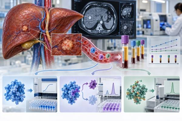
AFP, AFP-L3%, and DCP/PIVKA-II are blood biomarkers associated with hepatocellular carcinoma, but they do not function as a stand-alone “liver cancer panel” that can rule cancer in or out. AFP is the most familiar marker and is used with ultrasound in U.S. HCC surveillance for at-risk patients. AFP-L3% measures a specific glycoform of AFP, while des-gamma-carboxy prothrombin (DCP) and protein induced by vitamin K absence or antagonist-II (PIVKA-II) are two names for the same abnormal prothrombin biomarker. The latter two are used more extensively in parts of Asia and in multivariable risk models such as GALAD. Values can be affected by active hepatitis, pregnancy, germ-cell tumors, vitamin K deficiency, warfarin, cholestasis, and assay differences. A suspicious biomarker pattern should lead to liver imaging and clinical evaluation, not treatment based on blood tests alone. HCC can also occur with normal AFP and normal adjunct markers.
- DCP and PIVKA-II are the same biomarker, not two independent tests.
- AFP is used with ultrasound for HCC surveillance in many at-risk patients; a single elevated AFP does not diagnose HCC.
- AFP-L3% and DCP/PIVKA-II may improve risk stratification when combined with AFP but have limited sensitivity when used alone.
- Vitamin K deficiency and warfarin can raise DCP/PIVKA-II without HCC.
- Normal biomarkers do not exclude HCC; diagnostic imaging criteria or pathology remain central.
Table of Contents
- What AFP, AFP-L3, and DCP/PIVKA-II measure
- How biomarkers fit into HCC surveillance
- How to interpret abnormal values and trends
- Common non-cancer causes of abnormal liver tumor markers
- Role in HCC diagnosis and treatment monitoring
- Limitations of AFP-based biomarker panels
- Practical next steps after an abnormal HCC biomarker panel
What AFP, AFP-L3, and DCP/PIVKA-II measure
AFP is a fetal serum protein that normally falls to very low levels after birth. Some HCCs re-express AFP, but elevations also occur with active liver regeneration and other tumors. AFP-L3 is the fraction of AFP that binds Lens culinaris agglutinin and is reported as a percentage of total AFP. Higher fractions are associated with HCC biology but are difficult to interpret when total AFP is very low.
DCP is an abnormal form of prothrombin produced when vitamin K-dependent carboxylation is impaired. HCC cells can produce DCP even when systemic vitamin K is adequate. PIVKA-II is another name for DCP. This synonym is important because ordering both does not provide two independent data points.
How biomarkers fit into HCC surveillance
HCC surveillance is aimed at people with sufficiently high annual risk, most commonly those with cirrhosis and selected chronic hepatitis B populations. In the United States, ultrasound approximately every six months is the imaging backbone, and AFP is commonly added. Surveillance is different from diagnosis: its goal is to trigger timely diagnostic imaging when a new lesion or concerning marker change appears.
AFP-L3 and DCP/PIVKA-II are FDA cleared for risk stratification rather than routine stand-alone surveillance in the United States. They are incorporated more broadly in some Asian practices and into models such as GALAD, which combines age, sex, AFP, AFP-L3%, and DCP. Local guidelines therefore matter.
How to interpret abnormal values and trends
There is no universal AFP level that proves HCC. A cutoff around 20 ng/mL is often used to increase surveillance sensitivity, but specificity depends on the population and active liver disease. Larger or steadily rising values are more concerning, especially when hepatitis is controlled. AFP-L3% thresholds around 10% to 15% and DCP thresholds around 40 mAU/mL appear in research and clinical use, but assay-specific reference ranges should guide interpretation.
A single result is less useful than trend and context. A falling AFP after locoregional or systemic therapy can support treatment response in a patient whose tumor secreted AFP at baseline, but imaging remains essential. Marker-negative HCC is common enough that normal blood tests cannot reassure away a suspicious lesion.
Common non-cancer causes of abnormal liver tumor markers
AFP can rise with active viral hepatitis, acute hepatic injury, pregnancy, and certain germ-cell tumors. DCP/PIVKA-II can rise with vitamin K deficiency, warfarin or other vitamin K antagonism, malabsorption, prolonged antibiotic exposure, and severe cholestasis. Those confounders can be especially important in patients with advanced liver disease.
When a value is unexpected, clinicians may repeat it after correcting a reversible cause, review medication history and liver tests, and compare it with imaging. The goal is to avoid both false reassurance and unnecessary alarm. Biomarkers are most useful when the pre-test risk of HCC is already defined.
Role in HCC diagnosis and treatment monitoring
In many at-risk adults, HCC can be diagnosed noninvasively when multiphasic CT or MRI shows a characteristic arterial-enhancement and washout pattern in an appropriate lesion. Biomarkers can raise suspicion but are not part of the core imaging criteria that independently establish HCC. Biopsy is reserved for lesions without diagnostic imaging features or when histology will change management.
After treatment, AFP and sometimes DCP/PIVKA-II can be useful response markers if they were elevated before therapy. A biochemical response that conflicts with imaging should not be overinterpreted. Likewise, a rising marker can precede visible progression but should prompt evaluation rather than automatic treatment change.
Limitations of AFP-based biomarker panels
No biomarker works in isolation. A technically accurate result can still be clinically misleading if it is applied to the wrong cancer type, disease stage, specimen, or treatment question. Cutoffs may also differ by assay, drug label, guideline, and country. For that reason, the laboratory’s own interpretive criteria and the treating team’s current guideline should take priority over a generic internet threshold.
Tumors are heterogeneous, meaning different areas can carry different alterations or levels of protein expression. A small biopsy may miss a positive clone, while a blood-based assay can miss disease that sheds little DNA into the circulation. Conversely, a detectable alteration may be real but not be the main driver of the current disease. Pre-analytic issues such as delayed fixation, decalcification, low tumor content, recent transfusion, or poor plasma handling can also affect some assays.
The safest interpretation separates three questions: analytical validity—did the assay measure what it claims to measure; clinical validity—does the result correlate with the cancer feature of interest; and clinical utility—does acting on the result improve a meaningful decision for this patient. A result can be strong in one category and limited in another.
Practical next steps after an abnormal HCC biomarker panel
A biomarker result should be read beside the pathology report, stage, imaging, treatment history, and the exact specimen tested. A useful question for the oncology team is not simply “is this positive?” but “what decision does this result change now?” That keeps the result tied to a concrete action such as confirming a diagnosis, choosing a drug, deciding whether hereditary evaluation is needed, or setting a surveillance plan.
If a result seems inconsistent with the clinical picture, ask whether the sample had enough viable tumor, whether the method covered the relevant alteration, and whether a newer metastatic or recurrent specimen would be more representative. Repeating a test is most useful when there is a specific reason to think the original specimen was inadequate, old, or biologically different from the disease being treated today.
Patients should also keep a copy of the complete molecular or pathology report, not only a portal summary. The full report usually lists the method, specimen, tumor percentage, assay limitations, exact variant or staining score, and interpretive comments. Those details matter when seeking a second opinion, transferring care, or checking eligibility for a targeted therapy or clinical trial.
One practical way to avoid overreading Liver Cancer Biomarker Panel is to separate the laboratory finding from the clinical decision. The report may be analytically clear while the next step remains conditional on stage, prior therapy, other biomarkers, and patient goals. For example, a result that is highly relevant in metastatic disease may have no established treatment role after curative surgery. Likewise, a biomarker that predicts drug resistance is not necessarily a marker of worse overall prognosis. Keeping those categories separate makes the report easier to use and prevents a single word such as “positive” from carrying more meaning than the evidence supports.
The specimen date deserves attention. Cancer evolves under treatment, and the sample used for Liver Cancer Biomarker Panel may have been collected months or years before the current decision. Early driver alterations often remain stable, but protein expression, copy number, and acquired resistance mechanisms can change. Retesting is most valuable when there is a plausible biological reason for change and when a new result could alter management. Repeating testing merely because a value is available is less useful than choosing the specimen that best represents the disease being treated now.
Laboratory reports also vary in how much interpretation they provide. Some give only a final category; others show raw staining percentages, copy-number estimates, variant allele fractions, quality metrics, and assay limitations. For Liver Cancer Biomarker Panel, the detailed version is preferable because treatment criteria can evolve. A result that was not actionable when the tissue was first tested may become relevant later, and the original numerical or molecular detail may allow the oncology team to reassess eligibility without immediately repeating a biopsy.
Finally, biomarker testing works best as part of multidisciplinary care. Pathologists judge specimen quality and assay interpretation; medical oncologists connect the finding to treatment; surgeons and gastroenterologists provide disease context; genetic counselors address possible inherited risk when appropriate. Patients do not need to master every technical detail, but they benefit from knowing the purpose of the test, the exact result, what uncertainty remains, and what concrete decision follows. Those four questions turn a complex biomarker report into a usable plan.
One practical way to avoid overreading Liver Cancer Biomarker Panel is to separate the laboratory finding from the clinical decision. The report may be analytically clear while the next step remains conditional on stage, prior therapy, other biomarkers, and patient goals. For example, a result that is highly relevant in metastatic disease may have no established treatment role after curative surgery. Likewise, a biomarker that predicts drug resistance is not necessarily a marker of worse overall prognosis. Keeping those categories separate makes the report easier to use and prevents a single word such as “positive” from carrying more meaning than the evidence supports.
The specimen date deserves attention. Cancer evolves under treatment, and the sample used for Liver Cancer Biomarker Panel may have been collected months or years before the current decision. Early driver alterations often remain stable, but protein expression, copy number, and acquired resistance mechanisms can change. Retesting is most valuable when there is a plausible biological reason for change and when a new result could alter management. Repeating testing merely because a value is available is less useful than choosing the specimen that best represents the disease being treated now.
Laboratory reports also vary in how much interpretation they provide. Some give only a final category; others show raw staining percentages, copy-number estimates, variant allele fractions, quality metrics, and assay limitations. For Liver Cancer Biomarker Panel, the detailed version is preferable because treatment criteria can evolve. A result that was not actionable when the tissue was first tested may become relevant later, and the original numerical or molecular detail may allow the oncology team to reassess eligibility without immediately repeating a biopsy.
Finally, biomarker testing works best as part of multidisciplinary care. Pathologists judge specimen quality and assay interpretation; medical oncologists connect the finding to treatment; surgeons and gastroenterologists provide disease context; genetic counselors address possible inherited risk when appropriate. Patients do not need to master every technical detail, but they benefit from knowing the purpose of the test, the exact result, what uncertainty remains, and what concrete decision follows. Those four questions turn a complex biomarker report into a usable plan.
One practical way to avoid overreading Liver Cancer Biomarker Panel is to separate the laboratory finding from the clinical decision. The report may be analytically clear while the next step remains conditional on stage, prior therapy, other biomarkers, and patient goals. For example, a result that is highly relevant in metastatic disease may have no established treatment role after curative surgery. Likewise, a biomarker that predicts drug resistance is not necessarily a marker of worse overall prognosis. Keeping those categories separate makes the report easier to use and prevents a single word such as “positive” from carrying more meaning than the evidence supports.
The specimen date deserves attention. Cancer evolves under treatment, and the sample used for Liver Cancer Biomarker Panel may have been collected months or years before the current decision. Early driver alterations often remain stable, but protein expression, copy number, and acquired resistance mechanisms can change. Retesting is most valuable when there is a plausible biological reason for change and when a new result could alter management. Repeating testing merely because a value is available is less useful than choosing the specimen that best represents the disease being treated now.
Laboratory reports also vary in how much interpretation they provide. Some give only a final category; others show raw staining percentages, copy-number estimates, variant allele fractions, quality metrics, and assay limitations. For Liver Cancer Biomarker Panel, the detailed version is preferable because treatment criteria can evolve. A result that was not actionable when the tissue was first tested may become relevant later, and the original numerical or molecular detail may allow the oncology team to reassess eligibility without immediately repeating a biopsy.
Finally, biomarker testing works best as part of multidisciplinary care. Pathologists judge specimen quality and assay interpretation; medical oncologists connect the finding to treatment; surgeons and gastroenterologists provide disease context; genetic counselors address possible inherited risk when appropriate. Patients do not need to master every technical detail, but they benefit from knowing the purpose of the test, the exact result, what uncertainty remains, and what concrete decision follows. Those four questions turn a complex biomarker report into a usable plan.
One practical way to avoid overreading Liver Cancer Biomarker Panel is to separate the laboratory finding from the clinical decision. The report may be analytically clear while the next step remains conditional on stage, prior therapy, other biomarkers, and patient goals. For example, a result that is highly relevant in metastatic disease may have no established treatment role after curative surgery. Likewise, a biomarker that predicts drug resistance is not necessarily a marker of worse overall prognosis. Keeping those categories separate makes the report easier to use and prevents a single word such as “positive” from carrying more meaning than the evidence supports.
The specimen date deserves attention. Cancer evolves under treatment, and the sample used for Liver Cancer Biomarker Panel may have been collected months or years before the current decision. Early driver alterations often remain stable, but protein expression, copy number, and acquired resistance mechanisms can change. Retesting is most valuable when there is a plausible biological reason for change and when a new result could alter management. Repeating testing merely because a value is available is less useful than choosing the specimen that best represents the disease being treated now.
Laboratory reports also vary in how much interpretation they provide. Some give only a final category; others show raw staining percentages, copy-number estimates, variant allele fractions, quality metrics, and assay limitations. For Liver Cancer Biomarker Panel, the detailed version is preferable because treatment criteria can evolve. A result that was not actionable when the tissue was first tested may become relevant later, and the original numerical or molecular detail may allow the oncology team to reassess eligibility without immediately repeating a biopsy.
References
- AASLD Practice Guidance on prevention, diagnosis, and treatment of hepatocellular carcinoma 2023 (Guideline)
- Biomarkers for diagnosis and therapeutic options in hepatocellular carcinoma 2024 (Review)
- Diagnostic Performance of AFP, AFP-L3, or PIVKA-II for Hepatitis C Virus-Associated Hepatocellular Carcinoma: A Multicenter Analysis 2022 (Study)
- Validation and optimization of AFP-based biomarker panels for early HCC detection in Latin America and Europe 2023 (Study)
- The diagnostic performance of AFP and PIVKA-II models for non-B non-C hepatocellular carcinoma 2023 (Study)
Disclaimer
This article explains biomarker testing for educational purposes and is not a diagnosis or treatment plan. Cancer testing and treatment should be interpreted by the oncology and pathology teams using the complete medical record, current guidelines, and the specific laboratory method. Seek prompt medical care for new or rapidly worsening symptoms rather than relying on a biomarker result alone.