
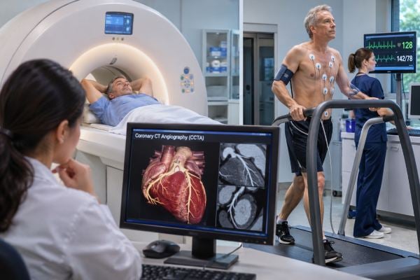
A heart CT scan and a stress test answer different questions, so the “better” test depends on what your clinician needs to know. Coronary CT angiography (CCTA) shows the coronary arteries directly and can identify both plaque and narrowing, while a stress test looks for evidence that the heart is not getting enough blood when it works harder. A separate CT test, the coronary artery calcium scan, measures calcified plaque for risk assessment and is not a substitute for CCTA or a stress test in someone with active concerning symptoms. For many people with stable chest pain and no known coronary disease, CCTA is a strong first test. Functional stress testing may be more useful when coronary disease is already known, heavy calcium or prior stents make CT harder to interpret, or the main question is whether a narrowing actually causes ischemia. Acute chest pain is different: emergency evaluation starts with an ECG and cardiac troponin, not with choosing a test from a menu.
Table of Contents
- What Heart CT and Stress Tests Show
- When Coronary CT Angiography Makes Sense
- When Stress Testing Makes Sense
- Test Types and How to Prepare
- Accuracy, Limitations, and Risks
- How Results Change the Next Step
- How to Choose the Right Test
What Heart CT and Stress Tests Show
The simplest distinction is anatomy versus function. Coronary CT angiography shows the structure of the coronary arteries. Stress testing asks how the heart behaves when demand rises.
| Test | Main question | What it can show | Common limitation |
|---|---|---|---|
| Coronary CT angiography | Is there coronary plaque or narrowing? | Calcified and noncalcified plaque, stenosis, coronary anatomy | Image quality can fall with heavy calcium, motion, some stents, or irregular rhythm |
| Coronary calcium scan | How much calcified plaque is present? | Calcium score for risk assessment | Does not show noncalcified plaque well or tell whether a narrowing reduces blood flow |
| Exercise ECG stress test | Does exercise provoke electrical evidence of ischemia? | Symptoms, exercise capacity, heart rate and blood-pressure response, ECG changes | Lower diagnostic accuracy than stress imaging in many patients |
| Stress imaging | Does increased demand reveal abnormal function or blood flow? | Wall-motion abnormalities or reduced perfusion, depending on the test | Cost, availability, and test-specific artifacts or contraindications |
A “heart CT” can mean several things, which is why the name alone is not enough. A coronary artery calcium (CAC) scan is a quick, noncontrast CT used mainly to refine preventive risk in selected people who do not have known coronary disease. A CCTA uses iodinated contrast and ECG-synchronized imaging to visualize the artery lumen and wall. If you are deciding whether preventive treatment should be intensified rather than investigating current symptoms, the role of a coronary artery calcium score is a separate question.
Stress testing also has several forms. A basic treadmill test records an ECG during exercise. Stress echocardiography uses ultrasound. Nuclear stress testing evaluates blood flow with a radioactive tracer. Stress cardiac MRI can assess perfusion and heart-muscle function without ionizing radiation. A pharmacologic stress agent can be used when a person cannot exercise adequately.
Neither category is universally superior. CCTA can be excellent for ruling out important anatomic coronary disease in the right patient. Stress imaging can be excellent for deciding whether known or suspected disease is functionally important.
When Coronary CT Angiography Makes Sense
CCTA is often a good choice for stable chest pain in a person with no known coronary artery disease when the clinician wants a direct look at plaque and stenosis. Modern chest-pain guidelines place CCTA among the main first-line options for appropriately selected patients at intermediate risk.
CCTA is especially attractive when:
- Symptoms are stable rather than an ongoing emergency
- There is no established coronary disease
- The probability of obstructive disease is not extremely high
- The heart rate and rhythm allow good image acquisition
- Kidney function and contrast allergy history permit iodinated contrast
- Knowing whether plaque is present would change preventive treatment
A major strength is that CCTA can show nonobstructive plaque, not just severe blockages. That matters because a person can have atherosclerosis that does not yet reduce blood flow enough to produce an abnormal stress test. Finding plaque can change the discussion about LDL lowering, smoking cessation, blood-pressure treatment, and other preventive steps.
CCTA can also show whether an apparent narrowing is mild, moderate, or severe. Some centers can add CT-derived fractional flow reserve or other advanced analysis to estimate whether an intermediate lesion is likely to limit blood flow, though availability varies.
CCTA in acute chest pain
In selected emergency-department patients who have had an initial ECG and high-sensitivity troponin assessment and remain at intermediate risk, CCTA can help clarify whether obstructive coronary disease is present. It is not the first action for someone with a clear ST-elevation heart attack, unstable vital signs, or another high-risk presentation that already requires urgent treatment.
If you are currently having new chest pressure, severe shortness of breath, fainting, or other emergency warning signs, the priority is urgent evaluation for dangerous chest pain, not scheduling an outpatient CT.
When CCTA is less attractive
Very heavy coronary calcification can create blooming artifact that makes a narrowing look worse or obscures the artery lumen. Fast or irregular heart rhythms, inability to cooperate with breath-holding, severe obesity depending on scanner capability, or extensive prior coronary stenting can also reduce image quality. People with significant kidney dysfunction or a serious iodinated-contrast reaction may need another strategy.
Prior bypass surgery is more nuanced. CT can image bypass grafts well in many cases, but assessing native vessels and deciding on the most useful test depends on the clinical question and local expertise.
When Stress Testing Makes Sense
A stress test is most useful when the key question is whether the heart develops ischemia under increased demand and how much functional limitation it causes.
Functional testing may be preferred when:
- Coronary artery disease is already known
- A prior CT or angiogram found an intermediate narrowing and its significance is uncertain
- Heavy coronary calcium is likely to make CCTA difficult to interpret
- Prior stents or bypass surgery complicate anatomic CT assessment
- Iodinated contrast is undesirable
- Exercise capacity itself is clinically important
- The local team has strong expertise in stress echo, PET, SPECT, or stress MRI
An exercise test can provide information that a resting CT cannot: how long you can exercise, whether symptoms occur at a reproducible workload, how heart rate and blood pressure respond, and whether arrhythmias appear with exertion. For someone with exertional chest pressure, that functional context can be useful.
But not all stress tests are equal. A treadmill ECG without imaging works best when the person can exercise adequately and the resting ECG is interpretable. Baseline ECG abnormalities, paced rhythm, left bundle branch block, use of certain medications, or inability to reach a useful exercise workload can make a simple treadmill test less informative.
Stress imaging adds more diagnostic information. Stress echocardiography looks for new wall-motion abnormalities when the heart works harder. Nuclear SPECT or PET looks for differences in blood flow between stress and rest. PET generally offers high image quality and can quantify myocardial blood flow in centers that have it. Stress cardiac MRI can evaluate perfusion, function, and scar without ionizing radiation.
The result should be interpreted in context. An “abnormal” stress test does not automatically mean a person needs a stent, and a “normal” test does not guarantee zero future cardiovascular risk. A guide to what cardiac stress test results mean can help separate ischemia findings from exercise capacity, rhythm changes, and artifacts.
Test Types and How to Prepare
Preparation depends heavily on which test you are having. Do not use generic internet instructions in place of the imaging center’s directions, especially for caffeine, medications, food, and diabetes treatment.
Preparing for CCTA
Many CCTA protocols aim for a relatively slow, steady heart rate because motion blur makes coronary arteries harder to see. You may receive a beta blocker before the scan if it is safe for you. Nitroglycerin is often given shortly before imaging to widen the coronary arteries and improve visualization.
Typical instructions may include:
- Avoiding caffeine for a specified period
- Taking usual medicines unless told otherwise
- Discussing erectile-dysfunction drugs because of potential interaction with nitrates
- Following instructions about eating and drinking
- Reporting previous iodinated-contrast reactions
- Providing recent kidney-function information when clinically indicated
During the scan, an IV delivers contrast. You lie on the CT table, ECG leads are placed on the chest, and you hold your breath briefly while images are acquired. The actual imaging is fast, although preparation can take longer.
Preparing for an exercise stress test
Wear clothing and shoes suitable for walking or running. You may be told to avoid a heavy meal beforehand. Some heart-rate-slowing medications can blunt the ability to reach a target workload, so the ordering clinician may adjust them for certain tests. Never stop a beta blocker or other prescription on your own; the desired preparation differs depending on whether the test is assessing ischemia, rhythm, symptoms, or treatment response.
Preparing for nuclear or pharmacologic stress testing
Caffeine can interfere with commonly used vasodilator stress agents, so nuclear and some MRI stress protocols may require strict caffeine avoidance for a specified period. That can include coffee, tea, cola, chocolate, energy products, and some medicines. Diabetes medications and insulin may need coordination if fasting is required.
Stress echocardiography uses ultrasound and no radioactive tracer. Nuclear testing uses a tracer and a specialized camera. Stress MRI uses a strong magnetic field and contrast in many protocols, so implanted devices, kidney function, metal exposure, and claustrophobia may need review.
Accuracy, Limitations, and Risks
No cardiac test has perfect accuracy. The best test is the one whose strengths fit the patient and whose limitations are least likely to distort the answer.
CCTA limitations and risks
CCTA exposes the patient to ionizing radiation, although modern protocols can keep the dose relatively low. It also uses iodinated contrast. Most people tolerate contrast well, but allergic-type reactions can occur, and kidney risk requires individual consideration, especially in people with significant renal impairment or acute illness.
Calcified plaque can cause “blooming,” making a lesion look larger than it is. Motion from a high or irregular heart rate can blur images. Small-vessel disease and coronary spasm may cause symptoms even when the major epicardial arteries do not show a severe fixed blockage.
CCTA is highly useful when it shows no significant plaque or stenosis in an appropriately selected patient, but an anatomic test cannot explain every cause of chest pain. It may also uncover incidental findings outside the heart that lead to further imaging.
Stress-test limitations and risks
Exercise ECG has modest diagnostic accuracy compared with modern imaging and can produce false-positive or false-negative results. Stress imaging improves accuracy but introduces its own artifacts. Body habitus can affect echocardiography and SPECT. Prior infarction or scar can complicate interpretation.
Exercise stress testing carries a small risk of serious arrhythmia, severe ischemia, or other complications, which is why trained staff monitor the test and stop it for concerning symptoms or changes. Pharmacologic stress agents can cause flushing, breathlessness, chest discomfort, headache, low blood pressure, bronchospasm in susceptible people, or rhythm effects. The team screens for contraindications and keeps reversal or emergency treatment available.
Radiation varies by modality. Stress echocardiography and stress MRI do not use ionizing radiation. Nuclear SPECT and PET do. CCTA does. The relevant question is not simply “which has radiation?” but whether the expected diagnostic benefit justifies the exposure and whether a non-radiation alternative would answer the same question well.
How Results Change the Next Step
A useful test is one that changes management. Before ordering CCTA or a stress test, clinicians should be able to say what they will do with a clearly normal, borderline, or clearly abnormal result.
If CCTA is normal or nearly normal
No plaque or only minimal plaque makes obstructive coronary disease an unlikely explanation for stable symptoms. The next step may shift toward non-cardiac causes, microvascular or vasospastic disease in selected patients, or simple follow-up depending on the presentation.
If CCTA shows nonobstructive plaque, the symptoms may still need another explanation, but the plaque itself matters for prevention. LDL goals, statin treatment, blood pressure, smoking, diabetes, exercise, and diet may deserve more attention.
Moderate stenosis can lead to functional assessment, advanced CT analysis, stress imaging, or invasive angiography depending on the artery involved, symptoms, and risk. Severe or high-risk anatomy may prompt invasive angiography and a discussion about revascularization.
If a stress test is normal
A normal high-quality stress test lowers the likelihood that a flow-limiting blockage is causing symptoms at the tested workload. It does not mean the coronary arteries are plaque-free. A person can have nonobstructive atherosclerosis with a normal functional test, so prevention still depends on overall risk factors.
If a stress test is abnormal
The significance depends on how much ischemia is present, where it occurs, exercise capacity, symptoms, blood-pressure response, ventricular function, and other findings. Mild or equivocal abnormalities may lead to another noninvasive test. High-risk findings can lead to invasive coronary angiography.
Results should also be matched to the original symptom. If the problem is exertional pressure suggestive of angina, a result that explains the symptom carries more weight than an incidental finding unrelated to the clinical pattern.
How to Choose the Right Test
For most people, the decision comes down to four questions: What is the symptom pattern? Is coronary disease already known? Which test will produce interpretable images in this person? What result would change treatment?
A simplified way to think about it is:
- Stable chest pain, no known coronary disease, suitable for CT: CCTA is often an excellent first choice.
- Known coronary disease or a need to assess ischemia: stress imaging is often more informative.
- Heavy coronary calcium, prior stents, or anatomy likely to be hard to read on CT: functional testing may be preferable.
- Good exercise capacity and interpretable resting ECG with an appropriate clinical question: exercise ECG may be useful, although many patients benefit from imaging.
- Need to avoid ionizing radiation: stress echo or stress MRI may fit if otherwise appropriate.
- Need to avoid iodinated contrast: a functional test may be better.
- Preventive risk question without active symptoms: a calcium scan may be the relevant CT test rather than CCTA.
Age can influence test choice, but it is not a rigid rule. Younger patients often have less coronary calcium and may obtain especially clear anatomic information from CCTA. Older patients are more likely to have heavy calcification or established disease, which can make functional testing attractive. Individual anatomy and history matter more than an age cutoff.
Cost and local expertise also matter. A technically excellent test interpreted by an experienced team is more valuable than a theoretically ideal test that is not available or cannot be performed well. Insurance rules can also affect the sequence.
If you have ongoing stable symptoms, ask the ordering clinician three practical questions: “Are we looking for plaque, a flow problem, or both?” “What could make this test hard to interpret in me?” and “What happens if the result is borderline?” Those questions usually reveal why one test is being chosen over another.
And if symptoms are acute or rapidly worsening, do not try to choose between CCTA and a stress test yourself. Emergency clinicians first determine whether you are having a heart attack or another dangerous condition. The right advanced test comes after that initial risk assessment.
References
- 2021 AHA/ACC/ASE/CHEST/SAEM/SCCT/SCMR Guideline for the Evaluation and Diagnosis of Chest Pain: A Report of the American College of Cardiology/American Heart Association Joint Committee on Clinical Practice Guidelines 2021 (Guideline)
- 2022 ACC Expert Consensus Decision Pathway on the Evaluation and Disposition of Acute Chest Pain in the Emergency Department: A Report of the American College of Cardiology Solution Set Oversight Committee 2022 (Position Statement)
- 2022 use of coronary computed tomographic angiography for patients presenting with acute chest pain to the emergency department: An expert consensus document of the Society of cardiovascular computed tomography (SCCT): Endorsed by the American College of Radiology (ACR) and North American Society for cardiovascular Imaging (NASCI) 2022 (Position Statement)
- CAD-RADS™ 2.0 – 2022 Coronary Artery Disease-Reporting and Data System: An Expert Consensus Document of the Society of Cardiovascular Computed Tomography (SCCT), the American College of Cardiology (ACC), the American College of Radiology (ACR), and the North America Society of Cardiovascular Imaging (NASCI) 2022 (Position Statement)
- CAR and CSTR Cardiac Computed Tomography (CT) Practice Guidelines: Part 1 Coronary CT Angiography (CCTA) 2024 (Guideline)
- ESR Essentials: imaging in stable chest pain – practice recommendations by ESCR 2024 (Guideline)
Disclaimer
This article is for general education and cannot determine which cardiac test is appropriate for an individual patient. Test choice depends on symptoms, prior coronary disease, ECG findings, kidney function, rhythm, medications, pregnancy status, contrast history, and local expertise. New or severe chest pressure, fainting, major shortness of breath, or rapidly worsening symptoms require urgent medical evaluation rather than outpatient test selection.