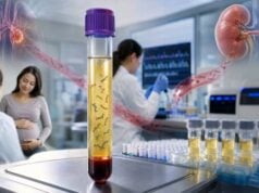
A liquid biopsy analyzes tumor-related material in blood or another body fluid. In routine precision oncology, the term most often refers to testing plasma for circulating tumor DNA, or ctDNA: short DNA fragments released from cancer cells into the bloodstream. The test can identify selected mutations, copy-number changes, rearrangements, or methylation patterns without removing a new tissue sample. It may help select treatment in advanced cancer, detect resistance, follow molecular response, or investigate residual disease after curative-intent therapy. Its convenience does not eliminate biological limitations. Some tumors release abundant ctDNA, while others shed very little, especially when disease volume is low or confined to certain sites. Blood also contains DNA from normal cells, including age-related blood-cell clones that can mimic tumor variants. A positive result may be highly informative, but a negative result cannot always exclude an actionable tumor alteration. Interpretation requires the assay’s scope, tumor context, variant level, collection timing, and intended clinical use.
- Liquid biopsy is a specimen approach; different tests measure different analytes and answer different questions.
- Plasma ctDNA can reveal tumor mutations and may capture DNA from multiple disease sites.
- Low tumor shedding is a major cause of false-negative or noninformative results.
- Variants from clonal hematopoiesis or germline DNA can be mistaken for tumor findings.
- Tissue testing may still be needed for diagnosis, histology, or confirmation after a negative plasma test.
- Molecular residual disease and cancer-screening applications require assay- and disease-specific evidence.
Table of Contents
- What liquid biopsy measures
- How ctDNA testing is used in cancer care
- Collection and laboratory analysis
- Understanding a positive result
- Understanding a negative or low-shedding result
- Molecular residual disease and serial monitoring
- Clonal hematopoiesis, germline findings, and other pitfalls
- Choosing next steps
What Liquid Biopsy Measures
“Liquid biopsy” is an umbrella term. Tests may analyze cell-free DNA, circulating tumor cells, cell-free RNA, extracellular vesicles, proteins, metabolites, or combinations of markers in blood, urine, cerebrospinal fluid, pleural fluid, or other specimens. A test labeled liquid biopsy should therefore be identified by its analyte and purpose rather than assumed to be a single standardized procedure [1].
Most clinical molecular liquid biopsies begin with plasma cell-free DNA. Cell-free DNA consists of short fragments released during normal cell turnover. In a person with cancer, a variable fraction originates from tumor cells and is called ctDNA. The proportion can range from undetectable to a substantial share of the total cell-free DNA. It is influenced by tumor burden, vascularity, cell death, disease site, treatment, and specimen handling.
A plasma assay may be broad or targeted. Broad next-generation sequencing panels evaluate many cancer genes for single-nucleotide variants, small insertions or deletions, selected copy-number changes, and some fusions. Targeted PCR or digital PCR assays look for one or a few known variants with high sensitivity. Tumor-informed assays first sequence a tissue tumor, select patient-specific variants, and then search for those variants in plasma. Tumor-naive or tumor-uninformed assays analyze a predefined set of features without requiring prior tumor tissue.
Liquid biopsy does not produce a physical piece of tumor. It cannot show tissue architecture, cell type, grade, invasion, or the tumor microenvironment. It also may not reveal where an abnormal signal originated. For most new cancers, tissue pathology remains necessary to establish the diagnosis. Plasma testing is most useful when it answers a focused molecular question, complements tissue, or provides information when a tissue biopsy is unsafe, delayed, insufficient, or no longer representative.
One potential advantage is that ctDNA can enter the bloodstream from more than one tumor site. A single tissue biopsy samples one location, whereas plasma may reflect several metastatic clones. However, this representation is uneven. A rapidly shedding liver metastasis may dominate the plasma signal while a low-shedding brain or bone lesion contributes little. Liquid biopsy is therefore a composite sample, not a complete map of every cancer cell.
How ctDNA Testing Is Used in Cancer Care
The strongest routine use is often genomic profiling in advanced cancer when the result can guide therapy. Plasma may identify an actionable driver such as an activating mutation, resistance alteration, amplification, or fusion. It can be especially helpful when tissue is scarce or obtaining another biopsy would be risky. The exact genes and variant types detected depend on the assay, and treatment relevance depends on tumor type and current clinical guidance.
In lung cancer, plasma profiling may rapidly identify targetable alterations. In breast cancer, it may detect acquired endocrine-resistance mutations. In prostate cancer, it may identify homologous-recombination pathway alterations or other treatment-relevant changes. Colorectal, biliary, ovarian, and additional cancers also have specific contexts in which plasma findings may contribute. These examples are not interchangeable: an alteration actionable in one cancer may have uncertain relevance in another.
Liquid biopsy can also evaluate resistance after progression. Tumors evolve under treatment pressure, and new variants can emerge that reduce drug binding, reactivate a pathway, or activate a bypass pathway. A blood draw may be easier to repeat than a tissue biopsy and can reveal several resistance clones. Yet tissue remains important when progression could reflect histologic transformation, because ctDNA cannot show a change in cell morphology.
Serial ctDNA measurements are being studied and used in selected settings to assess response. A falling tumor-derived signal after treatment may indicate molecular response, while a rising signal can precede radiographic progression. Timing is critical: transient release of DNA after treatment can cause an early increase, and different assays report quantity differently. A single value should not be interpreted as a universal tumor-burden measurement.
After surgery or other curative-intent treatment, highly sensitive assays may look for molecular residual disease. This application can stratify recurrence risk in some cancers, but whether changing treatment solely because of a result improves outcomes must be established for each disease, stage, assay, and intervention. Regulatory guidance emphasizes rigorous validation when ctDNA is used as a biomarker in early-stage drug development [2].
Multi-cancer early detection and population screening are separate applications. A screening test must perform well in people without a known cancer, where prevalence is low and false-positive consequences are substantial. Results from treatment-selection or residual-disease assays cannot be generalized to screening. Positive screening signals require a defined diagnostic pathway, and a negative test cannot replace established screening programs unless evidence and guidelines specifically support that use.
Collection and Laboratory Analysis
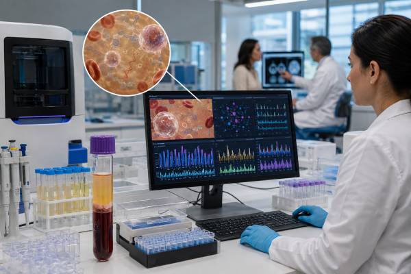
Plasma ctDNA testing begins with a blood draw into tubes designed either for rapid processing or for stabilizing blood cells during transport. Delayed separation or poor handling can cause white blood cells to rupture and release genomic DNA, diluting the tumor fraction. The laboratory separates plasma, extracts cell-free DNA, prepares the assay, and applies quality controls.
The amount of ctDNA is usually small, so methods must distinguish true low-level variants from sequencing and polymerase errors. Unique molecular identifiers can label original DNA fragments before amplification, allowing bioinformatics to collapse duplicate reads and suppress noise. Deep sequencing increases the opportunity to observe rare molecules, but depth alone cannot overcome absent tumor shedding or uncovered genomic regions.
Different assay designs create different detection limits:
| Assay approach | Typical strength | Important limitation |
|---|---|---|
| Broad plasma NGS panel | Surveys many genes and variant classes in one test | Sensitivity varies by alteration; low-level and complex events may be missed |
| Targeted digital PCR | Very sensitive for one known variant | Cannot discover unanticipated resistance or other drivers |
| Tumor-informed MRD assay | Tracks individualized tumor variants at very low levels | Requires adequate tumor tissue and extra development time |
| Tumor-naive MRD assay | Does not require prior tumor sequencing | May have less patient-specific discrimination and assay-dependent specificity |
| Methylation or fragment-pattern assay | Can use broader cancer-associated signals | Tissue-of-origin and clinical meaning require dedicated validation |
Preanalytic timing matters. A sample obtained soon after surgery or a major procedure may contain increased background cell-free DNA. Transfusion, inflammation, exercise, and other physiologic factors can affect total cell-free DNA, although their practical influence varies. The test requisition should include cancer type, stage, treatment history, and collection timing so the laboratory can interpret the result appropriately.
Reports may provide variant allele fraction, or VAF, the percentage of sequence reads or corrected molecules carrying a variant. Plasma VAF is not the same as the percentage of tumor cells. It reflects tumor fraction, copy number, clonality, DNA shedding, and technical processing. A variant with a VAF of 0.5% can be clinically real, but confidence depends on assay validation and controls.
A low or undetectable estimated tumor fraction limits the strength of a negative result. Some reports use maximum somatic allele fraction or another composite measure to estimate ctDNA content. Others do not provide a tumor-fraction estimate. The absence of this information does not invalidate the assay, but it makes the report’s quality and limitation statements especially important.
Understanding a Positive Result
A positive result indicates that the assay detected a reportable molecular feature in the fluid sample. Interpretation should answer four questions: Is the signal technically credible? Is it likely tumor derived? Is it clinically relevant in this cancer? Does it change management now?
For treatment selection, the report may classify variants by evidence level and list associated therapies or trials. An oncologist should confirm that the evidence applies to the patient’s tumor type, stage, and treatment setting. A drug approved for one molecular alteration may require a specific variant, mutation class, or companion diagnostic. A general statement that a gene is “targetable” is not enough.
Concordance with known tumor biology increases confidence. A plasma result that matches a previously documented tissue driver is usually straightforward. A newly detected resistance variant after exposure to the relevant therapy may also fit the clinical course. An unexpected variant at a low level deserves scrutiny for sequencing artifact, clonal hematopoiesis, or a germline origin.
A positive plasma result can sometimes avoid a repeat tissue biopsy when the alteration is well validated and treatment guidance accepts plasma testing. It does not necessarily replace pathology. If no tissue diagnosis exists, imaging and molecular findings alone may be insufficient to establish cancer type. Tissue can also be needed to assess protein expression, histologic transformation, or biomarkers not reliably measured in plasma.
Multiple variants can appear because tumors are heterogeneous. The report may show a dominant driver, subclonal resistance variants, passenger changes, and alterations from non-tumor blood cells. VAF differences can suggest but do not prove clonal relationships. Copy-number gains and losses are particularly dependent on tumor fraction, and complex fusion detection can be less sensitive in fragmented plasma DNA than in tissue RNA.
A positive residual-disease result has a different meaning from a positive treatment-selection panel. In the MRD setting, it generally indicates that tumor-derived molecules remain detectable after therapy and is often associated with a higher recurrence risk. It does not specify where disease is located, how much visible tumor is present, or exactly when recurrence will occur. Management should follow evidence for that disease and assay rather than a generic rule.
Understanding a Negative or Low-Shedding Result
A negative plasma test means no reportable alteration was detected within the test’s scope. It is not equivalent to “the cancer has no mutations” or “no cancer is present.” The result may be truly negative, or the tumor may not have released enough detectable DNA.
Low shedding is common in small-volume disease and varies by tumor biology. Some brain tumors, isolated central nervous system metastases, low-grade tumors, and disease limited to certain anatomic sites may contribute little ctDNA to plasma. An NCI discussion of liquid-biopsy sensitivity highlights the fundamental challenge that increasing the available ctDNA could improve detection [3]. No laboratory technique can identify a molecule that never entered the tested tube.
A negative treatment-selection panel should be interpreted alongside tumor fraction. If the sample contains adequate ctDNA and no alteration is found, the result is more informative. If tumor fraction is low or unknown, tissue testing is often recommended when feasible. ESMO expert recommendations have emphasized that a noninformative plasma result should prompt tissue analysis because of false-negative risk [4].
Assay scope is another limitation. A panel may not cover every exon, intron, fusion partner, copy-number event, or structural variant. Some biomarkers require tissue RNA, immunohistochemistry, or other methods. A negative DNA liquid biopsy may miss an expressed fusion that a tissue RNA assay could detect. Similarly, plasma cannot reliably determine all histologic or microenvironmental biomarkers.
A negative MRD result means the assay did not detect its targeted signal at that time. It lowers risk in some validated contexts but does not prove cure. Residual disease may be below the detection threshold, may not be shedding, or may lack the tracked variants because of clonal evolution. Serial negative tests can be more informative than one isolated result, but surveillance imaging and clinical follow-up should not be discontinued unless a validated protocol specifically supports that change.
The wording “not detected,” “negative,” “below limit of detection,” and “quantity not sufficient” can represent different situations. Patients and clinicians should review the technical comment and ask whether the laboratory considers the result valid, limited, or failed.
Molecular Residual Disease and Serial Monitoring
Molecular residual disease testing looks for tumor-derived material after treatment intended to eliminate all disease. It is often discussed after surgery for colorectal, breast, bladder, lung, and other solid tumors. The clinical promise is to identify patients who may need more treatment while sparing lower-risk patients unnecessary toxicity. Achieving that promise requires prospective evidence that a ctDNA-guided decision improves outcomes, not only that ctDNA predicts recurrence.
Tumor-informed assays select variants from the patient’s tumor and design a personalized plasma test. Tracking several variants can improve specificity and reduce the chance that a blood-cell variant is mistaken for residual cancer. Tumor-naive assays use standardized panels, methylation signatures, fragment patterns, or integrated signals. They may be faster or avoid tissue dependency, but performance characteristics differ.
The collection schedule affects interpretation. Postoperative samples drawn too early can be diluted by cell-free DNA released during tissue injury. Later samples may improve analytical sensitivity but delay a treatment decision. During systemic therapy, predefined landmarks and trends are more interpretable than opportunistic draws taken at inconsistent times.
Results may be qualitative—detected or not detected—or quantitative. Some assays report molecules per milliliter, mean tumor molecules, VAF, or another proprietary measure. Values from different platforms are not directly interchangeable. A decline on one assay should be interpreted using that assay’s validated method and the same specimen type.
For monitoring advanced disease, ctDNA kinetics can complement imaging. Molecular changes may appear earlier than radiographic changes, but flare phenomena, mixed response, and low-shedding lesions complicate the picture. A treatment should not generally be stopped solely because of an isolated ctDNA rise unless a validated clinical pathway supports that action. Contemporary reviews describe growing integration while emphasizing unresolved standardization and clinical-utility questions [5].
Serial testing also creates a risk of overreaction to small fluctuations near the detection limit. Laboratory variation, blood volume, total cell-free DNA, and stochastic sampling can affect very low results. Trends should be assessed with clinical symptoms, imaging, tumor markers, and treatment timing.
Clonal Hematopoiesis, Germline Findings, and Other Pitfalls
Most cell-free DNA in plasma comes from noncancer cells, especially blood-forming cells. With age, some hematopoietic stem cells acquire mutations and expand into clones. This clonal hematopoiesis can release variants into plasma that overlap with genes altered in cancer. Without paired white-blood-cell sequencing or careful bioinformatic interpretation, a blood-cell variant may be incorrectly attributed to the tumor.
Clonal hematopoiesis commonly affects genes such as DNMT3A, TET2, and ASXL1, but clinically confusing variants can occur in other cancer genes. Context matters: a low-level mutation inconsistent with the tumor type may be blood derived, while the same gene can be a genuine tumor alteration in another setting. In people with hematologic malignancies, distinguishing sources is even more complex.
A variant near approximately 50% VAF can suggest a germline change, but VAF alone is not diagnostic. Tumor fraction, copy number, loss of heterozygosity, and clonal hematopoiesis can produce similar values. Plasma tumor profiling is not a substitute for hereditary-cancer testing. When a potentially inherited pathogenic variant is detected, confirmation should use an appropriate non-tumor specimen through a clinical germline laboratory, with genetic counseling.
Other pitfalls include:
- Recent or concurrent cancers: ctDNA may originate from a different malignancy than the one under evaluation.
- Biological heterogeneity: a plasma alteration may represent one metastatic clone rather than all disease sites.
- False-positive low-level calls: rare technical errors can survive filtering, especially near the assay threshold.
- False-negative structural variants: fragmented DNA and incomplete breakpoint coverage can reduce fusion detection.
- Treatment-related changes: therapy can alter shedding and clonal composition, changing the profile without a simple relationship to tumor size.
- Uncertain variants: a detected change may have no established diagnostic or treatment significance.
These problems do not make liquid biopsy unreliable. They explain why result interpretation is an expert task and why paired tissue, leukocyte, or germline testing may be needed.
Choosing Next Steps
Before ordering, define the intended decision. A broad plasma panel for treatment selection is different from a targeted resistance assay, MRD test, recurrence-surveillance test, or early-detection test. The evidence, optimal timing, and consequences of a negative result differ for each.
After a positive treatment-selection result, ask whether the alteration is clearly tumor derived, whether it is actionable in this cancer, and whether confirmation is required. Review current treatment guidance and prior therapies. A molecular tumor board can help with unusual variants, multiple competing targets, or trial options.
After a negative result, ask whether tumor fraction was adequate and whether the assay covered the relevant alteration classes. When tissue is available and the result is noninformative, tissue profiling may reveal changes missed in plasma. A new biopsy can also assess histologic transformation. When biopsy is unsafe, repeating plasma at progression or using another body fluid may be considered in selected situations.
For MRD or monitoring, ask what prospective evidence supports testing at the planned time point and what action will follow each possible result. Testing is less useful when neither a positive nor negative result would change care. Insurance coverage and laboratory access may also depend on disease and indication.
The full report should be retained. Important details include specimen date, assay name, reportable genes and variant classes, detection limits, tumor-fraction estimate, variants and VAFs, therapy evidence level, and comments about clonal hematopoiesis or germline confirmation. Comparing serial tests is most reliable when the same validated method is used.
Liquid biopsy is best viewed as a dynamic complement to tissue, imaging, and clinical assessment. It can answer questions that tissue cannot answer easily, particularly about evolving tumor genetics. It also has failure modes unique to blood. The safest interpretation recognizes both sides: a detected, well-validated tumor variant may be immediately useful, while an undetected signal may simply reflect too little ctDNA in the sample.
References
- Liquid Biopsy Consortium. 2025. National Cancer Institute program overview.
- Use of Circulating Tumor Deoxyribonucleic Acid for Early-Stage Solid Tumor Drug Development. 2024. FDA guidance.
- Increasing ctDNA Volume to Improve Liquid Biopsy Sensitivity. 2024. National Cancer Institute research summary.
- ESMO recommendations on the use of circulating tumour DNA assays for patients with cancer: a report from the ESMO Precision Medicine Working Group. 2022. Professional recommendation.
- Circulating tumor DNA to monitor treatment response in solid tumors. 2025. Review article.
Disclaimer
This article provides general information and does not determine whether a liquid biopsy is appropriate or interpret an individual cancer result. Treatment, surveillance, and residual-disease decisions should be made with an oncology team using the complete report, tissue findings, imaging, and current disease-specific guidance. A plasma test should not delay urgent diagnostic biopsy or treatment when clinically required.