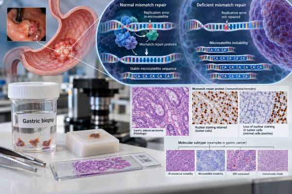
An MSI test for gastric cancer checks whether the tumor has a defective DNA mismatch repair system. When mismatch repair fails, repeated DNA sequences called microsatellites accumulate errors, producing a microsatellite instability-high (MSI-H) tumor. The same biology may be reported as deficient mismatch repair (dMMR) when pathology testing shows loss of one or more mismatch repair proteins. In stomach cancer, MSI/MMR status is useful because it helps define tumor biology and can influence treatment, especially the use of immune checkpoint inhibitors. It may also raise the question of an inherited mismatch repair disorder such as Lynch syndrome, although most MSI-H gastric cancers are not caused by Lynch syndrome. Testing is usually performed on biopsy or surgical tumor tissue, and it often sits beside HER2, PD-L1, and other biomarker tests in advanced gastric adenocarcinoma. The most useful interpretation comes from combining the MSI/MMR result with stage, histology, treatment setting, the exact assay used, and any hereditary cancer clues.
- MSI-H or dMMR means the gastric tumor has defective mismatch repair and may be especially sensitive to immune checkpoint blockade.
- MSS or pMMR means high-level microsatellite instability was not detected; most gastric cancers fall into this group.
- An MSI-H gastric tumor does not automatically mean Lynch syndrome because many cases arise from tumor-only MLH1 silencing.
- Testing can use MMR immunohistochemistry, MSI PCR, or validated molecular sequencing, depending on the laboratory and clinical purpose.
- MSI should be interpreted alongside HER2, PD-L1 CPS, stage, and other treatment biomarkers rather than used alone.
Table of Contents
- What MSI Means in Gastric Cancer
- How MSI and MMR Testing Is Done
- How to Interpret MSI-H, dMMR, MSS, and pMMR
- MSI as a Gastric Cancer Molecular Subtype
- How MSI Status Can Affect Treatment
- Lynch Syndrome and Hereditary Risk
- Limitations and Practical Follow-Up
What MSI Means in Gastric Cancer
Microsatellites are short repeated DNA sequences scattered throughout the genome. They are particularly prone to small copying errors when cells divide. A healthy mismatch repair system corrects many of these errors. The proteins most often evaluated in routine clinical testing are MLH1, PMS2, MSH2, and MSH6.
When this system fails, the tumor can accumulate many insertions and deletions in microsatellite regions. If instability reaches the laboratory’s threshold, the tumor is classified as MSI-H. The corresponding protein-level state is called dMMR. Although the two terms are often paired because they usually identify the same biology, one describes a DNA pattern and the other describes dysfunction of the repair proteins.
In gastric adenocarcinoma, this matters for more than classification. MSI-H tumors tend to carry a high number of mutations, which can generate abnormal proteins that make the cancer more visible to the immune system. This provides a biological explanation for the strong activity of immune checkpoint inhibitors seen in many MSI-H/dMMR tumors.
MSI-H is not a measurement of tumor burden. It cannot tell whether a tumor is 2 cm or 8 cm, whether lymph nodes contain cancer, or whether metastatic disease is present. Those questions require staging with pathology and imaging. MSI is instead a molecular characteristic that helps describe how the tumor developed and how it may respond to selected therapies.
The proportion of gastric cancers that are MSI-H varies by region, stage, age, and tumor location. MSI-H is generally more common in resectable or earlier-stage gastric cancer than in advanced metastatic cohorts. It is also more frequently seen in older patients and in certain intestinal-type tumors. These associations can raise suspicion, but no demographic or pathology feature is accurate enough to substitute for testing.
How MSI and MMR Testing Is Done
Testing is usually performed on formalin-fixed tumor tissue obtained by endoscopic biopsy or surgery. A patient normally does not need fasting, medication changes, or a second procedure solely for MSI testing if adequate tissue is already available.
MMR immunohistochemistry
Mismatch repair immunohistochemistry, or MMR IHC, stains tumor tissue for MLH1, PMS2, MSH2, and MSH6. Normal cells on the same slide act as internal positive controls. Pathologists evaluate whether the tumor-cell nuclei retain or lose staining.
Common patterns include:
- loss of MLH1 and PMS2;
- loss of MSH2 and MSH6;
- isolated loss of MSH6; or
- isolated loss of PMS2.
In sporadic gastric cancer, MLH1 loss is commonly linked to acquired MLH1 promoter hypermethylation within the tumor. That is one reason an abnormal MMR result is not synonymous with an inherited syndrome.
MSI PCR
PCR-based testing compares a set of microsatellite markers and looks for abnormal length changes. Depending on the assay, tumor DNA may be compared with normal DNA from the same person or analyzed using a panel designed to identify instability directly. The result is usually reported as MSI-H or microsatellite stable.
Next-generation sequencing
Some broad tumor sequencing panels calculate MSI status while simultaneously assessing mutations, amplifications, and other genomic changes. This can be efficient when a patient with advanced gastric cancer already needs comprehensive molecular profiling. However, professional guidance has emphasized that assay choice should match the tumor type and intended clinical use. For gastroesophageal cancers, MMR IHC and MSI PCR remain well-established approaches.
Sampling can matter in gastric cancer because small biopsies may contain limited viable tumor, inflammation, necrosis, or treatment-related change. If the tissue is insufficient or a result is technically indeterminate, a different block or later specimen may be tested. When results are discordant and the decision affects immunotherapy, confirmatory testing with a second validated method can be appropriate.
How to Interpret MSI-H, dMMR, MSS, and pMMR
An MSI-H result means the tumor demonstrates high-level microsatellite instability by a molecular assay. A dMMR result means the mismatch repair system is deficient, usually shown by loss of one or more MMR proteins on IHC. In routine gastric cancer care, these results often lead to the same broad clinical conclusion: the tumor has mismatch repair deficiency biology.
An MSS result means the tested microsatellites do not meet criteria for high instability. A pMMR result means MLH1, PMS2, MSH2, and MSH6 are retained on IHC. Most gastric adenocarcinomas are MSS/pMMR.
The important point is what these categories do and do not imply.
MSI-H/dMMR can be predictive. In advanced cancer, it predicts a higher probability of benefit from immune checkpoint blockade than would be expected in an unselected population. It is therefore a treatment-selection biomarker.
MSI-H can be prognostic. In localized gastric cancer, MSI-H biology has often been associated with a more favorable prognosis than some other molecular subtypes, although the size of that advantage differs by study and clinical setting.
MSI-H can be a hereditary clue. Loss of mismatch repair function can occur in Lynch syndrome, but in gastric cancer a tumor-only mechanism is common. Hereditary assessment therefore depends on the protein-loss pattern, age, personal history, family history, and sometimes additional tumor testing.
MSS/pMMR is not a “normal cancer” result. It only means that mismatch repair deficiency was not detected. An MSS tumor can still have HER2 amplification, high PD-L1 expression, CLDN18.2 expression, EBV-related biology, actionable gene fusions, or other clinically relevant features.
Results should also be distinguished from tumor mutational burden. MSI-H tumors often have high tumor mutational burden, but the tests are not interchangeable. A tumor can be TMB-high without being MSI-H, and assay thresholds differ.
MSI as a Gastric Cancer Molecular Subtype
Modern gastric cancer classification recognizes that tumors that look similar under a microscope can be biologically different. Large genomic studies helped establish several broad molecular patterns, including Epstein-Barr virus-associated tumors, MSI tumors, genomically stable tumors, and tumors with chromosomal instability.
The MSI subtype is characterized by defective mismatch repair and a high mutation burden. Many MSI gastric cancers show MLH1 promoter methylation rather than an inherited mismatch repair mutation. They also tend to have distinctive immune features, including increased immune-cell infiltration and expression of immune regulatory pathways.
This molecular-subtype concept is useful because it explains why a single pathology label such as “gastric adenocarcinoma” is no longer enough for many treatment decisions. Two patients with the same stage can have tumors with very different therapeutic vulnerabilities.
The molecular subtype does not replace standard pathology. Clinicians still need to know whether the tumor is intestinal or diffuse type, whether signet-ring features are present, how deeply the tumor invades, whether lymph nodes are involved, whether margins are clear after surgery, and whether metastatic disease is present.
Nor does MSI replace the other biomarker tests that may be required in advanced disease. Depending on tumor location and treatment setting, a gastric cancer workup may include:
- HER2 testing by IHC with confirmatory in situ hybridization when appropriate;
- PD-L1 testing using combined positive score (CPS);
- MSI or MMR testing;
- CLDN18.2 testing where relevant to available therapy; and
- broad genomic profiling in selected patients.
The practical value of molecular classification is therefore additive. It creates a more complete picture when combined with traditional staging and pathology.
How MSI Status Can Affect Treatment
MSI-H/dMMR status has its clearest treatment effect in advanced or metastatic gastric cancer because immune checkpoint inhibitors can be particularly effective in this biological subgroup.
Advanced disease
Treatment of advanced gastric or gastroesophageal junction adenocarcinoma increasingly depends on several biomarkers at once. HER2 status, PD-L1 CPS, MSI/MMR status, and other markers can influence the regimen. MSI-H/dMMR is especially important because checkpoint inhibitors can produce durable responses even when other conventional biomarkers are less favorable.
The exact immunotherapy regimen depends on line of therapy, HER2 status, PD-L1 expression, drug approvals, prior treatment, comorbidities, and local guidelines. MSI-H should therefore trigger a treatment discussion, not an automatic prescription without context.
It is also important not to confuse PD-L1 CPS with MSI status. A tumor can be MSI-H with a low CPS or MSS with a high CPS. The biomarkers measure different biological processes. When both are available, the oncologist integrates them rather than choosing one and ignoring the other.
Localized and resectable disease
The role of MSI in localized gastric cancer is more nuanced. Retrospective and pooled analyses have suggested that MSI-H localized gastric cancers may have favorable prognosis and may derive different levels of benefit from conventional perioperative chemotherapy than MSS tumors. At the same time, immunotherapy is rapidly changing the research landscape for localized dMMR/MSI-H disease.
Because evidence and approvals evolve, a patient with resectable MSI-H gastric cancer should have treatment planned in a multidisciplinary setting. Surgery, perioperative systemic therapy, clinical trials, tumor location, stage, and surgical fitness all matter. MSI status alone should not be used to skip established curative treatment without specialist guidance.
After progression
If a patient has already received treatment and the tumor was never tested for MSI/MMR, later testing can still be useful. Archival tissue may be adequate. If not, a new biopsy or a validated comprehensive molecular assay may provide the information. The value is highest when the result could open a treatment option that was not previously considered.
Lynch Syndrome and Hereditary Risk
Lynch syndrome is caused by an inherited pathogenic variant in a mismatch repair gene or EPCAM. Gastric cancer is part of the Lynch-associated cancer spectrum, although colorectal and endometrial cancers are more common manifestations.
An MSI-H/dMMR gastric cancer can therefore raise hereditary concern, especially when the patient is young, has multiple Lynch-associated cancers, or has a strong family history. However, most MSI-H gastric cancers are not Lynch syndrome. Many result from acquired MLH1 promoter hypermethylation in the tumor.
The pattern of protein loss can guide next steps. MLH1/PMS2 loss often leads clinicians to consider tumor methylation analysis and the overall clinical context. Loss of MSH2/MSH6, isolated MSH6 loss, or isolated PMS2 loss may create stronger concern for an inherited mismatch repair defect.
Germline testing is performed on normal DNA, usually from blood or saliva. Possible results include a pathogenic variant, a negative result, or a variant of uncertain significance. A VUS should not be used as proof of Lynch syndrome or as a reason to test relatives for a presumed disease-causing mutation.
If a pathogenic germline variant is found, relatives may benefit from targeted testing. The result can influence screening for colorectal and other Lynch-associated cancers. Recommendations depend on the specific gene because cancer risks differ between MLH1, MSH2, MSH6, and PMS2.
A normal MSI/MMR tumor screen reduces the likelihood of classic Lynch-associated mismatch repair deficiency but does not erase a highly suspicious family history. Genetics professionals may still recommend germline testing based on age, multiple cancers, or family pattern.
Limitations and Practical Follow-Up
MSI/MMR tests are robust, but they are not infallible. Small biopsies, poor fixation, low tumor-cell content, necrosis, and unusual staining patterns can make results difficult to interpret. Prior therapy can also change tissue appearance.
True discordance between IHC and molecular MSI testing is uncommon. When it occurs, the laboratory may review the slides, repeat stains, test another tumor block, or use an alternative method. A discordant result deserves particular attention if it determines eligibility for immunotherapy or prompts hereditary testing.
Common interpretation mistakes include:
- assuming MSI-H proves Lynch syndrome;
- assuming MSS means immunotherapy can never be used for any reason;
- using MSI as a marker of how much cancer is present;
- ignoring PD-L1, HER2, and other biomarkers when planning advanced-disease treatment;
- comparing tests from different laboratories without considering assay differences; and
- treating a VUS on germline testing as a pathogenic mutation.
After receiving a result, patients can ask their oncology team:
- Which method was used: IHC, PCR, NGS, or more than one?
- If IHC was abnormal, which proteins were lost?
- Does this result change my current treatment plan?
- Is PD-L1 CPS or HER2 status also available?
- Should I have genetic counseling or germline testing?
- Is the tissue result clear enough, or does it need confirmation?
- If my cancer is resectable, does MSI status affect the recommended perioperative strategy?
Keeping a copy of the full pathology and molecular report is useful. The precise wording often determines whether an oncologist or genetic counselor needs additional testing. A shorthand note such as “MSI positive” can leave out whether the tumor was MSI-H by PCR, dMMR by IHC, or abnormal by a sequencing algorithm.
The central takeaway is that MSI/MMR testing in gastric cancer has three different possible roles: defining molecular subtype, predicting treatment response, and flagging possible hereditary risk. The relative importance of each role changes with the patient’s stage and history. Interpreting all three correctly is more useful than treating MSI as a single yes-or-no cancer marker.
For patients whose tumor was tested on a very small pretreatment biopsy, it is worth confirming that the pathology report documents adequate internal controls and enough viable cancer cells. A technically satisfactory result usually remains clinically useful after surgery, but a failed or equivocal biopsy test can often be repeated on the larger resection specimen. This is especially important when MSI/MMR status will determine an immunotherapy option or hereditary evaluation.
References
- Mismatch Repair and Microsatellite Instability Testing for Immune Checkpoint Inhibitor Therapy: Guideline From the College of American Pathologists in Collaboration With the Association for Molecular Pathology and Fight Colorectal Cancer 2022 (Guideline)
- Mismatch Repair and Microsatellite Instability Testing for Immune Checkpoint Inhibitor Therapy: ASCO Endorsement of College of American Pathologists Guideline 2023 (Guideline)
- Gastric cancer: ESMO Clinical Practice Guideline for diagnosis, treatment and follow-up 2022 (Guideline)
- Potent therapeutic strategy in gastric cancer with microsatellite instability-high and/or deficient mismatch repair 2024 (Review)
- Analysis of Concordance Between Next-Generation Sequencing Assessment of Microsatellite Instability and Immunohistochemistry-Mismatch Repair From Solid Tumors 2024 (Study)
Disclaimer
This article provides general educational information and is not a substitute for individualized cancer care. MSI/MMR results should be interpreted by the treating oncology and pathology teams alongside stage, HER2, PD-L1, treatment history, and hereditary-risk information. Do not change cancer treatment based only on an online explanation of a biomarker result.