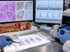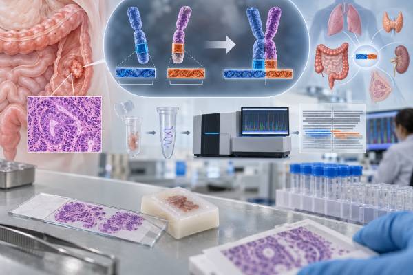
An NTRK fusion test for colon cancer looks for an abnormal joining of an NTRK gene with another gene, creating a continuously active TRK signaling protein that can drive cancer growth. True NTRK1, NTRK2, or NTRK3 gene fusions are rare in colorectal cancer, but they matter because they can make a tumor eligible for TRK-targeted therapy regardless of where the cancer started. That is why NTRK fusion is called a tumor-agnostic biomarker. The key word is fusion: an NTRK point mutation, amplification, or nonspecific TRK protein stain is not automatically equivalent to an actionable NTRK gene fusion. Testing may be performed with RNA- or DNA-based next-generation sequencing, fluorescence in situ hybridization, or immunohistochemistry used as a screening method in selected settings. A positive result should identify the fusion clearly enough for the oncology team to judge whether it is pathogenic and targetable. In colon cancer, NTRK testing is usually part of broader molecular profiling, especially in advanced disease.
- A confirmed NTRK1, NTRK2, or NTRK3 gene fusion can provide a tumor-agnostic treatment target in advanced colon cancer.
- NTRK fusions are very rare in colorectal cancer, so testing is often performed as part of a broad genomic panel rather than as a stand-alone test.
- An NTRK mutation or amplification is not the same as an NTRK fusion and does not automatically predict benefit from a TRK inhibitor.
- RNA-based sequencing is often especially useful for fusion detection because it can show the expressed fusion transcript.
- A negative result is only as complete as the assay’s fusion coverage, specimen quality, and ability to detect rare partners.
Table of Contents
- What an NTRK Fusion Is
- Why NTRK Testing Matters in Colon Cancer
- How NTRK Fusions Are Tested
- How to Read Positive, Negative, and Uncertain Results
- TRK Inhibitors and Tumor-Agnostic Treatment
- NTRK Fusions With Other Colon Cancer Biomarkers
- Limitations, Resistance, and Practical Next Steps
What an NTRK Fusion Is
The NTRK gene family contains NTRK1, NTRK2, and NTRK3. These genes encode the TRKA, TRKB, and TRKC receptor tyrosine kinases. In normal tissue, TRK proteins help regulate development and nervous-system signaling. They are normally activated in controlled circumstances by specific neurotrophins.
A cancer-driving NTRK fusion occurs when part of an NTRK gene becomes joined to part of another gene. The new hybrid gene can produce a fusion protein in which the TRK kinase domain is switched on continuously. That constant signal can activate downstream pathways involved in growth, survival, and proliferation.
This is different from several findings that may also appear on a molecular report:
- NTRK point mutation: a change in one or a few DNA letters. Most such variants are not established TRK-inhibitor biomarkers.
- NTRK amplification: extra copies of the gene. Amplification alone is not equivalent to a fusion.
- TRK protein expression: protein staining may be present for reasons other than an oncogenic fusion.
- NTRK fusion: a structural rearrangement that creates a functional NTRK fusion gene and is the established tumor-agnostic target.
The fusion partner can vary. In some cancers, classic recurrent partners are common; in colorectal cancer, partner genes can be diverse. The important feature is preservation of the NTRK kinase domain in a configuration that drives signaling.
Because true NTRK fusions are uncommon in colorectal cancer, their prevalence is generally well below 1% in unselected cases. They may be enriched in selected molecular subgroups, including some tumors that are wild-type for more common MAPK pathway drivers. Rare frequency is exactly why broad next-generation sequencing can be efficient: one assay can look for NTRK fusions while also evaluating other actionable alterations.
Why NTRK Testing Matters in Colon Cancer
The clinical importance of an NTRK fusion is disproportionate to how rarely it occurs. A confirmed fusion can identify a patient for a class of highly specific TRK inhibitors that were developed across tumor types rather than for one organ alone.
This is known as tumor-agnostic treatment. Instead of saying “this drug treats only colon cancer,” the indication is based on the molecular driver: an NTRK fusion-positive solid tumor that meets the treatment criteria. Colon, thyroid, salivary, lung, sarcoma, and other cancers can therefore share the same targeted-treatment logic when the fusion is present.
In metastatic colorectal cancer, molecular testing is already used to evaluate RAS, BRAF, MSI/MMR, HER2, and other alterations. NTRK testing fits naturally into this precision-oncology workup, especially when a comprehensive sequencing panel is available.
The result is most actionable when the cancer is locally advanced or metastatic, surgery would cause major morbidity, standard options have been exhausted or are unsatisfactory, or a labeled tumor-agnostic therapy otherwise applies. The exact sequence of therapy depends on current approvals, prior treatment, disease pace, symptoms, and other molecular targets.
For an early-stage colon cancer removed by surgery, finding an NTRK fusion does not automatically mean a TRK inhibitor should be given as adjuvant therapy. The strongest established role is in advanced disease. A biomarker can be actionable in one treatment setting without being validated for every stage.
NTRK status is also not a conventional prognostic score. A fusion can drive tumor biology, but the test is primarily ordered because it can identify a therapeutic target. Stage, metastatic sites, resectability, performance status, and response to prior therapy remain essential for prognosis.
How NTRK Fusions Are Tested
Several laboratory methods can detect NTRK fusions, but they do not have identical strengths.
RNA-based next-generation sequencing
RNA sequencing is often a strong method for fusion detection because it examines expressed transcripts. If an abnormal NTRK fusion is actively transcribed, RNA testing can identify the two joined genes and often the fusion breakpoint. This approach can detect diverse fusion partners without requiring the laboratory to guess each partner in advance.
RNA quality can be a limitation in older formalin-fixed tissue. If RNA is badly degraded, the assay may fail even when enough DNA remains for other testing.
DNA-based next-generation sequencing
DNA NGS can detect many structural rearrangements while also profiling mutations and copy-number changes. Its performance for NTRK fusions depends heavily on panel design, including whether relevant intronic regions are covered. Some NTRK genes have large introns that are technically difficult to capture, so a negative DNA result may occasionally need RNA follow-up when suspicion remains.
Pan-TRK immunohistochemistry
Pan-TRK IHC detects TRK protein expression. It can be used as a relatively quick screening method, particularly in settings where fusions are more common or sequencing resources are limited. However, protein expression is not perfectly specific for a fusion. Positive staining generally requires molecular confirmation before treatment.
Fluorescence in situ hybridization
FISH can identify rearrangements involving a specific NTRK gene. It can be useful when a particular gene is suspected, but separate probes may be needed for NTRK1, NTRK2, and NTRK3. It may also show that a rearrangement exists without identifying the exact fusion partner.
In colon cancer, broad RNA-capable NGS is often attractive because NTRK is only one of several rare actionable events. The best method depends on available tissue, laboratory expertise, cost, turnaround time, and whether the test is being used to screen or confirm.
How to Read Positive, Negative, and Uncertain Results
A positive actionable result should state that an NTRK1, NTRK2, or NTRK3 fusion was detected and, when possible, name the fusion partner. The report may describe the alteration as pathogenic, oncogenic, likely oncogenic, or therapeutically actionable.
The oncologist should confirm that the finding is a true gene fusion expected to retain the TRK kinase domain. A report containing only an NTRK missense variant should not be treated as equivalent.
A negative result means no reportable NTRK fusion was found by that assay. It does not have the same absolute meaning across every testing platform. A negative RNA fusion panel with good quality metrics can be more reassuring than a DNA-only panel with limited intronic coverage.
An indeterminate or failed result is different from negative. Examples include insufficient tumor, degraded RNA, low sequencing coverage, or technical quality-control failure. In that situation, another tissue block, a metastatic specimen, or a different method may be needed.
A variant of uncertain significance in NTRK is not the same as a confirmed fusion. VUS findings lack enough evidence to establish biological or treatment meaning. They should not be used alone to justify a TRK inhibitor.
The report may also state allele frequency, read support, breakpoint details, or assay confidence. These technical details help a molecular tumor board resolve unusual findings.
When the result comes from liquid biopsy, a positive fusion can be useful, but detection depends on how much tumor DNA is circulating. A negative plasma test can miss an alteration when ctDNA shedding is low. Tissue confirmation may therefore be reasonable if no driver is found and identifying an NTRK fusion would change therapy.
TRK Inhibitors and Tumor-Agnostic Treatment
TRK inhibitors block the abnormal kinase signal created by an NTRK fusion. The first generation of drugs established that tumors from many organs can respond when they share this driver.
Larotrectinib was developed as a selective TRK inhibitor and has shown high response rates across multiple NTRK fusion-positive solid tumors. Entrectinib also targets TRK proteins and has activity in fusion-positive tumors, with additional kinase targets. More recently, repotrectinib received an FDA tumor-agnostic approval for eligible adult and pediatric patients aged 12 years and older with NTRK gene fusion-positive solid tumors under the conditions specified in its label.
Drug choice depends on age, prior TRK inhibitor exposure, central nervous system disease, adverse-effect profile, availability, and regulatory indications. A patient who has never received a TRK inhibitor is different from one whose tumor has progressed after larotrectinib or entrectinib.
Responses can be dramatic, but they are not always permanent. Tumors may acquire on-target resistance mutations in the TRK kinase domain that reduce drug binding, or they may activate alternative signaling pathways. Newer TRK inhibitors have been designed partly to overcome some resistance mechanisms.
The existence of resistance does not weaken the value of finding the original fusion. It illustrates a broader principle of precision oncology: a driver can remain highly actionable while the tumor evolves under selective pressure.
Side effects vary by drug and can include dizziness, neurologic symptoms, weight gain, gastrointestinal effects, liver-enzyme abnormalities, fatigue, and other toxicities. Prescribing information and oncology monitoring are essential because TRK signaling also has normal physiologic roles.
NTRK Fusions With Other Colon Cancer Biomarkers
A complete colorectal cancer profile puts NTRK into context with more common markers.
RAS: KRAS and NRAS mutations are common drivers and guide anti-EGFR treatment. NTRK fusions are more often found in tumors lacking common RAS pathway mutations, although unusual co-alterations can occur.
BRAF: BRAF V600E defines another targetable subgroup. A classic BRAF V600E driver and a strong NTRK fusion driver are not typically expected together.
MSI/MMR: Some NTRK fusion-positive colorectal cancers have been reported in MSI-H/dMMR contexts. If both are present, the treatment team may have more than one biologically rational option, including immunotherapy and TRK inhibition, and must decide sequencing based on the clinical setting.
HER2: HER2 amplification can guide HER2-directed therapy in selected metastatic colorectal cancers. Like NTRK, it is part of the broader search for actionable drivers after RAS/BRAF testing.
Gene-fusion landscape: Colorectal cancer can rarely harbor other fusions such as ALK, ROS1, RET, or FGFR-related rearrangements. Their clinical relevance varies. Modern comprehensive profiling can detect multiple rare events in one test.
This is why NTRK should not be interpreted as a stand-alone “positive cancer marker.” The most useful question is: Does this fusion provide a validated treatment target in this patient’s stage and treatment setting, and how does it rank against other actionable biomarkers?
Limitations, Resistance, and Practical Next Steps
The biggest limitation of NTRK testing is not that the biomarker lacks value; it is that the event is rare and technically more difficult to detect than a simple hotspot mutation.
DNA panels can miss certain rearrangements because large introns are incompletely covered. RNA can degrade in fixed tissue. IHC can produce false-positive staining because normal TRK proteins are expressed in some tissues. FISH may require multiple gene-specific probes. Each method therefore answers the question differently.
When a report is unclear, useful follow-up questions include:
- Is the finding a true NTRK fusion or another type of NTRK alteration?
- Which gene is involved: NTRK1, NTRK2, or NTRK3?
- What is the fusion partner?
- Was the fusion detected by RNA, DNA, FISH, or IHC?
- If it was detected only by IHC, was molecular confirmation performed?
- If DNA testing was negative, did the assay have strong NTRK fusion coverage?
- Are my RAS, BRAF, MSI/MMR, and HER2 results also available?
- Does my current disease setting meet criteria for a TRK inhibitor?
- Have I previously received a TRK inhibitor, and could resistance testing matter now?
Patients should keep the complete molecular report rather than only a summary. Rare fusions can be easier to evaluate later if the exact partner, breakpoint, test method, and specimen are documented.
A final practical distinction is between screening and confirmation. A pan-TRK stain may be a screening result. A sequencing report that identifies a functional NTRK fusion is much closer to a treatment-defining result. Confusing those steps can lead either to missed therapy or to inappropriate treatment.
For the small fraction of colon cancers that truly harbor an NTRK fusion, accurate testing can uncover one of the clearest examples of tumor-agnostic precision medicine: a rare molecular alteration that matters more than the organ label for selection of a specific targeted drug class.
Another practical issue is when to retest. If a comprehensive RNA-capable assay on adequate tissue was clearly negative, repeating the same test immediately is unlikely to add value. Retesting becomes more reasonable when the original panel had limited fusion coverage, the assay failed quality metrics, only a small DNA panel was performed, or new metastatic tissue becomes available after the cancer has evolved. A molecular tumor board can help decide whether a second method is justified.
The pathology context can also change pretest probability. NTRK fusions are rare overall, so a positive result from a low-specificity screening method has a meaningful chance of being false positive unless confirmed. Conversely, a colorectal cancer that is wild-type for common drivers and has unusual molecular features may justify a more sensitive fusion search even after limited testing was unrevealing.
Patients sometimes ask whether relatives need NTRK testing. In the usual colorectal cancer setting, an oncogenic NTRK fusion is a somatic structural event in the tumor, not an inherited familial mutation. It therefore does not ordinarily lead to cascade testing of healthy relatives. Hereditary risk should be assessed separately through family history, MSI/MMR status, and germline testing when indicated.
When a fusion is confirmed, the report should remain in the patient’s permanent oncology record even if a TRK inhibitor is not needed immediately. Targeted options can become relevant later as disease and treatment history change, and repeating an old tissue search may be unnecessary if the original fusion was well characterized. Preserving the exact molecular result can therefore save time when a later-line decision becomes urgent.
References
- Molecular features and clinical actionability of gene fusions in colorectal cancer. 2025 (Review)
- FDA grants accelerated approval to repotrectinib for adult and pediatric patients with NTRK gene fusion-positive solid tumors 2024 (Official FDA Page)
- Treatment of Metastatic Colorectal Cancer: ASCO Guideline 2023 (Guideline)
- Metastatic colorectal cancer: ESMO Clinical Practice Guideline for diagnosis, treatment and follow-up 2023 (Guideline)
- NTRK fusions in colorectal cancer: clinical meaning and future perspective 2021 (Review)
Disclaimer
This article is for general education and is not a substitute for molecular pathology or oncology advice. NTRK findings should be interpreted using the exact alteration type, assay, disease stage, prior treatment, and current drug indications. Do not start or change targeted therapy without review by the treating cancer team.