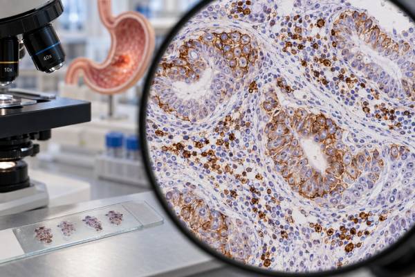
A PD-L1 test for gastric cancer measures programmed death-ligand 1 expression in tumor tissue, usually with immunohistochemistry, to help estimate whether an immune checkpoint inhibitor should be part of treatment. In gastric and gastroesophageal junction adenocarcinoma, the most commonly used scoring system is the combined positive score (CPS). CPS counts PD-L1-staining tumor cells and certain immune cells, divides that number by the total number of viable tumor cells, and multiplies by 100. A higher CPS generally indicates more PD-L1 expression and can strengthen the case for immunotherapy in specific treatment settings, but it is not a simple yes-or-no measure of whether immunotherapy will work. The clinically relevant cutoff depends on the drug, HER2 status, line of therapy, regulatory indication, and guideline. PD-L1 is also only one biomarker. MSI/MMR, HER2, CLDN18.2, tumor stage, prior therapy, and overall health can be equally or more important. The exact assay and scoring method on the pathology report should therefore be reviewed before treatment is selected.
- PD-L1 CPS is calculated from staining in tumor cells plus selected immune cells relative to the number of viable tumor cells.
- Common clinical cutoffs include CPS ≥1 and CPS ≥5, but the relevant threshold depends on the treatment regimen and current indication.
- A low CPS does not prove immunotherapy cannot work, and a high CPS does not guarantee response.
- For HER2-positive advanced gastric/GEJ adenocarcinoma, the current FDA pembrolizumab combination indication requires PD-L1 CPS ≥1.
- PD-L1 testing should be interpreted alongside HER2 and MSI/MMR status because these biomarkers can change first-line treatment choices.
Table of Contents
- What PD-L1 Is and Why It Is Tested
- How the CPS Score Is Calculated
- What CPS 1, CPS 5, and Higher Scores Mean
- How PD-L1 Affects Immunotherapy Decisions
- PD-L1 With HER2, MSI, and Other Biomarkers
- Testing Methods, Sampling, and Limitations
- Questions to Ask About a PD-L1 Result
What PD-L1 Is and Why It Is Tested
PD-L1 is a protein that can appear on tumor cells and immune cells in the tumor microenvironment. It binds to the PD-1 receptor on T cells and can suppress immune activity. Cancers can exploit this pathway to reduce immune attack.
Immune checkpoint inhibitors such as pembrolizumab and nivolumab block PD-1 signaling, releasing part of this immune brake. PD-L1 expression is therefore used as a predictive biomarker: it helps estimate how likely a patient is to benefit from adding a checkpoint inhibitor in certain advanced gastric cancer settings.
The test is performed on tumor tissue, not as a routine blood marker. A pathology laboratory applies a validated antibody assay to biopsy or surgical tissue and evaluates staining under the microscope.
PD-L1 is not a cancer stage and does not measure tumor quantity. A CPS of 20 does not mean 20% of the body contains cancer, nor does it mean the tumor is “20% immune responsive.” It is a scoring system for local protein expression in the tested tissue.
PD-L1 is also dynamic. Expression can differ between the primary gastric tumor and a metastasis, between separate areas of the same tumor, and before versus after systemic therapy. This biological variability helps explain why the score is useful but imperfect.
In gastric cancer, PD-L1 has become especially important because randomized trials have shown greater average benefit from adding PD-1 blockade to chemotherapy in groups with higher CPS values, while some approvals and recommendations use specific CPS thresholds. The threshold is treatment-specific rather than a universal definition of “PD-L1 positive.”
How the CPS Score Is Calculated
The combined positive score includes more than tumor cells. The numerator counts PD-L1-staining tumor cells, lymphocytes, and macrophages that meet assay criteria. The denominator is the total number of viable tumor cells. The ratio is multiplied by 100.
In simplified form:
CPS = (PD-L1-positive tumor cells + lymphocytes + macrophages) ÷ total viable tumor cells × 100
CPS is generally capped at 100. Because immune cells are included in the numerator, CPS can be higher than the percentage of tumor cells that stain.
This is different from tumor proportion score (TPS), which focuses on the percentage of viable tumor cells with membranous PD-L1 staining. TPS is widely used in some other cancers, especially lung cancer, but CPS is the common scoring framework in gastric and gastroesophageal adenocarcinoma.
The exact assay matters. Different antibody clones and companion diagnostic platforms have been used in major trials. Examples include the PD-L1 IHC 22C3 pharmDx assay associated with pembrolizumab indications and the 28-8 assay used in nivolumab studies. Although scores can correlate, they should not be assumed to be perfectly interchangeable in every laboratory setting.
Pathologists need enough viable tumor cells and adequate tissue quality. Tiny biopsies, extensive necrosis, crush artifact, or limited immune-cell representation can make scoring less reliable. If the sample is inadequate, another block or biopsy may be required.
A report should ideally include the scoring method, CPS value, assay or antibody, specimen site, and whether the tissue was adequate for interpretation.
What CPS 1, CPS 5, and Higher Scores Mean
A CPS result is best interpreted against the treatment decision being considered.
CPS below 1 means very little qualifying PD-L1 staining was identified in the tested specimen. This may reduce the expected benefit from some immunotherapy combinations, but it is not an absolute biological prohibition. Other biomarkers, especially MSI-H/dMMR, can independently support checkpoint inhibitor therapy.
CPS ≥1 is an important threshold in some gastric cancer indications. As of 2025, the FDA’s traditional approval of pembrolizumab with trastuzumab plus fluoropyrimidine- and platinum-containing chemotherapy for first-line HER2-positive locally advanced unresectable or metastatic gastric/GEJ adenocarcinoma requires tumors to express PD-L1 at CPS ≥1.
CPS ≥5 was a key population in the CheckMate 649 trial of nivolumab plus chemotherapy. Long-term follow-up continued to show survival benefit in this group. Many clinical recommendations therefore pay special attention to CPS 5 as an enrichment threshold for benefit from nivolumab-based first-line treatment.
Higher values such as CPS ≥10 have been examined in multiple gastric cancer immunotherapy trials and often correlate with a larger average treatment effect. However, there is no single universal “high PD-L1” cutoff that applies to every drug and regimen.
A result should therefore be read as a continuum. On average, the probability and magnitude of benefit from checkpoint inhibition tend to increase with stronger PD-L1 expression in appropriate clinical settings, but individual responses overlap substantially between CPS groups.
A person with CPS 0 can respond, while a person with CPS 20 can progress. Biomarkers improve decision-making probabilities; they do not predict an individual outcome with certainty.
How PD-L1 Affects Immunotherapy Decisions
PD-L1 matters most in advanced unresectable or metastatic gastric or gastroesophageal junction adenocarcinoma.
For HER2-negative disease, randomized trials have established first-line checkpoint inhibitor plus chemotherapy strategies. CheckMate 649 showed durable benefit with nivolumab plus chemotherapy, with the strongest prespecified evidence in PD-L1 CPS ≥5. KEYNOTE-859 showed improved outcomes with pembrolizumab plus chemotherapy in HER2-negative advanced gastric/GEJ cancer, with benefit evaluated across PD-L1 subgroups.
For HER2-positive disease, treatment combines HER2 targeting with chemotherapy and, in appropriately selected patients, immunotherapy. The current FDA pembrolizumab regimen with trastuzumab and chemotherapy requires PD-L1 CPS ≥1.
These examples show why the pathology report should not simply say “PD-L1 positive.” The oncologist needs the actual CPS value because a cutoff that matters for one regimen may not match another.
PD-L1 testing can also influence later-line discussions, but current first-line use is where it has its greatest routine impact. As treatment approvals evolve, an older pathology report should be interpreted under the current drug label and guideline rather than the rules that existed when the specimen was tested.
Checkpoint inhibitors have a different toxicity profile from chemotherapy. They can cause immune-related inflammation of the thyroid, colon, liver, lungs, skin, pituitary, kidneys, and other organs. A favorable CPS does not eliminate these risks. Patients with autoimmune disease, organ transplants, or other special conditions may require individualized assessment.
The treatment goal also matters. A biomarker that supports immunotherapy in metastatic disease does not automatically define perioperative treatment for resectable gastric cancer. Clinical trials are rapidly changing the localized-disease landscape, but stage-specific evidence remains essential.
PD-L1 With HER2, MSI, and Other Biomarkers
PD-L1 should be interpreted as part of a multi-biomarker profile.
HER2 identifies tumors that may benefit from HER2-directed therapy. In first-line metastatic HER2-positive disease, PD-L1 CPS determines whether the FDA-approved pembrolizumab/trastuzumab/chemotherapy combination applies.
MSI/MMR can be even more biologically powerful for immunotherapy. MSI-H/dMMR gastric cancers are highly immunogenic and may respond strongly to checkpoint blockade regardless of an otherwise modest PD-L1 score. MSI therefore should not be inferred from CPS, and CPS should not be inferred from MSI.
CLDN18.2 is a cell-surface marker that can identify eligibility for claudin-directed therapy in HER2-negative advanced disease, depending on the approved setting and test. It is independent of PD-L1.
EBV status has biological and research relevance because Epstein-Barr virus-associated gastric cancers can have immune-rich features and PD-L1 expression. EBV testing is not interchangeable with a treatment-linked CPS assay.
Tumor mutational burden may correlate with immunogenicity but is a separate genomic measurement. MSI-H tumors often have high TMB, yet the markers are not identical.
The practical first-line question in advanced gastric adenocarcinoma is therefore not “Is PD-L1 positive?” but “What is the full biomarker profile, and which approved regimen best matches HER2, CPS, MSI/MMR, CLDN18.2, disease burden, and patient health?”
This integrated approach prevents one biomarker from overshadowing another with stronger or more specific treatment implications.
Testing Methods, Sampling, and Limitations
PD-L1 IHC is technically straightforward but biologically noisy. Several limitations matter.
Tumor heterogeneity: PD-L1 staining can be patchy. A small endoscopic biopsy may sample an area with less expression than the rest of the tumor.
Primary-metastatic differences: A liver metastasis and the primary stomach tumor may have different immune environments and different CPS values.
Treatment effects: Chemotherapy, radiation, targeted therapy, and immunotherapy can alter the immune microenvironment. A specimen obtained years earlier may not perfectly represent the current tumor.
Assay differences: Different antibody clones, staining platforms, and scoring training can produce modestly different results. Companion diagnostic requirements should be respected when a drug label specifies an approved test.
Observer variability: CPS requires a pathologist to identify and count qualifying stained cells. Training and quality assurance improve consistency, but borderline values near a cutoff can be difficult.
Limited tissue: Gastric biopsies are often small and may also be needed for HER2, MSI/MMR, NGS, and CLDN18.2. Careful tissue management is important.
When a CPS result is unexpectedly low or technically questionable and treatment hinges on a cutoff, pathology review or retesting another adequate specimen may be reasonable. Routine repeated biopsies are not necessary for every patient, but the option can matter when the result appears inconsistent with specimen quality or current disease biology.
Questions to Ask About a PD-L1 Result
Useful questions include:
- What is my exact PD-L1 CPS value?
- Which assay or antibody was used?
- Was the specimen from the primary tumor or a metastasis?
- Was there enough viable tumor for a reliable score?
- Is my tumor HER2-positive or HER2-negative?
- What is my MSI/MMR status?
- Has CLDN18.2 been tested if relevant?
- Which current treatment indication uses my CPS cutoff?
- Does a borderline score need pathology review or repeat testing?
- What immune-related side effects should I understand before treatment?
Patients should keep the complete pathology report because the exact CPS and assay matter. “PD-L1 positive” can be too vague for future treatment decisions.
It is also useful to distinguish predictive strength from eligibility threshold. A cutoff may define who qualifies under a drug label, while the magnitude of expected benefit can still vary continuously above and below that cutoff. Regulatory thresholds simplify decisions; tumor biology is more gradual.
The best interpretation is therefore specific: PD-L1 CPS estimates the immune-expression environment of the tested gastric cancer and helps select checkpoint inhibitor strategies, but it must be combined with the regimen-specific cutoff and the rest of the biomarker profile.
CPS interpretation becomes especially important near a regulatory threshold. A score of 0, 1, 4, 5, or 10 may place a patient into different trial-defined or label-defined groups even though the underlying biology changes gradually rather than abruptly. When a value sits immediately below or above a clinically decisive cutoff, clinicians may ask pathology to confirm specimen adequacy and scoring. This is not the same as “shopping” for a higher score; it is quality control when a small scoring difference could alter an approved regimen.
The site and timing of biopsy can also explain apparently inconsistent results. Gastric cancers are heterogeneous, and immune-cell density can vary from superficial mucosal tissue to deeper invasive tumor and from the primary site to lymph-node or liver metastases. A pretreatment endoscopic biopsy may therefore produce a different CPS from a later metastatic biopsy. Neither result is automatically wrong. The oncology team decides which specimen best represents the current treatment setting and whether repeat testing is likely to change care.
PD-L1 should also be separated from response monitoring. Once immunotherapy starts, clinicians do not repeat CPS every few weeks to see whether treatment is working. Response is judged with symptoms, physical findings, imaging, and sometimes tumor markers. A baseline CPS is primarily a treatment-selection biomarker. Changes in PD-L1 expression during treatment remain biologically interesting but are not a standard serial monitoring tool.
Imaging response with immunotherapy can occasionally be complicated by inflammatory changes. Although true pseudoprogression is uncommon in gastric cancer, immune-cell infiltration can sometimes make lesions appear larger before later improvement. Treatment teams use established radiographic criteria, clinical stability, and repeat imaging when appropriate rather than using the original CPS to decide whether an enlarging lesion is immune inflammation or real progression.
For localized gastric cancer, PD-L1 CPS is not interpreted by simply importing metastatic cutoffs. Perioperative immunotherapy trials are changing practice and guideline recommendations, but localized disease has different goals, endpoints, and treatment combinations. MSI-H/dMMR status may have particularly strong relevance in this setting. A patient with resectable disease should therefore have biomarker results reviewed in a multidisciplinary discussion rather than assuming that a CPS threshold from a metastatic FDA label dictates curative treatment.
The pathology report can be more useful when it identifies the specimen date and prior treatment status. If a tumor was tested after chemotherapy, the immune microenvironment may differ from treatment-naive tissue. When archived tissue is used years later, clinicians should ask whether a newer adequate specimen exists and whether retesting could materially change the regimen.
Finally, PD-L1 is a probability marker, not a promise. Trial results describe average outcomes in groups of patients. They cannot tell one person how many months of benefit they will receive or whether they will develop an immune-related adverse event. The most useful CPS conversation combines three pieces of information: whether the score meets the regimen’s eligibility threshold, how strongly the trial population with that score benefited on average, and what competing treatment options fit the rest of the tumor profile.
PD-L1 results also need to be read against treatment intent and patient fitness. A person with rapidly progressive metastatic disease may need a regimen that can begin immediately while remaining biomarker results are finalized. Conversely, when the patient is clinically stable, waiting briefly for HER2, CPS, and MSI/MMR can prevent starting a regimen that would soon need to be changed. Good tissue coordination at diagnosis reduces this problem by ordering the key biomarkers in parallel.
A score can also affect shared decision-making when expected benefit is modest. Patients may weigh the potential survival advantage of adding immunotherapy against extra clinic visits, immune toxicities, cost, and uncertainty. The CPS does not make that value judgment; it supplies evidence that helps the patient and oncologist make it together.
If the initial report predates current indications, the tissue may still be usable without a new biopsy. The pathology laboratory can sometimes perform an additional validated PD-L1 assay on the archived block if enough tumor remains. This can be preferable to an invasive biopsy, provided the old specimen still represents the disease well enough for the decision. The oncology team balances tissue age, treatment since collection, and procedural risk before requesting a new sample.
References
- FDA approves pembrolizumab for HER2 positive gastric or gastroesophageal junction adenocarcinoma expressing PD-L1 (CPS ≥1) 2025 (Official FDA Page)
- First-Line Nivolumab Plus Chemotherapy for Advanced Gastric, Gastroesophageal Junction, and Esophageal Adenocarcinoma: 3-Year Follow-Up of the Phase III CheckMate 649 Trial 2024 (Clinical Trial Update)
- Pembrolizumab plus chemotherapy versus placebo plus chemotherapy for HER2-negative advanced gastric cancer (KEYNOTE-859): a multicentre, randomised, double-blind, phase 3 trial 2023 (RCT)
- Gastric cancer: ESMO Clinical Practice Guideline for diagnosis, treatment and follow-up 2022 (Guideline)
- Immune biomarkers and predictive signatures in gastric cancer: Optimizing immunotherapy responses 2024 (Review)
Disclaimer
This article is for general education and does not replace individualized oncology or pathology advice. PD-L1 CPS should be interpreted with the exact assay, current drug indication, HER2 status, MSI/MMR status, stage, and treatment history. Do not start or change immunotherapy based on a CPS number without review by the treating cancer team.