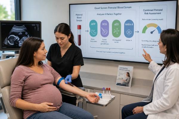
The quad screen is a second-trimester maternal blood test that estimates the chance of certain fetal chromosome conditions and open neural tube defects. It measures four substances: alpha-fetoprotein (AFP), human chorionic gonadotropin (hCG), unconjugated estriol (uE3), and inhibin A. A laboratory combines these markers with gestational age and patient-specific factors to calculate risks for trisomy 21, trisomy 18, and, through AFP, open neural tube defects such as spina bifida. The test is a screening test, not a diagnosis. Most screen-positive results do not mean the fetus has the condition, and a screen-negative result cannot eliminate every possibility. Accurate dating is especially important because all four markers change during pregnancy. Follow-up may include review of dates, a detailed ultrasound, cell-free DNA screening, genetic counseling, or diagnostic amniocentesis. The best next step depends on the exact pattern, ultrasound findings, prior screening, gestational age, and the patient’s preferences.
- The quad screen measures AFP, hCG, unconjugated estriol, and inhibin A in maternal blood.
- It is usually offered between 15 and 22 weeks, with many programs favoring 16 to 18 weeks.
- Results are converted to multiples of the median and combined into condition-specific probabilities.
- A positive screen indicates increased chance, not a confirmed fetal diagnosis.
- Incorrect gestational age, twins, weight, diabetes, and a vanishing twin can alter the calculation.
Table of Contents
- What the Quad Screen Measures
- Who May Be Offered the Test
- Timing, Preparation, and Accuracy
- How MoM Values Create a Risk Result
- Patterns for Trisomy 21 and Trisomy 18
- AFP and Open Neural Tube Defects
- Follow-Up After a Positive Screen
- Limitations, Special Situations, and Next Steps
What the Quad Screen Measures
The quad screen evaluates four biochemical markers that originate from the fetus, placenta, or both. The measured concentrations are not interpreted independently. Their combined pattern provides more screening information than any single marker.
Alpha-fetoprotein (AFP) is made mainly by the fetal liver. It enters amniotic fluid and then maternal blood. Maternal serum AFP is the marker used to estimate risk for open neural tube defects and certain abdominal wall defects. It also contributes to chromosome-risk calculations.
Human chorionic gonadotropin (hCG) is produced by placental trophoblast cells. The assay may measure total hCG or a defined form depending on the screening platform. Second-trimester hCG patterns differ from the early quantitative hCG tests used to assess pregnancy viability. A quad-screen hCG result should not be read using first-trimester doubling rules.
Unconjugated estriol (uE3) is an estrogen generated through cooperation among the fetal adrenal glands, fetal liver, and placenta. Because several parts of the fetoplacental unit contribute, low uE3 can occur in chromosome conditions and in a small number of other fetal or placental disorders.
Inhibin A is a placental and ovarian glycoprotein hormone. Adding it to the older triple screen improves screening performance for trisomy 21. Higher inhibin A is part of the classic trisomy 21 pattern.
The laboratory converts each raw concentration to a multiple of the median, or MoM, for the exact gestational age. It then adjusts the values using information such as maternal age, weight, diabetes, smoking status, ethnicity or ancestry variables used by that program, number of fetuses, and method of conception.
The final report usually gives separate probabilities rather than one overall “quad score.” A patient might receive one risk for trisomy 21, another for trisomy 18, and an AFP-related result for open neural tube defects. These outcomes require different follow-up pathways.
Who May Be Offered the Test
Prenatal screening and diagnostic options should be discussed with every pregnant patient, regardless of age or baseline risk. The quad screen may be especially relevant when first-trimester screening was not completed, cell-free DNA screening is unavailable or declined, or a regional prenatal program uses sequential or integrated serum screening.
The test can be used as a stand-alone second-trimester screen. It can also form the second part of an integrated screen that combines first-trimester PAPP-A and nuchal translucency with second-trimester markers. In a sequential approach, early results may be released before the quad component. The laboratory must know which protocol applies because risk calculations differ.
A quad screen is not usually added simply to repeat chromosome screening after a low-risk cell-free DNA result. Running several independent screens can produce conflicting probabilities and unnecessary anxiety. However, second-trimester AFP screening or detailed ultrasound may still be offered to assess open neural tube defects because cell-free DNA does not screen for those structural conditions.
Patients who present for prenatal care in the second trimester may value the quad screen because it can be completed with one blood draw and does not pose a procedure-related risk to the pregnancy. It is also less expensive than some newer tests in certain health systems.
The test may be less informative in multifetal pregnancy, after a vanishing twin, or when fetal reduction occurred. Screening algorithms for twins differ, and performance is lower than in singleton pregnancy. In higher-order multiples, some programs do not offer a standard risk calculation.
Before testing, patients should understand the possible outcomes. A low-risk result may reduce concern but not guarantee an unaffected fetus. A positive result creates choices: ultrasound, cell-free DNA as a secondary screen in selected cases, or diagnostic testing. Knowing in advance how the result could affect decisions makes consent more meaningful.
Timing, Preparation, and Accuracy
Most laboratories accept quad screening from 15 weeks through 22 weeks of pregnancy. Many programs prefer approximately 16 to 18 weeks because marker performance and time for follow-up are favorable. The exact window on the order form takes priority, and some systems use narrower limits.
No fasting is usually required. The test uses a standard venous blood sample. Patients can generally eat, drink, take prenatal vitamins, and continue normal activity. Mild bruising, soreness, or lightheadedness can occur after the draw.
Accurate gestational dating is essential. AFP, hCG, uE3, and inhibin A change substantially week by week. A pregnancy dated too early or too late can create an apparent abnormal pattern. Ultrasound dating is often more reliable than an uncertain last menstrual period. If a later ultrasound changes the gestational age enough, the laboratory may be able to recalculate the result without repeating the blood draw, provided the corrected age remains within its validated range.
The order should accurately record:
- estimated due date and dating method;
- maternal date of birth or egg age when required;
- current weight;
- singleton, twin, or higher-order pregnancy;
- insulin-treated diabetes, if used by the program;
- smoking status;
- IVF, donor egg, or other assisted conception;
- known vanishing twin or fetal reduction;
- prior first-trimester or cell-free DNA screening.
Maternal weight affects concentration through blood-volume dilution. Diabetes and smoking can shift marker distributions. Multiple placentas contribute additional analytes. If these details are entered incorrectly, the corrected MoM and final risk may be wrong even though the laboratory measurement itself is accurate.
Results commonly take several days. Portal reports may show four raw values before the final interpretation is available. Raw concentrations should not be compared with internet ranges because laboratories use different assays and gestational medians. The clinically meaningful report includes corrected MoMs, condition-specific risks, cutoff definitions, and recommendations.
How MoM Values Create a Risk Result
MoM means multiple of the median. A corrected value of 1.0 MoM is at the median for pregnancies of the same gestational age after the laboratory’s adjustments. A value of 2.0 MoM is twice the median, and 0.5 MoM is half the median.
Each marker’s distribution overlaps widely between affected and unaffected pregnancies. The laboratory therefore uses likelihood ratios from all four markers rather than declaring one analyte normal or abnormal. Maternal or egg age contributes a prior probability, which the marker pattern modifies.
A simplified overview is:
| Screening target | AFP | hCG | uE3 | Inhibin A |
|---|---|---|---|---|
| Trisomy 21 | Often low | Often high | Often low | Often high |
| Trisomy 18 | Often low | Often low | Often low | Usually not the dominant marker |
| Open neural tube defect | Often high | Not the primary marker | Not the primary marker | Not the primary marker |
The actual software uses continuous values, not just arrows. It may also incorporate gestational age, weight, diabetes, ethnicity adjustment, and other variables. Two patients with the same hCG MoM can receive different risks because their other markers and prior probabilities differ.
A report may state “1 in 100” or “1:100.” That means an estimated 1% chance in patients with a similar profile, not a 100% diagnosis. A result of 1 in 2,000 corresponds to 0.05% residual risk. Laboratories define a screen-positive cutoff, such as 1 in 250 or another threshold, according to the program. The numerical probability is more informative than the word positive.
The AFP portion may be reported separately using a cutoff such as 2.0 or 2.5 MoM for singleton pregnancy. Cutoffs differ, and twin pregnancies use different expectations. A flagged AFP result is not interpreted through the trisomy cutoff.
Screening performance depends on the population and protocol. The quad screen detects many, but not all, pregnancies with trisomy 21 and trisomy 18. Cell-free DNA has higher screening accuracy for common trisomies, while diagnostic testing is required for certainty.
Patterns for Trisomy 21 and Trisomy 18
In trisomy 21, the classic quad pattern is lower AFP and uE3 with higher hCG and inhibin A. The markers do not need to follow that pattern perfectly. The algorithm evaluates how likely the observed combination is in affected versus unaffected pregnancies.
A screen-positive trisomy 21 result does not mean that four separate tests are positive. It means the combined probability crossed the program’s threshold. Most positive screens are false positives because the conditions are relatively uncommon, especially in lower-risk age groups.
For trisomy 18, AFP, hCG, and uE3 are often lower. Inhibin A contributes less consistently. A markedly low uE3 or several low markers may prompt careful ultrasound review because dating error, fetal growth concerns, or other rare conditions can produce similar findings.
Trisomy 13 is not always included as a separately validated quad-screen output. Programs vary. The report should specify exactly which conditions were assessed. A normal quad screen also does not screen for every chromosome difference, microdeletion, single-gene condition, or structural anomaly.
After a positive chromosome screen, options commonly include:
- genetic counseling to review the actual probability and choices;
- a detailed ultrasound for structural markers and dating;
- cell-free DNA screening as a more accurate secondary screen in selected patients;
- amniocentesis for diagnostic chromosome analysis;
- no additional testing, if that aligns with informed preferences.
Cell-free DNA remains a screening test. It can substantially refine risk but does not provide the same certainty as amniocentesis. A normal ultrasound also cannot fully exclude a chromosome condition. The choice between secondary screening and diagnosis depends on gestational age, how much certainty is desired, and whether results would change pregnancy or delivery planning.
Patients should avoid interpreting analyte values one at a time. For example, a mildly high hCG does not diagnose trisomy 21, and a low estriol does not independently diagnose trisomy 18. The integrated probability and clinical follow-up are what matter.
AFP and Open Neural Tube Defects
Maternal serum AFP is the quad-screen component used to estimate risk for open neural tube defects such as spina bifida and anencephaly. AFP can leak from exposed fetal tissues into amniotic fluid and maternal circulation. It may also rise with open abdominal wall defects.
High AFP has many explanations besides a fetal defect. The most common first step is to confirm gestational age and fetus number. Underestimated gestational age, twins, fetal demise, placental bleeding, or a dating error can raise the measured MoM. Maternal liver disease and rare tumors are unusual alternative causes.
A high AFP result generally leads to a targeted ultrasound. Sonography can confirm dates, identify multiple gestation, evaluate the fetal spine and skull, assess the abdominal wall, and look for placental or growth findings. Modern detailed ultrasound detects many open neural tube defects. Amniocentesis for amniotic-fluid AFP and acetylcholinesterase may be considered when ultrasound is uncertain or diagnostic confirmation is desired.
A low AFP value contributes to the chromosome-risk calculation but is not used to diagnose a problem on its own. The quad screen does not assess closed neural tube defects reliably because skin covers the lesion and AFP may not leak into amniotic fluid.
Even after a low-risk quad result, the routine second-trimester anatomy ultrasound remains important. Serum screening and ultrasound answer different questions. The blood test estimates risk from marker patterns; ultrasound directly examines fetal structures within the limits of imaging.
An unexplained high AFP with normal anatomy can still be associated with placental complications, fetal growth restriction, preterm birth, or stillbirth. The strength of association depends on the degree of elevation and other findings. Some obstetric teams recommend additional growth surveillance, but management should be individualized rather than based on AFP alone.
AFP screening identifies pregnancies needing closer evaluation. It does not establish that the fetus has spina bifida, nor does a normal AFP guarantee that every neural tube or structural condition is absent.
Follow-Up After a Positive Screen
A positive result should trigger a structured review, not an immediate conclusion. First, verify the demographic and pregnancy data used in the calculation. An incorrect due date, weight, fetus number, or diabetes status may justify recalculation. The laboratory or prenatal screening program can advise whether the original sample remains valid.
Second, distinguish the reason for positivity. A trisomy 21 risk, trisomy 18 risk, and high-AFP result require different counseling. Ask for the numerical probability, corrected MoMs, cutoff, and whether more than one category was flagged.
Third, review prior testing. A previous first-trimester screen, cell-free DNA result, ultrasound finding, or known vanishing twin changes interpretation. Repeating an unrelated screening test without a plan can prolong uncertainty.
A detailed ultrasound is often central. It can confirm gestational age and fetus number, identify structural anomalies, evaluate placental findings, and help determine whether diagnostic testing is appropriate. Ultrasound cannot replace chromosome analysis when certainty is needed.
Amniocentesis is a diagnostic procedure usually performed from about 15 weeks onward. Under ultrasound guidance, a clinician removes a small amount of amniotic fluid for fetal chromosome or genetic testing. It provides definitive information for the tested conditions but carries a small procedure-related risk that should be discussed in the local clinical context.
Cell-free DNA may be offered after a positive serum screen to refine risk for common trisomies, particularly when a patient wants to avoid an invasive procedure. However, this approach can delay diagnosis and may not be ideal when ultrasound is abnormal or when broader chromosome analysis is needed.
The decision is personal. Some patients want maximum certainty, others prefer noninvasive information, and some decline further testing. Genetic counseling should present probabilities, benefits, limitations, timing, and possible outcomes without assuming one correct choice.
Limitations, Special Situations, and Next Steps
The quad screen has important limitations. It estimates risk only for specified conditions, depends heavily on accurate dating, and has lower screening accuracy for common trisomies than cell-free DNA. It cannot diagnose a condition, assess every structural anomaly, or predict overall fetal health.
Special situations require modified interpretation:
- Twins: Marker levels reflect both fetuses and placentas, so screening performance is reduced and the result cannot identify which twin is affected.
- Vanishing twin: Residual placental tissue may alter serum markers for weeks and produce an unreliable calculation.
- Donor egg: The program may need donor age rather than the pregnant patient’s age for chromosome prior risk.
- IVF: Conception method and dating should be entered accurately; embryo-transfer dates often provide precise gestational age.
- Diabetes or high maternal weight: Adjustment factors can materially change MoMs.
- Abnormal ultrasound: A structural anomaly may justify diagnostic testing even when the serum screen is low risk.
The test also may reveal marker patterns associated with placental dysfunction, but it was not designed as a definitive placental health test. An unexplained high AFP, high inhibin A, or other extreme analyte can lead to closer blood-pressure, growth, or fetal surveillance based on the whole pregnancy profile.
When reviewing the report, ask: Was the gestational age ultrasound-confirmed? Are the MoMs corrected? What exact condition crossed the cutoff? What is the numerical chance? Does the result affect neural tube defect screening, chromosome screening, or both? What follow-up can still be completed at the current gestational age?
A result is most useful when it leads to an informed next step rather than panic. Confirm the data, obtain expert counseling, and choose between additional screening and diagnostic testing according to the desired level of certainty. Continue routine anatomy ultrasound and prenatal care regardless of whether the quad screen is low risk.
A positive screen may create time-sensitive choices, particularly as gestation advances. Genetic counseling should be arranged promptly enough to allow detailed ultrasound, cell-free DNA when appropriate, or amniocentesis without unnecessary delay. Promptness does not mean rushing into an irreversible decision; it means preserving the full range of informed options.
A negative screen should also be framed correctly. It lowers the estimated chance for the conditions included in the algorithm but does not evaluate every chromosome change, genetic syndrome, heart defect, brain anomaly, or developmental condition. The routine anatomy scan and other indicated prenatal care remain necessary.
When the quad screen follows an earlier serum screen, confirm whether the program is integrated or sequential. A laboratory should combine the stages using its validated algorithm. Independently interpreting two risk reports or selecting whichever looks more reassuring can produce an inaccurate conclusion.
Before declining or choosing further testing, patients may ask how long results will take and what each option can answer. Cell-free DNA refines risk for selected chromosome conditions; amniocentesis diagnoses the tested fetal chromosome complement; detailed ultrasound evaluates anatomy. Their scopes are different, so the choice should match the information the patient wants.
Keep the original laboratory report rather than only the “positive” or “negative” label. The condition-specific risk, MoMs, dating, and cutoff are needed for meaningful counseling and comparison with later testing.
References
- Prenatal Genetic Screening 2025 (Review)
- Biochemical Screening for Fetal Trisomy 21: Pathophysiology of Maternal Serum Markers and Involvement of the Placenta 2023 (Review)
- Performance of Serum Quad Test in Screening for Fetal Down Syndrome in a Large-Scale Unselected Population in a Developing Country 2023 (Study)
- Abnormal maternal serum markers and adverse pregnancy outcomes 2025 (Technical Update)
- Alpha-Fetoprotein Analysis 2024 (Review)
Disclaimer
This article provides general education and does not interpret an individual prenatal screening result. Quad-screen probabilities should be reviewed with an obstetric clinician or genetic counselor using accurate dating, ultrasound, prior screening, and personal preferences. A positive screen is not a diagnosis, and urgent pregnancy symptoms require direct medical evaluation.