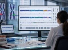
Whole-exome sequencing, or WES, examines most of the protein-coding regions of thousands of genes at once. These regions, called exons, make up only a small portion of the genome but contain many variants known to cause rare inherited disorders. WES is often used when symptoms could be explained by many genes, when earlier targeted tests were negative, or when a person has a complex combination of findings that does not point to one clear diagnosis.
WES is broad, but it is not a complete reading of the genome. Capture methods do not cover every exon equally, and standard analysis is strongest for single-nucleotide variants and small insertions or deletions. Repeat expansions, balanced rearrangements, many deep intronic changes, methylation disorders, and low-level mosaic variants can be missed. Results can include a diagnosis, a variant of uncertain significance, secondary findings unrelated to the original question, or no explanation. Parental samples and later reanalysis can substantially improve interpretation.
- WES surveys most coding exons across thousands of genes rather than testing one gene at a time.
- It is commonly used for unexplained developmental, neurologic, metabolic, immune, cardiac, and multisystem disorders.
- Trio testing with both biological parents can help identify de novo, recessive, and inherited variants.
- A negative exome does not rule out a genetic condition or variants outside coding regions.
- Reanalysis may find a diagnosis later as gene-disease knowledge and analytic methods improve.
Table of Contents
- Why the Exome Is a Useful Diagnostic Target
- How WES Is Performed
- Singleton, Duo, and Trio Analysis
- What Types of Variants WES Can Find
- How Exome Results Are Reported
- What WES Can Miss
- Reanalysis, Follow-Up, and Family Implications
Why the Exome Is a Useful Diagnostic Target
The exome is the collection of exons that contribute to protein-coding transcripts. Although coding sequence represents roughly 1% to 2% of the human genome, many established Mendelian disease variants occur there. This concentration makes the exome a practical compromise between a focused gene panel and whole-genome sequencing.
A gene panel analyzes a predefined list selected for a particular phenotype. WES captures a much broader set and can be analyzed using a virtual panel at first, then expanded if the initial review is negative. This is useful when clinical features overlap several disease categories or evolve over time.
WES may be considered for:
- developmental delay, intellectual disability, congenital anomalies, or autism with additional medical findings;
- unexplained epilepsy, movement disorders, neuropathy, muscle disease, or hearing and vision disorders;
- suspected metabolic, mitochondrial, renal, cardiac, endocrine, skeletal, or immune conditions;
- severe or unusual disease beginning in infancy or childhood;
- multiple affected organ systems without a unifying diagnosis;
- a family pattern suggesting a rare inherited condition;
- negative or inconclusive prior single-gene or panel testing.
The test is often valuable when genetic heterogeneity is high—meaning variants in many different genes can cause a similar phenotype. Instead of ordering genes one after another, WES can assess them simultaneously. It may also identify an unexpected diagnosis that would not have been included on a narrow panel.
WES is not always the best first test. A distinctive condition caused by a repeat expansion, methylation abnormality, chromosome imbalance, or common copy-number change may be better evaluated with a targeted assay or chromosomal microarray. When a known family variant exists, targeted testing is usually simpler and more appropriate for relatives.
Clinical utility extends beyond naming a condition. A molecular diagnosis can change surveillance, medication, diet, surgery, transplant decisions, reproductive counseling, or eligibility for a trial. It can also prevent repeated low-yield investigations and connect families with condition-specific resources. However, not every diagnosis changes treatment, and the value of ending uncertainty may differ among families.
Diagnostic yield varies widely by referral group. It tends to be higher when the phenotype is well documented, the disorder is likely monogenic, and both parents are tested. A quoted percentage from one study should not be treated as a personal prediction. Age, prior testing, family structure, ancestry, and laboratory reporting policy all influence yield.
How WES Is Performed
Most clinical WES begins with DNA from blood. Saliva, cheek swabs, cultured cells, prenatal specimens, or tissue may be accepted. Blood is often preferred because it yields consistent DNA and reduces some contamination risks. A different tissue may be necessary when mosaicism is suspected or blood is affected by a transplant or hematologic condition.
The laboratory workflow includes several stages.
DNA extraction and library preparation
DNA is purified and fragmented into smaller pieces. Short adapter sequences are attached so fragments can be amplified and read by the sequencing instrument. Each sample receives identifiers that allow it to be tracked when multiple libraries are processed together.
Exome capture
Laboratories use probes designed to bind coding exons and selected nearby regions. Bound fragments are separated from most noncoding DNA. Capture kits differ in which transcripts, untranslated regions, mitochondrial sequences, and clinically important noncoding sites they include.
Capture creates unevenness. Some exons bind efficiently and receive deep coverage; others have low coverage because of high GC content, repetitive sequence, pseudogenes, or probe design. A report may provide the percentage of targeted bases covered above a minimum read depth.
Sequencing
Millions of captured fragments are sequenced in parallel. Most clinical WES uses short paired-end reads. Each position is ideally read many times. Depth helps distinguish a true variant from random error and may support limited detection of mosaicism or copy-number change.
Alignment and variant calling
Software aligns reads to a human reference genome. It identifies positions that differ and assigns quality measures. Separate algorithms may search for single-nucleotide variants, small insertions and deletions, copy-number variants, mitochondrial changes, or selected repeat expansions.
Not every laboratory performs all of these analyses. “Whole-exome sequencing” describes the captured data, but the reportable variant types depend on the laboratory’s validated pipeline.
Annotation and filtering
A raw exome contains tens of thousands of variants. Software annotates population frequency, predicted consequence, gene function, inheritance, previous clinical classifications, and published evidence. Analysts prioritize variants that fit the patient’s features and expected inheritance pattern.
Clinical information is essential. Laboratories may translate symptoms into standardized Human Phenotype Ontology terms and compare them with known gene-disease profiles. A vague requisition can reduce the chance that the correct variant is prioritized.
Expert review and reporting
Scientists and medical directors review candidate findings, inspect sequence data, evaluate evidence, and classify variants. Some calls are confirmed with another method when quality, variant type, or laboratory policy requires it. The final report includes results relevant to the indication and may include elected secondary findings.
Turnaround is often several weeks, though rapid exome sequencing can be completed much faster for critically ill patients. Rapid testing requires coordinated consent, specimen collection, analysis, and clinical response. A fast result is useful only if the care team can act on it promptly.
Singleton, Duo, and Trio Analysis
WES can be performed on the affected person alone, with one relative, or with both biological parents. These designs generate the same type of sequence data but differ in interpretive power.
Singleton exome
A singleton analyzes only the patient. It can identify pathogenic variants, but determining inheritance may require later parental testing. Many rare variants remain candidates because the laboratory cannot immediately tell whether they are de novo, inherited, or on opposite chromosomes.
Singleton testing may be necessary when parents are unavailable, deceased, adopted, or unwilling to test. A detailed family history becomes especially important.
Duo exome
A duo includes the patient and one informative relative, often one parent or an affected sibling. It can clarify some inheritance but cannot fully evaluate de novo status or phase in many cases. The choice of relative should match the hypothesis.
Trio exome
A trio analyzes the affected person and both biological parents together. It helps identify:
- de novo variants present in the child but absent from both parents;
- compound heterozygous variants inherited one from each parent in a recessive gene;
- homozygous variants and whether each parent is a carrier;
- X-linked inheritance;
- parental mosaicism or unexpected allele balance;
- variants inconsistent with the reported family relationship or sample identity.
Trio analysis reduces the number of candidate variants and can increase diagnostic efficiency, especially for severe early-onset or neurodevelopmental disorders. It does not eliminate uncertainty. Low-level parental mosaicism can be missed, and a de novo variant is not automatically pathogenic.
Testing parents raises consent and privacy issues. Their data may reveal medically important secondary findings or unexpected biological relationships. Laboratories differ in whether they analyze parental secondary findings independently or only use parental data to interpret the child.
Affected siblings or multiple relatives can be even more informative in some families. Shared variants may support dominant or recessive inheritance, while differences can reveal phenocopies or more than one diagnosis. Large pedigrees may require linkage or specialized segregation analysis rather than a standard trio workflow.
Phase describes whether two variants lie on the same chromosome or opposite chromosomes. In a recessive condition, two pathogenic variants usually need to be in trans. Trio data often establish phase through inheritance, but de novo or complex variants may require long-read sequencing, cloning, or other methods.
What Types of Variants WES Can Find
WES is strongest for variants within well-covered coding exons and nearby splice junctions.
Single-nucleotide variants
Single-base substitutions can create missense, nonsense, splice, synonymous, or regulatory effects. Coverage and mapping quality determine sensitivity. Variants in genes with pseudogenes or repetitive exons may need gene-specific confirmation.
Small insertions and deletions
WES can detect many small indels, especially when they occur in unique, well-covered sequence. Homopolymers, repeats, and larger indels are more difficult. A frameshift can disrupt the protein, but classification depends on whether loss of function is a known disease mechanism.
Splice-region variants
Capture usually extends a limited distance into introns. Canonical splice-site variants can be detected, and selected nearby changes may be reported. Deep intronic variants are generally outside the target. RNA sequencing may demonstrate whether an uncertain variant alters splicing.
Copy-number variants
Read-depth analysis can infer deletions or duplications of one or more exons. Performance varies with capture uniformity, event size, and laboratory validation. Exome-based copy-number calls may require confirmation by microarray, MLPA, or another method.
WES may identify a deletion spanning several genes, but chromosomal microarray can provide more consistent genome-wide copy-number assessment. A normal exome copy-number analysis does not exclude every exon-level event.
Mitochondrial variants
Some exome tests include mitochondrial DNA analysis, while others do not. Incidental off-target reads may provide coverage but are not automatically validated for diagnosis. Heteroplasmy detection, large mitochondrial deletions, and tissue selection require specific methods.
Selected mosaic variants
A variant present in a fraction of blood cells may be detectable when depth is high and allele fraction exceeds the laboratory threshold. Standard germline pipelines may filter low-level calls. Tissue-limited mosaicism will be missed if absent from the sample.
Candidate and novel gene findings
Some laboratories report variants in genes not yet definitively linked to human disease as candidates, often through research rather than a clinical report. Establishing a new gene-disease relationship requires additional unrelated cases, functional evidence, and expert review. A plausible variant in a candidate gene is not equivalent to a clinical diagnosis.
How Exome Results Are Reported
A WES report should separate findings that explain the indication from secondary or uncertain information. Common result categories include positive, uncertain, negative, and secondary findings.
Positive or diagnostic result
A positive result identifies one or more pathogenic or likely pathogenic variants that fit the patient’s phenotype and inheritance. The report should explain whether the finding confirms a dominant, recessive, X-linked, mitochondrial, or mosaic condition.
A diagnosis may be complete or partial. One variant can explain some features while leaving others unresolved. Dual diagnoses occur when pathogenic variants in two genes account for different parts of a complex presentation.
Clinical correlation remains necessary. The same gene can cause several phenotypes, and penetrance may be incomplete. Management should follow condition-specific evidence rather than the label “positive” alone.
Variant of uncertain significance
A variant of uncertain significance lacks enough evidence for a pathogenic or benign classification. A report may include a VUS when the gene strongly matches the phenotype. It should not be used alone to make irreversible decisions or predictive tests in healthy relatives.
Parental testing, segregation, RNA analysis, biochemical studies, or later reinterpretation may help. Not every VUS warrants additional work; the laboratory or genetics team can identify which studies are likely to be informative.
Negative result
A negative exome means no reportable explanation was found in the analyzed data. It does not prove the condition is not genetic. The cause may be outside the exome, in a poorly covered gene, a variant type not analyzed, a gene not yet linked to disease, or an acquired or multifactorial process.
The report may list no variants even though many benign variants were detected and filtered. “Negative” is a conclusion about clinically relevant findings within the test’s scope.
Secondary findings
Secondary findings are pathogenic or likely pathogenic variants in genes unrelated to the reason for testing but associated with preventable or medically actionable conditions. Professional organizations maintain lists for deliberate analysis in clinical exome and genome testing. Policies and opt-in or opt-out choices vary by country, laboratory, and age.
A secondary finding is not the same as an incidental VUS. Laboratories generally seek only defined variant types in selected genes. A confirmed result may prompt surveillance and family testing.
Carrier and pharmacogenetic findings
Some laboratories report carrier status or pharmacogenetic variants; others do not. These analyses require explicit validation and consent. The absence of such findings on an exome report does not mean the person is not a carrier or has no medication-related variants.
What WES Can Miss
The largest limitation is that WES does not comprehensively examine noncoding DNA. Regulatory variants, deep intronic changes, and structural breakpoints can lie far from captured exons.
Other important blind spots include:
- repeat expansions and many tandem-repeat disorders;
- balanced translocations and inversions;
- complex structural variants and mobile element insertions;
- methylation and imprinting abnormalities;
- regions with pseudogenes, segmental duplications, or extreme GC content;
- low-level or tissue-specific mosaicism;
- large mitochondrial deletions and low heteroplasmy unless specifically validated;
- variants in genes or exons absent from the capture design;
- noncoding disease mechanisms not included in analysis;
- some exon-level deletions, duplications, and complex indels.
Coverage is not uniform. A laboratory may report that 95% or more of targets met a depth threshold, yet the clinically important variant could lie in the uncovered fraction. When one gene remains strongly suspected, review its exon-by-exon coverage and consider supplemental Sanger or long-range testing.
Capture kits become outdated as transcripts and genes change. Reanalysis can reinterpret stored variants, but it cannot create reads for a region that was never captured. Re-sequencing with a newer exome or genome method may be needed.
A negative blood exome can miss somatic mosaicism confined to skin, brain, tumor, vascular tissue, or another organ. Testing affected tissue with deep sequencing may be more informative. Cultured fibroblasts, buccal cells, urine, or surgical material can be considered depending on the disorder.
Short-read alignment is difficult in genes with highly similar pseudogenes. A call may be falsely assigned or a true variant may be filtered. Specialized assays use long-range PCR, unique anchors, or long reads to separate these regions.
WES can also miss a diagnosis because of interpretation rather than technology. The gene-disease link may not yet be known, the phenotype may be incomplete, or an atypical presentation may cause the variant to be deprioritized. Reanalysis addresses some of these problems.
Reanalysis, Follow-Up, and Family Implications
Exome data can remain useful after the first report. New disease genes are discovered, variant classifications change, phenotype databases improve, and pipelines gain the ability to detect additional event types. Reanalysis applies updated knowledge to existing data.
A reasonable interval is often one to several years, but there is no universal schedule. Earlier review may be appropriate when major new symptoms appear, a sibling becomes affected, a candidate gene is published, or the laboratory releases a substantially improved pipeline. Ask whether reanalysis is automatic, clinician requested, or billed as a new service.
Reanalysis should include updated clinical features. A child’s phenotype can become more specific with age, allowing better gene prioritization. New biochemical, imaging, or pathology findings may be decisive.
When the exome remains negative, follow-up should target the leading residual possibilities:
- chromosomal microarray for copy-number changes;
- repeat-expansion testing for a compatible neurologic or neuromuscular phenotype;
- methylation testing for an imprinting disorder;
- genome sequencing for noncoding and structural variants;
- RNA sequencing for splicing or expression defects;
- long-read sequencing for repeats, complex variants, or pseudogenes;
- high-depth testing of affected tissue for mosaicism;
- biochemical or functional studies for a suspected pathway.
After a positive result, parental confirmation can establish inheritance and recurrence risk. At-risk relatives usually receive focused testing for the identified variant rather than WES. Reproductive options depend on inheritance and may include prenatal or preimplantation testing.
A de novo finding lowers but does not eliminate recurrence risk because a parent can have germline mosaicism. A recessive diagnosis usually means each parent is a carrier and future pregnancies have a 25% chance of being affected. X-linked, mitochondrial, and complex mechanisms require different counseling.
The family should keep the complete report, including laboratory name, variant nomenclature, transcript, and genome build. Sharing only a gene name is insufficient for accurate relative testing. If the result is reclassified, management and prior family tests may need review.
WES is most effective as part of an ongoing diagnostic process rather than a one-time answer. Its broad coding coverage can reveal unexpected diagnoses, while careful consent, phenotype documentation, parental analysis, and planned follow-up turn sequence data into clinically useful information.
References
- Reanalysis of Exome Sequencing Data in the Indian Undiagnosed Diseases Program — 2025 Study.
- Reanalysis of whole-exome sequencing data of children with neurodevelopmental disorders in a standard patient care context — 2024 Study.
- Clinician-Driven Reanalysis of Exome Sequencing Data From Patients With Inherited Retinal Diseases — 2024 Cohort Study.
- A systematic review of the assessment of the clinical utility of genomic sequencing: Implications of the lack of standard definitions and measures of clinical utility — 2024 Systematic Review.
- Exome and genome sequencing for rare genetic disease diagnosis: a scoping review — 2023 Review.
- Exome and genome sequencing for pediatric patients with congenital anomalies or intellectual disability: an evidence-based clinical guideline of the American College of Medical Genetics and Genomics — 2021 Guideline.
Disclaimer
This article is for general education and does not replace evaluation by a clinical geneticist, genetic counselor, or testing laboratory. WES content, coverage, secondary-finding policies, and detectable variant types vary among laboratories. Results should be interpreted using the full clinical history, family data, and current professional guidance.