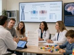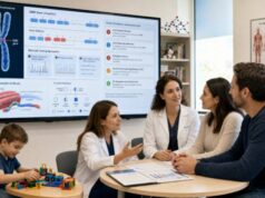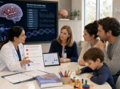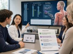
Prader-Willi syndrome testing looks for loss of activity from paternally expressed genes in chromosome region 15q11.2-q13. The recommended first step is usually DNA methylation analysis, often combined with a chromosome copy-number method. An abnormal maternal-only methylation pattern can confirm Prader-Willi syndrome in more than 99% of affected people, but a basic methylation test does not explain why the paternal genes are inactive. Additional testing determines whether the cause is a paternal deletion, maternal uniparental disomy, or an imprinting defect. That distinction matters for recurrence risk, prenatal options, and sometimes clinical expectations. Chromosomal microarray, parent-of-origin studies, and imprinting-center analysis answer different parts of the question. A normal test does not end the evaluation when the clinical picture remains concerning, because rare PWS-like disorders and unusual chromosome changes may require other methods.
- Prader-Willi syndrome results from absent expression of critical genes normally active on the paternal chromosome 15.
- DNA methylation testing is the key diagnostic test because it detects the abnormal parent-specific imprinting pattern.
- Methylation alone usually cannot distinguish deletion, maternal UPD, and imprinting defects.
- A SNP-based chromosomal microarray can identify most deletions and some forms of UPD.
- Recurrence risk is usually low but can reach 50% with an inherited imprinting-center deletion.
- Early molecular diagnosis supports prompt nutrition, endocrine, developmental, sleep, and behavioral care.
Table of Contents
- Why parent of origin matters on chromosome 15
- When Prader-Willi testing is considered
- The first-tier testing strategy
- Deletion, UPD, and imprinting-defect results
- Recurrence risk and family testing
- Prenatal testing, newborns, and sample issues
- Normal, inconclusive, and atypical results
- Medical care after a confirmed diagnosis
Why parent of origin matters on chromosome 15
Most genes are active from both the maternal and paternal chromosome copies. Imprinted genes are different: chemical marks established in egg or sperm cause one parental copy to be active and the other to be silent. In the Prader-Willi region of chromosome 15, several genes and noncoding RNAs are normally expressed from the paternal copy. The maternal copy carries a silenced imprint for these genes.
Prader-Willi syndrome occurs when the functional paternal contribution is missing. This can happen in three main ways:
- The paternal 15q11.2-q13 region is deleted.
- Both chromosome 15 copies come from the mother, called maternal uniparental disomy 15.
- A paternal chromosome is present but has an incorrect maternal-type imprint, called an imprinting defect.
All three mechanisms produce a similar molecular endpoint: only the maternal methylation pattern is detected at the tested imprinting locus. This is why methylation analysis is so sensitive for diagnosis and why it cannot, by itself, identify the mechanism.
The same chromosome region is involved in Angelman syndrome, but the parent-of-origin problem is different. Angelman syndrome generally involves loss of the maternally expressed UBE3A function in the brain. A laboratory must interpret methylation and copy-number findings with the parent of origin and clinical phenotype in mind.
The critical Prader-Willi region includes the SNURF-SNRPN locus, the imprinting center, and a cluster of small nucleolar RNAs, particularly SNORD116. The disorder is not usually caused by a conventional single-letter variant in one protein-coding gene. This is why routine exome sequencing can be normal even when Prader-Willi syndrome is present. Exome sequencing reads many coding regions but generally does not provide the methylation and parent-of-origin information needed for diagnosis.
Genomic imprinting also explains why the same deletion can have different effects depending on whether it arose on the paternal or maternal chromosome. A deletion of 15q11.2-q13 on the paternal chromosome causes Prader-Willi syndrome; a comparable deletion on the maternal chromosome causes Angelman syndrome. The physical DNA loss may be similar, but the active genes differ by parent of origin.
A confirmed molecular mechanism gives more than a label. It helps estimate recurrence, determines which relatives need testing, and can reveal a chromosome rearrangement that affects future pregnancies. It may also help interpret differences among individuals, although genotype does not predict every feature or degree of severity.
When Prader-Willi testing is considered
Clinical signs change with age. In the newborn period, severe hypotonia is often the leading clue. Affected infants may have a weak cry, reduced spontaneous movement, poor suck, feeding difficulty, and failure to gain weight. Boys may have undescended testes or a small penis, and girls may have underdeveloped genitalia. The combination of unexplained neonatal hypotonia and feeding problems should prompt early methylation testing even before later features appear.
During infancy and childhood, feeding usually improves, and excessive weight gain can begin before obvious hyperphagia. Developmental delay, short stature, small hands and feet, scoliosis, sleep-disordered breathing, reduced pain sensitivity, and characteristic behavior may emerge. Hyperphagia—persistent drive to eat with impaired satiety—becomes a major safety concern for many children.
In adolescents and adults, clues can include intellectual or learning differences, growth hormone deficiency, hypogonadism, incomplete puberty, infertility, obesity, sleep apnea, skin picking, anxiety, rigidity, repetitive questioning, and episodes of severe behavioral or psychiatric change. Not every person has every feature, and early treatment can alter the physical appearance and growth pattern.
Testing should not depend on a clinical score when the key signs are present. Molecular confirmation is reliable and changes care. It can prevent prolonged diagnostic uncertainty and support access to condition-specific nutrition, hormone, sleep, developmental, and behavioral management.
Prenatal clues are less specific. Reduced fetal movement, abnormal fetal position, excess amniotic fluid, and growth differences may raise concern, especially after an abnormal chromosome test or when there is a known family risk. These findings do not diagnose Prader-Willi syndrome without molecular testing.
A person with some PWS-like features but normal methylation may have another condition. Differential diagnoses include Schaaf-Yang syndrome, Temple syndrome, congenital myopathies, spinal muscular atrophy, chromosomal disorders, monogenic obesity syndromes, hypothalamic disorders, and other causes of hypotonia or developmental delay. The next test should be guided by the dominant features rather than assuming that every combination of obesity and developmental differences is PWS.
Early diagnosis matters even when hyperphagia has not started. Families can establish safe feeding practices, monitor growth, begin therapies, evaluate endocrine function, and assess sleep and breathing before complications become severe.
The first-tier testing strategy
The most efficient approach pairs a test that confirms the imprinting abnormality with a test that looks for the molecular mechanism. Laboratories may package these analyses differently, so the order name should be checked carefully.
DNA methylation analysis: Methylation-specific PCR examines an imprinted locus, commonly in the SNRPN region. A normal person shows both paternal and maternal patterns. A person with Prader-Willi syndrome shows a maternal-only pattern. This result establishes the diagnosis in nearly all typical cases. However, basic methylation-specific PCR does not reveal whether the paternal contribution was deleted, replaced by maternal copies, or incorrectly imprinted.
Methylation-sensitive MLPA: Methylation-sensitive multiplex ligation-dependent probe amplification can assess methylation and copy number across selected sites in the region. It can confirm an abnormal methylation pattern and detect many common deletions in one assay. It does not identify every UPD or every complex rearrangement, so additional testing may still be needed when no deletion is found.
SNP-based chromosomal microarray: A single-nucleotide polymorphism array evaluates chromosome copy number and long stretches of homozygosity. It can identify the common paternal deletions and define their size. It may also show maternal isodisomy, in which two identical copies of a maternal chromosome segment are present. A standard copy-number-only array without SNP probes may detect deletion but not provide evidence of UPD.
Maternal UPD can occur as isodisomy, heterodisomy, or a mixture. Heterodisomy means the child inherited both different chromosome 15 copies from the mother. Because the DNA is not necessarily homozygous, SNP array may look normal. When methylation is abnormal and no deletion is detected, parent-of-origin testing is needed to distinguish maternal UPD from an imprinting defect.
Parent-of-origin testing: DNA markers from the child and both parents are compared across chromosome 15. Maternal UPD is supported when both of the child’s chromosome 15 contributions are maternal. Testing both parents is important for a definitive analysis and for detecting sample or relationship issues that could affect interpretation.
Imprinting-center testing: If methylation is abnormal, no deletion is present, and UPD is excluded, the likely mechanism is an imprinting defect. The laboratory may perform deletion/duplication analysis and sequence analysis of the imprinting center. A small imprinting-center deletion can be inherited and carry a much higher recurrence risk than an epigenetic imprinting error without a DNA deletion.
A typical workflow can be summarized as follows:
| Step | Possible finding | What it establishes |
|---|---|---|
| Methylation analysis | Maternal-only methylation | Confirms Prader-Willi syndrome but may not identify cause |
| SNP array or dosage analysis | Paternal 15q11.2-q13 deletion | Defines deletion mechanism and size |
| SNP array plus parental markers | Maternal UPD 15 | Shows both chromosome 15 copies are maternal |
| UPD excluded; imprinting-center study abnormal | Imprinting-center deletion or sequence change | Defines a potentially heritable imprinting defect |
| UPD excluded; no DNA change found | Epigenetic imprinting defect | Explains abnormal imprinting with generally low recurrence |
No single test name should be assumed to include every step. The final report should state whether the diagnosis and the mechanism were both established.
Deletion, UPD, and imprinting-defect results
A paternal deletion is the most common mechanism. The report may describe a typical type 1 or type 2 deletion or an atypical deletion with different breakpoints. The size can include genes outside the smallest critical region, which may add clinical features. Copy-number analysis should determine whether the deletion is isolated or part of a larger chromosome rearrangement.
Most deletions arise as new events in the child. Parental chromosome studies may be recommended if the deletion is unusual, if a balanced rearrangement is possible, or if the family history is concerning. A parent can rarely carry a balanced chromosome rearrangement without having Prader-Willi syndrome but have an increased chance of an affected pregnancy.
Maternal uniparental disomy 15 means both chromosome 15 copies were inherited from the mother and no functional paternal imprint is present. UPD often results from correction of an early chromosome-number error. For example, an embryo may initially have three chromosome 15 copies and lose the paternal copy. Advanced maternal age can increase the chance of chromosome nondisjunction, but most families have no action or exposure that caused the event.
UPD results require attention to isodisomy. If a region contains two identical maternal copies, a child can become homozygous for a recessive pathogenic variant carried by the mother. This does not occur in most cases, but the SNP array may reveal long homozygous segments that justify review for relevant recessive conditions. The clinical team should not assume that every additional symptom is part of Prader-Willi syndrome.
An imprinting defect means the paternal chromosome is present but carries an inappropriate maternal-type imprint. Some imprinting defects are caused by a small deletion or sequence change in the imprinting center. Others are epimutations with no detectable DNA change. This distinction is central to recurrence risk.
The report may say “molecular class not determined” when methylation confirms PWS but the available studies do not separate UPD from imprinting defect. That is a valid diagnostic positive result, but it is incomplete for family counseling. Additional parental marker and imprinting-center testing should be considered.
The molecular class may be associated with average differences in pigmentation, verbal skills, autism features, or psychosis risk across groups. These associations cannot predict one person’s development or mental health. Care should be based on actual symptoms, age, and established PWS surveillance rather than assumptions from subtype.
An abnormal array without abnormal methylation requires careful review. A deletion near the PWS region may not involve the imprinted critical genes, may be on the maternal chromosome, or may represent another chromosome disorder. Copy number and methylation answer different questions; one should not substitute for the other.
Recurrence risk and family testing
Most families with Prader-Willi syndrome have a recurrence risk below 1%, but that general statement should not be given until the molecular mechanism is known. Rare mechanisms can substantially increase the chance in another pregnancy.
For a typical de novo paternal deletion with normal parental chromosomes, recurrence is generally below 1%. The small residual risk reflects rare germline mosaicism or an undetected parental rearrangement. If a parent carries a chromosome 15 rearrangement, risk depends on the specific structure and can be much higher. A chromosome specialist should calculate it from the family’s results.
For maternal UPD caused by a sporadic chromosome-number error, recurrence is also usually below 1%. Maternal age and the specific UPD mechanism may be discussed. Rare parental chromosome rearrangements or predisposing situations can alter risk, so the laboratory findings must be reviewed rather than assuming every UPD case is identical.
For an imprinting defect without an identifiable DNA deletion, recurrence is generally low. In contrast, a small imprinting-center deletion on the paternal chromosome may be inherited from the father. If the father carries the deletion on the chromosome 15 he inherited from his mother, he can be unaffected because that chromosome is normally maternally imprinted in him. When he transmits the same chromosome as a paternal chromosome, it may fail to establish the correct paternal imprint. The recurrence risk can then be as high as 50% in each pregnancy.
This counterintuitive pattern is why parental testing is essential after an imprinting-center deletion. A clinically unaffected parent can carry a change with major reproductive consequences. The precise parental origin of the altered chromosome matters.
Family testing should be targeted to the mechanism. Relatives do not need broad methylation testing merely because one family member has a typical de novo deletion. They may need testing when an imprinting-center deletion, balanced rearrangement, or other heritable chromosome finding is identified.
A genetics professional can provide an individualized recurrence table and discuss options such as prenatal diagnosis or preimplantation genetic testing. General information about predictive genetic testing is less applicable here than mechanism-specific reproductive testing, because PWS diagnosis depends on parent-of-origin expression rather than simple carrier status.
Keep copies of the original methylation, microarray, UPD, and imprinting-center reports. A statement such as “positive for PWS” is not enough to select a prenatal assay in a later pregnancy.
Prenatal testing, newborns, and sample issues
Prenatal testing is most straightforward when the family’s molecular mechanism is known. For a prior paternal deletion, testing can assess fetal copy number and methylation or parent of origin as appropriate. For maternal UPD risk related to a chromosome rearrangement, chromosome and parent-of-origin studies may be required. For a familial imprinting-center deletion, targeted analysis can identify whether the fetus inherited the deletion, while methylation testing helps determine functional imprinting.
Chorionic villus sampling and amniocentesis obtain different fetal tissues at different gestational ages. Placental methylation and mosaicism can complicate interpretation of some samples. The testing laboratory should confirm that its assay is validated for the proposed prenatal specimen. A method validated only for blood should not be assumed to work identically on chorionic villi.
Cell culture can introduce selection or delay results. Maternal-cell contamination should be assessed because maternal DNA can distort parent-of-origin or methylation findings. Prenatal testing should be coordinated through a genetics and maternal-fetal medicine team rather than ordered as an isolated laboratory test.
Noninvasive prenatal screening based on cell-free DNA is a screening test, not a general diagnostic test for Prader-Willi syndrome. Some commercial screens advertise detection of selected microdeletions, but performance for rare 15q11.2-q13 events and the other mechanisms of PWS is limited. A negative screen cannot exclude PWS, and a positive screen requires diagnostic confirmation.
In newborns, blood methylation testing is usually reliable and should not be delayed while waiting for the later hunger phenotype. A rapid diagnosis can guide feeding support, reduce aspiration risk, and begin endocrine and developmental evaluation. Newborn screening programs do not universally include PWS, though methylation-based screening approaches are being studied.
Blood is the standard diagnostic specimen. A prior allogeneic stem-cell or bone-marrow transplant can make blood DNA reflect the donor and lead to an inaccurate result. The laboratory should be told about transplantation, transfusion, or sample concerns and may recommend another tissue such as buccal cells or cultured skin cells.
Low-level mosaicism is uncommon but can complicate testing. Mosaic methylation or copy-number findings may produce milder, atypical, or mixed results. The laboratory should quantify what its method can detect and may need to test a second tissue.
Normal, inconclusive, and atypical results
A normal methylation result makes typical Prader-Willi syndrome very unlikely. It does not explain the symptoms, and it does not exclude every rare alteration involving nearby genes. The next step should be a phenotype-driven evaluation rather than repeating the same methylation test without a technical reason.
A very small paternal deletion involving the SNORD116 cluster can produce a PWS-like phenotype and may not create the classic methylation abnormality detected at SNRPN. Chromosomal microarray or genome-based copy-number analysis may identify such a deletion. Other genes in the region can also produce overlapping neurodevelopmental syndromes.
Schaaf-Yang syndrome, caused by pathogenic MAGEL2 variants, can include neonatal hypotonia, feeding problems, developmental delay, sleep apnea, behavioral differences, and later excessive weight gain. Joint contractures are an important clue in some affected infants. Because MAGEL2 is imprinted, the effect depends on whether the pathogenic variant is on the active paternal copy. A standard PWS methylation test is usually normal.
Temple syndrome involves altered imprinting at chromosome 14 and may resemble parts of PWS, including hypotonia, feeding problems, short stature, and early puberty. Other chromosome disorders, congenital neuromuscular diseases, endocrine conditions, and monogenic obesity disorders should be considered according to age and findings.
An inconclusive methylation result can arise from poor sample quality, mosaicism, a borderline assay signal, or a technical limitation. Repeat testing with a fresh specimen or a different validated method may be appropriate. The report should not be simplified to “negative” if the laboratory states that the result was uninterpretable.
A positive methylation result with normal copy number and uncertain UPD analysis still confirms PWS, but the mechanism remains unresolved. Parental samples may complete the study. If parents are unavailable, some laboratories can use genome-wide SNP patterns or specialized methylation approaches, although limitations should be explained.
Exome sequencing is not a substitute for methylation testing. It may help after normal PWS testing when another monogenic syndrome is suspected, or it may identify an additional diagnosis in a person whose features extend beyond PWS. A broad whole-genome test can assess more variant classes than exome, but routine genome analysis still may not provide validated imprinting interpretation unless methylation and parent-of-origin methods are included.
Medical care after a confirmed diagnosis
A confirmed result should trigger coordinated care rather than a search for repeated confirmation. Prader-Willi syndrome affects growth, metabolism, breathing, sleep, development, behavior, bone health, and reproductive hormones. Management changes across the lifespan.
In infancy, priorities include safe feeding, adequate nutrition without overfeeding, swallowing assessment when indicated, physical therapy for hypotonia, and endocrine evaluation. Growth hormone treatment is often considered after appropriate assessment because it can improve growth, body composition, and physical function. Sleep and breathing evaluation are important before and during therapy.
As appetite and weight risk increase, families need a structured food environment. Hyperphagia is biologically driven, not a failure of discipline. Plans often include controlled food access, predictable meals, calorie supervision, physical activity, school coordination, and emergency planning. Sudden access to large amounts of food can cause choking, gastric dilation, or other medical emergencies.
Regular care may include monitoring of height, weight, body composition, thyroid function, glucose metabolism, scoliosis, bone density, vision, dental health, sleep apnea, and sex-hormone development. Cryptorchidism, delayed or incomplete puberty, and infertility require age-appropriate endocrine and urologic or gynecologic care.
Behavioral and mental-health support should anticipate anxiety, rigidity, skin picking, compulsive behavior, and vulnerability to psychiatric illness. A sudden change in behavior, sleep, eating, or function warrants medical assessment rather than being attributed automatically to the syndrome. Pain may be underreported, and serious illness can present subtly.
The molecular subtype may inform counseling but does not replace surveillance. Every person requires individualized assessment. Families should receive an emergency information plan and condition-specific guidance about anesthesia, gastrointestinal symptoms, temperature regulation, medication sensitivity, and respiratory risk.
The diagnostic report also enables accurate family counseling and access to research. If the mechanism was not completed, return to the genetics team to determine deletion, UPD, or imprinting defect. That follow-up is most useful when performed once, carefully, with the appropriate parental samples.
References
- Prader-Willi Syndrome (2024, GeneReviews)
- Genetics of Prader-Willi and Angelman Syndromes: 2024 Update (2025, American Journal of Medical Genetics Part A)
- Prader-Willi Syndrome: Guidance for Children and Transition into Adulthood (2024, Endocrine Connections)
- Overview of Genetic Testing in Prader-Willi Syndrome (2023, The Application of Clinical Genetics)
- Clinical Presentation, Genetics, and Laboratory Testing for Prader-Willi and Angelman Syndromes (2026, International Journal of Molecular Sciences)
- Recommendations for the Diagnosis and Management of Prader-Willi Syndrome (2022, Orphanet Journal of Rare Diseases)
Disclaimer
This article is for general education and does not replace evaluation by a clinical geneticist, genetic counselor, pediatrician, endocrinologist, or other qualified clinician. Testing strategy and recurrence risk depend on the exact methylation, copy-number, UPD, chromosome, and imprinting-center findings. Families should not make pregnancy or medical decisions from a general description without review of the original laboratory reports.