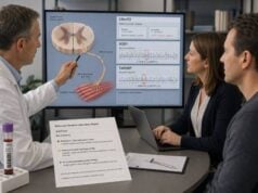
An epilepsy genetic panel test analyzes many genes associated with recurrent seizures and epilepsy syndromes at the same time. It is most likely to find a cause when seizures begin in infancy or early childhood, occur with developmental delay or intellectual disability, resist treatment, follow a recognizable genetic syndrome, or affect several relatives. A positive result can identify the epilepsy type, reveal risks beyond seizures, guide medication or dietary choices, end unnecessary testing, and clarify family recurrence risk. The result is not interpreted by the gene name alone. Different variants in the same gene can cause opposite effects on an ion channel and may call for different treatment approaches. A negative panel does not rule out a genetic cause because panels differ in gene content, coverage, copy-number analysis, mosaic sensitivity, and ability to detect repeat expansions, mitochondrial variants, structural changes, or variants in newly discovered genes. Clinical interpretation requires the seizure history, electroencephalogram, development, imaging, family history, and exact laboratory method.
- Epilepsy panels are most informative in early-onset, developmental, treatment-resistant, or familial epilepsies.
- A pathogenic result may change treatment, but only when the variant mechanism and the person’s epilepsy phenotype agree.
- A variant of uncertain significance is not a diagnosis and should not direct high-risk or irreversible treatment by itself.
- Trio testing with both biological parents can clarify de novo variants and reduce uncertainty.
- Negative results may justify exome or genome sequencing, targeted repeat or mitochondrial testing, or later reanalysis.
Table of Contents
- Who May Benefit From an Epilepsy Gene Panel
- What the Panel Tests and What It Can Miss
- Choosing Between a Panel, Exome, and Genome
- Interpreting Pathogenic, Uncertain, and Negative Results
- How a Genetic Diagnosis Can Change Care
- Inheritance, Mosaicism, and Family Testing
- Common Testing Mistakes and Limitations
- Preparing for Results and Next Steps
Who May Benefit From an Epilepsy Gene Panel
Epilepsy has many causes. Some arise from a structural brain lesion, infection, immune disorder, stroke, trauma, or metabolic problem. Others result from genetic variants that affect ion channels, synaptic signaling, brain development, energy metabolism, or the regulation of gene expression. “Genetic epilepsy” does not necessarily mean inherited; many causative variants occur de novo in the affected person.
Testing is especially valuable when the probability of a single-gene or genomic cause is high. Strong indications include seizures beginning in the neonatal period or infancy, developmental and epileptic encephalopathy, intellectual disability, autism or regression, multiple seizure types, drug-resistant epilepsy, congenital anomalies, movement disorder, abnormal muscle tone, or a family history of similar epilepsy.
A developmental and epileptic encephalopathy is a condition in which both the underlying biology and frequent epileptic activity can impair development. Early diagnosis may identify a treatable metabolic disorder, prevent use of an aggravating medication, or direct care toward a syndrome-specific therapy. Testing should not wait until every antiseizure medicine has failed when the initial presentation is highly suggestive.
Genetic testing can also help people with focal epilepsy. A normal brain magnetic resonance imaging scan does not exclude a genetic cause, and variants in genes such as DEPDC5, NPRL2, NPRL3, KCNT1, or SCN1A can present with focal seizures. A family with seizures arising from different brain regions may still share one inherited variant.
Adults should not be excluded because of age. Many adults with childhood-onset epilepsy, developmental disability, or an old nonspecific diagnosis never had access to modern testing. A diagnosis can still change medication, surveillance, reproductive counseling, and eligibility for research. Testing may also be useful in adult-onset familial epilepsy or when the phenotype points to a known genetic syndrome.
An epilepsy panel should not replace urgent evaluation for a first seizure, status epilepticus, infection, toxic exposure, low glucose, electrolyte disturbance, or acute brain injury. Electroencephalography and imaging answer different questions and often remain essential even when genetic testing is ordered.
What the Panel Tests and What It Can Miss
A panel uses next-generation sequencing to analyze a selected group of epilepsy-related genes. Small panels may focus on a syndrome, such as neonatal epilepsy. Large panels may include hundreds of genes spanning developmental epileptic encephalopathy, metabolic epilepsy, malformations of cortical development, neurodegeneration, and mitochondrial disease.
Most panels detect single-nucleotide variants and small insertions or deletions in coding exons and nearby splice sites. Many laboratories also report exon-level deletions and duplications, called copy-number variants. The report should state whether copy-number analysis was validated, because not every panel includes it and sensitivity varies by gene.
Coverage is not uniform. Some genes contain pseudogenes, repeated sequences, GC-rich exons, or regions that short-read sequencing handles poorly. The laboratory should disclose low-coverage regions and may fill them with another method. A “negative” result is less reassuring when clinically important exons were not adequately analyzed.
Panels can miss several important variant types:
- Repeat expansions that require repeat-primed polymerase chain reaction or another specialized method.
- Methylation and imprinting abnormalities, such as those involved in Angelman syndrome.
- Balanced chromosome rearrangements and some complex structural variants.
- Deep intronic or regulatory variants outside the tested region.
- Low-level mosaic variants present in only a small fraction of blood cells.
- Mitochondrial DNA variants when mitochondrial analysis is not included.
- Variants in genes discovered after the panel’s gene list was frozen.
Some laboratories use a broad sequencing backbone but analyze only a virtual panel of selected genes. This allows later reanalysis of additional genes without resequencing, provided the consent and laboratory policy permit it. Other panels physically capture only the listed genes, so expanding the analysis requires a new test.
Sample type is usually blood, saliva, or a cheek swab. Blood often provides reliable DNA and may be preferred for copy-number and mosaic analysis. A negative blood test cannot exclude a variant confined to brain or present at a very low level. Skin, buccal cells, or surgical brain tissue may be considered in selected mosaic malformation syndromes.
A high-quality order includes detailed phenotype information. “Seizures” is less useful than age at onset, seizure types, triggers, electroencephalographic pattern, imaging, developmental course, medication responses, movement features, head growth, and organ involvement. Laboratories use these details to prioritize variants and decide whether a gene fits the presentation.
Choosing Between a Panel, Exome, and Genome
A panel can provide focused, rapid analysis with deep coverage and fewer unrelated findings. It is often practical when the phenotype is clearly epileptic and the laboratory’s panel is broad, current, and includes copy-number analysis. The narrower scope may reduce uncertain findings and simplify consent.
Exome sequencing analyzes most protein-coding genes, including many not traditionally labeled as epilepsy genes. It is useful when seizures occur with congenital anomalies, unusual neurologic features, multisystem disease, or a wide differential diagnosis. Exome sequencing can also find newly recognized causes if the laboratory periodically reanalyzes the data.
Genome sequencing covers coding and noncoding DNA more evenly and can detect a wider range of structural and copy-number variants. It may combine functions that otherwise require a panel, microarray, and selected additional tests. Genome sequencing still has blind spots and does not automatically detect every repeat expansion, methylation abnormality, mitochondrial variant, or low-level mosaic change.
Trio testing with the affected person and both biological parents often increases interpretive power. It can quickly identify de novo variants, determine whether two recessive variants are on opposite gene copies, and show whether a candidate variant was inherited from a clinically unaffected parent. A duo or singleton analysis remains worthwhile when parental samples are unavailable.
The choice should account for turnaround time. Rapid exome or genome sequencing may be favored in a critically ill newborn with refractory seizures, metabolic instability, or suspected treatable genetic disease. A focused panel may return quickly in other settings, but advertised turnaround time should include confirmation and clinical reporting, not only sequencing.
Cost and insurance rules vary. A less expensive panel can become costly if it is followed by microarray, exome, and genome testing. Conversely, a broad test may generate more uncertain or secondary findings and may not have the depth of a specialized assay for a difficult gene. The best first test is the one most likely to capture the suspected variant types with reliable interpretation.
| Test | Common advantage | Common limitation |
|---|---|---|
| Epilepsy panel | Focused genes, strong coverage, often fast | Gene list can become outdated; variant-type coverage differs |
| Exome sequencing | Broad coding analysis and reanalysis potential | Uneven coverage; many noncoding and structural variants missed |
| Genome sequencing | Broad small, copy-number, and structural variant detection | Interpretation, repeat detection, and mosaic sensitivity vary |
| Targeted test | Best method for a specific repeat, methylation change, or familial variant | Does not evaluate alternative causes |
Interpreting Pathogenic, Uncertain, and Negative Results
The report should list the gene, exact DNA and protein change, zygosity, classification, inheritance if known, transcript, and evidence. A medication recommendation without the variant details is incomplete.
Pathogenic or likely pathogenic result
A pathogenic or likely pathogenic variant may establish a diagnosis when it matches the expected inheritance and phenotype. A single variant may be sufficient for an autosomal dominant condition. Two variants usually are needed for an autosomal recessive disorder, and they must affect opposite copies of the gene. X-linked and mitochondrial results require their own inheritance analysis.
“Likely pathogenic” means the evidence strongly supports disease causation and is generally actionable in the same way as a pathogenic result. The clinician still checks whether the gene’s known syndrome fits. A person can have more than one diagnosis, and a result may explain seizures but not every developmental or medical feature.
The mechanism is critical. Loss-of-function and gain-of-function variants in the same ion-channel gene can produce different seizure ages, movement disorders, and drug responses. A report that simply states “SCN2A-related epilepsy,” for example, is not enough to choose a sodium-channel medication without considering variant function and clinical onset.
Variant of uncertain significance
A variant of uncertain significance, or VUS, has insufficient or conflicting evidence. It is not proof of a genetic epilepsy. Parental testing, testing affected relatives, careful phenotype matching, functional studies, and time may clarify it.
A de novo VUS may become more suspicious, but new variants occur in healthy people too. An inherited VUS may still matter when the parent has subtle seizures, migraine, learning differences, reduced penetrance, or mosaicism. Neither result should be interpreted by a simple rule.
Do not change to a high-risk therapy, avoid a standard treatment indefinitely, or make reproductive decisions solely because of a VUS. Clinical management can still follow the observed epilepsy syndrome while the variant remains uncertain.
Negative or nondiagnostic result
A negative panel means no reportable explanation was found in the genes and regions analyzed. It does not rule out genetic epilepsy. The cause may involve an untested gene, structural variant, repeat expansion, mitochondrial change, low-level mosaicism, or a variant not yet recognized as pathogenic.
The next step depends on what the panel included. Exome or genome sequencing may be appropriate, along with chromosomal microarray, Fragile X testing, mitochondrial analysis, metabolic testing, or a syndrome-specific assay. Reanalysis can be valuable after 1–3 years or sooner when new features or a new family diagnosis appears.
Secondary and carrier findings
A broad test may identify medically actionable variants unrelated to epilepsy or carrier status for a recessive condition. Consent should address whether such findings will be reported. A carrier result usually does not explain dominant epilepsy, but it may have reproductive importance and occasionally prompts a search for a second variant.
How a Genetic Diagnosis Can Change Care
A molecular diagnosis can affect treatment, but precision medicine is not always a one-gene–one-drug rule. The strongest changes occur when a known metabolic pathway or ion-channel mechanism has a reliable intervention.
SLC2A1-related glucose transporter type 1 deficiency is a classic example. The brain cannot transport glucose normally, and ketogenic dietary therapy can provide an alternative fuel. Early recognition can improve seizures and movement symptoms. A genetic result should be integrated with cerebrospinal fluid findings and clinical features when needed.
ALDH7A1-related pyridoxine-dependent epilepsy and PNPO-related epilepsy can respond to vitamin B6–related therapy. Treatment may be started before genetic confirmation in a critically ill infant because delay can be harmful. The result then confirms the pathway, guides long-term management, and prompts monitoring for treatment-specific risks.
SCN1A loss-of-function Dravet syndrome often worsens with chronic sodium-channel-blocking medicines, while broad-spectrum and syndrome-specific therapies may be preferred. In contrast, selected gain-of-function variants in SCN2A or SCN8A with early-onset epilepsy may respond to sodium-channel blockers. Later-onset loss-of-function phenotypes may not. Variant mechanism and age at onset are therefore essential.
KCNQ2 and KCNA2 disorders also illustrate why function matters. Some variants reduce channel activity; others increase it. A drug choice based only on the gene can be ineffective or harmful. Functional evidence may be unavailable for a newly identified missense variant, so treatment must remain guided by the person’s observed response.
TSC1 or TSC2 findings can establish tuberous sclerosis complex and trigger kidney, heart, skin, eye, and brain surveillance. They may also support use of an mTOR inhibitor in appropriate clinical settings. DEPDC5, NPRL2, and NPRL3 variants can point to mTOR-pathway focal epilepsy and may influence imaging review, surgical evaluation, and family screening, although routine targeted drug treatment is not established for every case.
A diagnosis can prevent unnecessary tests, identify cardiac or metabolic risks, and improve seizure classification. It may explain why a magnetic resonance imaging lesion is part of a broader syndrome or why an apparently normal scan should be repeated with an epilepsy protocol.
Genetic information can also improve emergency planning. Some syndromes carry high risk of prolonged seizures, fever sensitivity, medication-induced worsening, arrhythmia, or metabolic decompensation during fasting. A written plan can specify rescue medication, illness management, and when to seek emergency care.
Not every diagnosis changes medication. It may still provide a prognosis range, connect families with syndrome-specific resources, enable clinical trial participation, and clarify recurrence risk. The value should be measured by informed care, not only by whether a new drug is prescribed.
Inheritance, Mosaicism, and Family Testing
Many severe early-onset epilepsies result from de novo dominant variants. Neither parent has the variant in routine blood testing, and the condition was not caused by anything they did. Recurrence risk is often low but not zero because a parent can have germline mosaicism.
If a parent carries an autosomal dominant variant, each pregnancy may have a 50% chance of inheritance. Severity can differ greatly between relatives because of variable expression and reduced penetrance. A parent with mild febrile seizures can have a child with a more severe epilepsy, and the reverse can also occur.
Autosomal recessive disorders usually require two pathogenic variants. When both parents are carriers, each pregnancy generally has a 25% chance of an affected child, a 50% chance of a carrier child, and a 25% chance of inheriting neither familial variant. The two variants must be confirmed to be in trans—on opposite gene copies—when phase is not obvious.
X-linked conditions affect families differently according to the gene, sex chromosomes, and X-inactivation. Females may be unaffected, mildly affected, or severely affected; the word “carrier” should not be used to dismiss symptoms. PCDH19-related epilepsy commonly affects heterozygous females and mosaic males through a distinctive cellular mechanism.
Mitochondrial DNA variants are usually transmitted through the egg, but severity depends on heteroplasmy—the proportion of altered mitochondrial DNA—and tissue distribution. A blood level may not predict brain involvement or recurrence precisely.
Somatic mosaicism means the variant arose after fertilization and is present in only some cells. Brain-limited mosaic variants are an important cause of focal cortical malformations and epilepsy. Blood, saliva, and skin may all be negative. Testing resected brain tissue can reveal a diagnosis and may inform recurrence counseling, although it is performed only when surgery is clinically indicated.
Once a familial pathogenic variant is known, relatives can receive targeted testing rather than repeating a large panel. Testing an unaffected child for an adult-onset or incompletely penetrant condition requires careful counseling. Reproductive options may include prenatal diagnosis or preimplantation genetic testing, but these decisions are personal.
Common Testing Mistakes and Limitations
One frequent mistake is treating all panels as equivalent. A 20-gene neonatal panel and a 500-gene comprehensive panel answer different questions. Even panels with similar size may differ in coverage, copy-number detection, mitochondrial analysis, update policy, and variant interpretation.
Another mistake is ordering a narrow panel after the phenotype has become broad and multisystemic. A child with epilepsy, congenital anomalies, growth disturbance, and organ disease may be better served by trio exome or genome sequencing. Conversely, broad sequencing may miss a repeat expansion or methylation disorder that a focused test detects well.
Commercial “treatment guidance” can overstate evidence. A laboratory may list drugs associated with a gene even when the patient’s variant mechanism is unknown or when evidence comes from a small number of cases. Prescribers should distinguish established guideline-level recommendations from experimental observations.
Outdated reports are another problem. Gene–disease relationships and variant classifications change quickly. A panel performed years ago may not have included a gene now recognized as a common cause. Reanalysis should use updated phenotype information and not merely rerun the same filter.
An uncertain result can distract from a more likely non-genetic cause or from a second genetic diagnosis. The entire clinical picture must be revisited when the result does not fit. Segregation in the family, electroencephalogram pattern, imaging, biochemical testing, and treatment response can all challenge an initial interpretation.
A genetic diagnosis does not replace standard epilepsy care. Medication adherence, sleep, mental health, bone health, pregnancy counseling, driving rules, rescue planning, and sudden unexpected death in epilepsy risk still require attention. Genetics adds information; it does not remove the need for ongoing clinical observation.
Preparing for Results and Next Steps
Before testing, collect seizure descriptions, videos when safely obtained, electroencephalogram reports, imaging, medication history, developmental milestones, and prior laboratory results. Record which medicines helped, worsened seizures, or caused serious adverse effects. A three-generation family history should include seizures, febrile seizures, developmental disability, migraine, movement disorder, sudden unexplained death, miscarriages, and known genetic conditions.
Ask the laboratory or clinician how many genes are included, whether the list is updated, whether deletion/duplication and mitochondrial analysis are included, what mosaic level can be detected, and whether parental samples are needed. Clarify the policy for secondary findings, data storage, reanalysis, and cost.
At the results visit, request the exact variant and classification in writing. Ask whether the proposed diagnosis explains the seizure type, development, imaging, and non-neurologic features; whether the mechanism is loss or gain of function; and what evidence supports any medication recommendation.
For a positive result, the care plan may include syndrome-specific surveillance, family testing, and review by metabolic, cardiac, renal, ophthalmologic, rehabilitation, or other specialists. A genetic counselor can provide recurrence information and a family letter.
For a VUS, identify what evidence could resolve it and who will monitor reclassification. For a negative result, review the panel’s gaps and decide whether exome, genome, repeat-expansion, methylation, mitochondrial, or mosaic testing is appropriate. Set a reanalysis date rather than allowing the report to disappear into the record.
Do not stop antiseizure medication abruptly after any result. Sudden withdrawal can provoke seizure clusters or status epilepticus. Seek emergency care for a convulsive seizure lasting 5 minutes or longer, repeated seizures without recovery, breathing difficulty, serious injury, or an event that differs substantially from the person’s usual seizures.
References
- Current practice in diagnostic genetic testing of the epilepsies 2022 (Practice Review).
- Genetic Testing in Epilepsy: Improving Outcomes and Informing Gaps in Research 2024 (Review).
- Genetic Testing to Inform Epilepsy Treatment Management From an International Study of Clinical Practice 2022 (Cohort Study).
- A Review of Advances in Epilepsy Genomics 2023 (Review).
- Impact of Genetic Testing Using Gene Panels, Exomes, and Genomes in 140 Patients With Epilepsy 2025 (Cohort Study).
- Effectiveness of genetic testing in determining the aetiology of epilepsy 2022 (Evidence Review).
Disclaimer
This article is general education and does not diagnose an epilepsy syndrome or provide individualized treatment advice. Genetic findings and medication implications should be reviewed by an epilepsy specialist and genetics professional using the complete clinical history and laboratory report. Do not stop antiseizure medication abruptly, and follow the prescribed emergency plan for prolonged or repeated seizures.