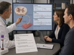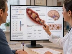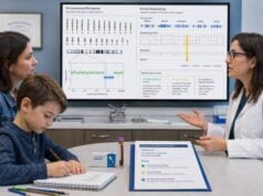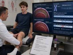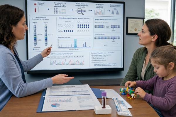
Angelman syndrome testing does more than look for a change in the UBE3A gene. The condition results from loss of the active maternal UBE3A contribution in brain cells, and several different genetic mechanisms can produce that loss. A child may have a maternal deletion of chromosome 15q11.2-q13, paternal uniparental disomy, an imprinting defect, or a pathogenic UBE3A sequence variant. Because these mechanisms leave different laboratory signatures, testing usually begins with DNA methylation analysis and then moves to copy-number, parental-origin, imprinting-center, or UBE3A sequencing studies as needed. The final subtype matters. It can explain why one test was positive while another was normal, help estimate the chance of Angelman syndrome recurring in a future pregnancy, and guide testing for relatives. Most affected children have severe developmental delay, limited speech, movement or balance problems, sleep disturbance, and seizures, but the presentation can vary. A normal first test does not always end the evaluation, especially when the clinical features remain strongly suggestive.
- DNA methylation testing detects most Angelman syndrome cases, including common deletions, paternal uniparental disomy, and many imprinting defects.
- UBE3A sequencing is usually considered when methylation is normal, because sequence variants may not disturb the methylation pattern.
- A positive methylation result confirms abnormal parent-specific imprinting but does not identify the exact mechanism by itself.
- The recurrence risk ranges from usually less than 1% to as high as 50%, depending on the molecular subtype and whether a parent carries a variant or imprinting-center deletion.
- Chromosomal microarray can define a 15q11.2-q13 deletion and show whether other genes are involved, which may help explain severity.
- Testing generally uses blood and does not require fasting, though parental samples may be needed to complete interpretation.
Table of Contents
- Why Angelman Syndrome Needs More Than One Test
- The Four Main Genetic Mechanisms
- A Stepwise Testing Strategy
- Reading Each Kind of Result
- How the Subtype Can Shape Care
- Recurrence Risk and Family Testing
- When Results Are Negative or Incomplete
Why Angelman Syndrome Needs More Than One Test
Angelman syndrome is an imprinting disorder. Imprinting means that some genes are used differently depending on whether they came from the mother or the father. In most tissues, both copies of UBE3A are active. In many neurons, however, the paternal copy is largely silenced by a long antisense RNA. The maternal copy therefore provides most functional UBE3A protein in the brain.
Angelman syndrome develops when the maternal contribution is absent, disrupted, or incorrectly marked. That can happen through a missing chromosome segment, inheritance of two paternal chromosome 15 copies, failure to establish or maintain the maternal imprint, or a damaging change within maternal UBE3A itself.
This biology explains why ordinary gene sequencing alone is not enough. Sequencing can find many UBE3A spelling changes, but it cannot reliably show whether a chromosome segment is missing, whether both chromosome 15 copies came from the father, or whether the imprinting marks are abnormal. Conversely, methylation testing can detect an abnormal parent-of-origin pattern but may miss an isolated UBE3A sequence variant.
Testing is often considered in a child with a combination of:
- Global developmental delay or intellectual disability
- Very limited or absent spoken language
- An unusually happy or excitable demeanor with frequent smiling or laughter
- Ataxic gait, tremulous movements, or poor balance
- Seizures or a characteristic electroencephalogram pattern
- Sleep disturbance
- Microcephaly that develops over time
- Feeding difficulty in infancy
- Wide-based walking, hand-flapping, or fascination with water
No single feature proves the diagnosis. Some infants have nonspecific developmental delay before the more recognizable pattern emerges. Other genetic conditions, cerebral palsy, autism, Rett syndrome, Pitt-Hopkins syndrome, Mowat-Wilson syndrome, and metabolic disorders may overlap. A molecular diagnosis can shorten that differential and direct syndrome-specific care.
Because Angelman syndrome involves parent-specific gene expression, it is also a useful example of why a methylation genetic test can answer a question that standard sequencing cannot.
The Four Main Genetic Mechanisms
The same clinical syndrome can result from several molecular routes. Their relative frequencies vary slightly among studies and laboratories, but the following categories account for most confirmed cases.
Maternal 15q11.2-q13 deletion
The most common mechanism is a deletion on the chromosome 15 inherited from the mother. The deleted region usually includes UBE3A and several neighboring genes. Because more than one gene is missing, children with a larger typical deletion may have more severe seizures, smaller head size, lighter skin or hair pigmentation, and greater developmental impairment than some children with other subtypes. Individual outcomes still vary widely.
A methylation test usually detects the abnormal paternal-only pattern, while chromosomal microarray defines the deletion. Microarray can also show approximate size and whether the deletion is one of the common classes. FISH can detect certain deletions but provides less genomic detail and may not be the preferred first copy-number method in many centers.
Most common deletions occur as new events, so recurrence is generally low. A small number result from a parental chromosome rearrangement or an inherited change that predisposes to deletion, making parental studies important when the laboratory or genetics team recommends them.
Paternal uniparental disomy
In paternal uniparental disomy, or UPD, both chromosome 15 copies come from the father and no maternal copy is present. The child therefore has two paternal imprinting patterns and lacks active maternal UBE3A in neurons.
Methylation is abnormal, but microarray may not show a deletion. SNP-based microarray or parental marker studies can identify regions of homozygosity and determine parental origin. UPD may involve two identical copies of one paternal chromosome, called isodisomy, two different paternal copies, called heterodisomy, or a mixture.
Recurrence is usually very low because UPD often results from a chance error in chromosome segregation followed by “rescue” of an abnormal embryo. Rare parental chromosome rearrangements can change that estimate.
Imprinting defect
An imprinting defect means the maternal chromosome 15 is present, but it carries a paternal-type epigenetic mark in the relevant region. Some defects are caused by a small deletion in the imprinting center; others are epimutations with no detectable DNA deletion.
Methylation testing is abnormal. Follow-up imprinting-center deletion analysis helps distinguish a structural imprinting defect from an epimutation. This distinction matters because an imprinting-center deletion may be inherited from an unaffected mother and can create a high recurrence risk, while an isolated epimutation usually has a low recurrence risk.
The mother can be unaffected because the same deletion may sit on her paternally inherited chromosome 15, where it does not disrupt the maternal imprinting program in her own brain. When she passes that chromosome through the egg, however, it may fail to acquire the required maternal imprint.
UBE3A pathogenic variant
A pathogenic sequence variant in the maternal UBE3A copy accounts for a smaller but important group of cases. Examples include nonsense, frameshift, splice, and certain missense variants that reduce or eliminate protein function. Because the imprinting marks across 15q11.2-q13 remain normal, routine methylation testing can be normal.
UBE3A sequencing is therefore a logical next step when clinical suspicion remains high after normal methylation. Deletion/duplication analysis of UBE3A may also be needed because sequence analysis alone may miss exon-level copy-number changes.
A UBE3A pathogenic variant may occur de novo or be inherited from the mother. An unaffected mother can carry the variant on the UBE3A copy she inherited from her father, where neuronal expression was largely silenced. If she transmits that variant maternally to a child, it becomes the active neuronal copy and can cause Angelman syndrome. That parent-of-origin effect is central to recurrence counseling.
| Mechanism | Methylation result | Useful follow-up test | Typical recurrence pattern |
|---|---|---|---|
| Maternal 15q11.2-q13 deletion | Abnormal paternal-only pattern | Chromosomal microarray; parental studies when indicated | Usually low, but depends on parental rearrangements |
| Paternal UPD 15 | Abnormal paternal-only pattern | SNP array or parental polymorphism analysis | Usually low |
| Imprinting defect | Abnormal paternal-only pattern | Imprinting-center deletion analysis | Low for many epimutations; may be high with inherited imprinting-center deletion |
| UBE3A pathogenic variant | Usually normal | UBE3A sequencing and deletion/duplication analysis | Low if de novo; up to 50% if the mother carries the relevant variant |
A Stepwise Testing Strategy
The exact order varies by laboratory and by what testing has already been done. A practical evaluation often follows this sequence.
- Begin with DNA methylation analysis of the 15q11.2-q13 imprinting region. Methylation-sensitive MLPA, methylation-specific PCR, or another validated method can detect the paternal-only pattern present in most Angelman syndrome cases. Some methods can also estimate copy number.
- Define the mechanism when methylation is abnormal. Chromosomal microarray can identify a deletion. If no deletion is found, parental-origin testing can assess UPD. If UPD is excluded, imprinting-center analysis may identify a deletion or support an epimutation.
- Sequence UBE3A when methylation is normal and suspicion remains strong. The test should include deletion/duplication analysis if the sequencing method does not reliably detect exon-level copy changes.
- Broaden the evaluation if UBE3A testing is negative. A neurodevelopmental panel, exome sequencing, genome sequencing, or targeted tests for overlapping conditions may be appropriate.
Some clinicians order chromosomal microarray and methylation testing together in a child with unexplained developmental delay. Microarray can detect the common deletion and unrelated copy-number conditions, while methylation detects UPD and imprinting defects that a standard array may not fully resolve. A chromosomal microarray test is particularly useful when the child has congenital anomalies or a broader phenotype.
Blood is the usual specimen. Buccal cells or other tissues may be considered in unusual mosaic cases, but specialized interpretation is required. Parental samples are not always needed for the first test, yet they may be essential for distinguishing de novo from inherited findings and calculating recurrence risk.
Turnaround may range from one to several weeks per stage. Sequential testing can take longer than a combined panel, but it may reduce unnecessary analysis and produce a clearer mechanistic answer. Families should ask whether the laboratory reflexes automatically from methylation to copy-number or sequence studies or whether each stage requires a new order.
Reading Each Kind of Result
An Angelman syndrome report should state both the finding and the method. “Positive” alone is not enough because the next medical and reproductive steps depend on the mechanism.
Abnormal methylation pattern
An abnormal paternal-only methylation pattern strongly supports Angelman syndrome in a person with compatible features. It generally captures maternal deletion, paternal UPD, and imprinting defects. It does not, by itself, show which of those three occurred.
Follow-up tests are not optional details. A deletion has different recurrence implications from UPD, and an imprinting-center deletion differs from an epimutation. Ask whether copy number was assessed in the same assay and whether parental samples are recommended.
Deletion result
A report may describe a deletion such as 15q11.2-q13 and list its genomic coordinates. The laboratory may classify it as a typical class I or class II deletion or another size. The missing interval can include UBE3A, GABRB3, OCA2, and other genes. The genotype can inform broad expectations but cannot forecast an individual child’s exact seizure burden, communication ability, sleep pattern, or adult independence.
SNP microarray can sometimes reveal mosaicism or additional copy-number findings. Low-level mosaic deletions may require confirmation with a second method or tissue.
UPD result
A paternal UPD result should state whether both chromosome 15 copies are paternal and, when possible, whether the pattern is heterodisomy, isodisomy, or mixed. Long stretches of isodisomy can unmask a recessive condition if the father carries a pathogenic variant in another chromosome 15 gene. That possibility is uncommon but may matter when the child has features not explained by Angelman syndrome alone.
Imprinting defect result
The report should distinguish a detectable imprinting-center deletion from an imprinting epimutation. A parental study may reveal whether the mother carries the same deletion. The phrase “imprinting defect” should never be interpreted as meaning the parents caused the condition through behavior, diet, stress, or pregnancy choices. It is a molecular marking error, not a parenting event.
UBE3A pathogenic or likely pathogenic variant
A pathogenic or likely pathogenic variant supports the diagnosis when it affects the maternal allele or when parent-of-origin analysis is consistent with disease. The report may not initially know which parent transmitted the variant. Testing the parents can clarify whether it is de novo, inherited from the mother, or located on the paternal allele.
A variant of uncertain significance is not diagnostic. The laboratory may request parental samples, clinical details, RNA studies, or follow-up over time. An uncertain missense change should not be treated as equivalent to a known loss-of-function variant. The broader genetic variant result categories apply here as well.
Normal result
A normal methylation test reduces the likelihood of a deletion, UPD, or imprinting defect but does not exclude a UBE3A sequence variant. A normal methylation-plus-UBE3A sequence result lowers the likelihood further, yet a small percentage of clinically diagnosed individuals remain without a molecular explanation. Technical limitations, mosaicism, noncoding variants, structural changes, or a different condition may account for the result.
How the Subtype Can Shape Care
Every child with Angelman syndrome needs individualized developmental and medical care regardless of subtype. Genetic findings help organize that care but do not replace observation of the child.
Seizures are common and may begin in early childhood. EEG abnormalities can occur even without obvious seizures. Neurology follow-up, a seizure action plan, and medicine selection based on seizure type are important. Sudden prolonged seizures, breathing difficulty, repeated vomiting with lethargy, or failure to recover normally requires urgent care.
Communication support should begin early. Many children understand more than they can express. Augmentative and alternative communication can include picture systems, signs, eye-gaze devices, tablets, and partner-assisted scanning. Waiting for spoken language before introducing these tools can delay meaningful communication.
Physical, occupational, and feeding therapies address balance, gait, tremor, joint range, swallowing, and daily skills. Sleep problems may reflect altered circadian signaling, seizures, reflux, obstructive sleep apnea, medicines, or learned patterns. Treatment should start with a careful sleep history rather than assuming every disturbance is intrinsic to the syndrome.
Deletion-positive children may have lighter pigmentation because the deletion can include OCA2. Some also have a more severe seizure or developmental phenotype on average. UPD or imprinting-defect groups may show somewhat better growth, communication, or motor outcomes in group studies, while obesity or food-seeking behavior may be more prominent in some. These are tendencies, not rules.
The molecular subtype can also affect eligibility for clinical trials. Experimental strategies include reactivating the normally silent paternal UBE3A copy, replacing UBE3A function, or modifying downstream pathways. Trial inclusion may depend on age, genotype, seizure stability, developmental level, and previous treatments. Families should distinguish registered research from commercial clinics selling unproven “stem cell” or gene treatments.
Recurrence Risk and Family Testing
Recurrence counseling is one of the strongest reasons to complete subtype testing. A general statement that Angelman syndrome is “usually not inherited” is too vague for family planning.
For a typical de novo maternal deletion, recurrence is generally below 1%, but parental chromosome studies may be recommended if the deletion pattern suggests a rearrangement. Germline mosaicism means the risk is not always zero even when parental blood tests are normal.
For paternal UPD, recurrence is also usually below 1%. If a parent has a Robertsonian translocation or another chromosome 15 rearrangement, the estimate may be higher and should be calculated from the specific karyotype.
For an imprinting epimutation without a deletion, recurrence is generally low. For an imprinting-center deletion, risk can be much higher—potentially up to 50% when the mother carries the deletion in the relevant parent-of-origin context.
For a UBE3A pathogenic variant, the central question is whether the mother carries it. A de novo variant usually carries a low but nonzero recurrence risk because of possible germline mosaicism. If the mother carries the variant, each pregnancy has a 50% chance of inheriting it, though the outcome depends on parent of origin. Extended maternal relatives may also benefit from counseling.
Reproductive options may include:
- Natural conception with or without prenatal diagnosis
- Chorionic villus sampling or amniocentesis for a known familial mechanism
- In vitro fertilization with preimplantation genetic testing when technically feasible
- Donor egg or sperm, depending on the familial finding
- Adoption
- Choosing not to pursue pregnancy
Prenatal testing must match the known mechanism. A UBE3A sequence test cannot rule out UPD or an imprinting defect, and a routine chromosome count may miss a small imprinting-center deletion. Families should use the exact prior laboratory report when arranging testing.
When Results Are Negative or Incomplete
A negative result should trigger a review of what was actually tested. Ask whether methylation, UBE3A sequencing, UBE3A deletion/duplication analysis, and microarray were all completed. Confirm that the report considered mosaicism and whether the laboratory method could detect low-level abnormal cells.
When the Angelman phenotype remains convincing, possible next steps include:
- Repeating or confirming methylation testing at a laboratory with imprinting expertise
- Testing a second tissue in selected mosaic cases
- Trio exome or genome sequencing
- A neurodevelopmental gene panel that includes genes associated with Angelman-like features
- Testing for Rett syndrome, Pitt-Hopkins syndrome, Mowat-Wilson syndrome, Christianson syndrome, Phelan-McDermid syndrome, or other conditions suggested by the examination
- Metabolic evaluation when regression, episodic illness, unusual movement, or organ involvement suggests it
Exome sequencing may identify a different diagnosis, but it does not automatically replace methylation analysis. Standard exome pipelines often do not assess parent-specific methylation and may miss repeat, structural, or noncoding changes. A whole-exome sequencing test is most useful when its limitations are understood and parental samples are included.
Families should keep copies of every report, including negative ones. Laboratories may reclassify variants or improve detection methods. A genetics clinic can revisit the evaluation as the child’s features evolve or new testing becomes available.
A molecular diagnosis can bring relief, but it may also bring grief, guilt, or uncertainty about future children. Neither parent caused Angelman syndrome through ordinary actions. Genetic counseling should address emotional meaning alongside technical facts and connect the family with developmental, neurologic, educational, and community resources.
References
- Angelman Syndrome 2025 (Review)
- Molecular aspects of Angelman syndrome: Defining the new perspectives of genetic etiology 2025 (Review)
- A multidisciplinary approach and consensus statement to establish standards of care for Angelman syndrome 2022 (Consensus Statement)
- Angelman Syndrome 2023 (Review)
- Angelman Syndrome: Pediatric Primary Care Guide 2024 (Clinical Guide)
- Genetic Evaluation of the Child With Intellectual Disability or Global Developmental Delay: Clinical Report 2025 (Clinical Report)
Disclaimer
This article provides general information and cannot interpret an individual child’s test result or replace care from a clinical geneticist, neurologist, pediatrician, or genetic counselor. Angelman syndrome recurrence risk depends on the exact molecular mechanism and parental studies. Seek urgent medical care for prolonged seizures, breathing difficulty, severe lethargy, sudden loss of skills, or other acute neurologic changes.