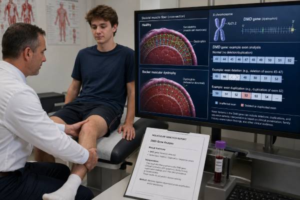
Becker muscular dystrophy testing looks for a pathogenic change in the DMD gene, which provides instructions for making dystrophin. Becker muscular dystrophy usually occurs when a DMD variant allows some shorter or partly functional dystrophin to be produced, leading to later-onset and more slowly progressive weakness than classic Duchenne muscular dystrophy. The distinction is not absolute: dystrophinopathies form a spectrum, and the same broad variant class can produce different severity in different people. A complete laboratory evaluation generally begins with deletion and duplication analysis because large exon changes are common, followed by DMD sequencing when copy-number testing is negative. Identifying the exact variant confirms the diagnosis, guides testing for relatives, helps estimate cardiac risk, and may affect eligibility for mutation-specific trials. It also identifies women and girls who may carry the variant and need cardiac surveillance even if they have little or no muscle weakness. A genetic result should be interpreted with creatine kinase, examination findings, family history, heart assessment, and, when needed, muscle dystrophin studies.
- DMD deletion/duplication analysis is usually the first genetic test, followed by full DMD sequencing if no copy-number variant is found.
- An in-frame variant often causes Becker muscular dystrophy, but the reading-frame rule has important exceptions.
- Cardiomyopathy may be severe even when skeletal muscle weakness is mild, so heart follow-up should begin after diagnosis.
- DMD is X-linked; a carrier mother has a 50% chance of passing the variant in each pregnancy.
- Women with a DMD pathogenic variant can develop cardiomyopathy or muscle symptoms and should not be described as medically unaffected by default.
- A negative blood test does not completely exclude a dystrophinopathy if the phenotype and creatine kinase remain strongly suggestive.
Table of Contents
- Recognizing a Becker Dystrophinopathy
- How DMD Variants Change Dystrophin
- The Testing Sequence
- Reading the Laboratory Report
- What the Result Means for Medical Care
- Carrier Testing and Reproductive Risk
- When the Genetic Answer Is Not Clear
Recognizing a Becker Dystrophinopathy
Becker muscular dystrophy, or BMD, is part of the dystrophinopathy spectrum caused by pathogenic variants in DMD. The gene lies on the X chromosome and is one of the largest human genes. Dystrophin links the internal muscle cell structure to the surrounding membrane complex. When dystrophin is absent or severely disrupted, repeated contraction damages muscle fibers. When some functional dystrophin remains, disease is often milder or later in onset.
BMD may first appear in childhood, adolescence, or adulthood. Some people remain independently mobile into late adulthood, while others develop substantial weakness much earlier. Common clues include:
- Difficulty running, climbing stairs, jumping, or rising from the floor
- Frequent falls or a waddling gait
- Calf enlargement caused by fat and connective tissue replacing muscle
- Tight heel cords or walking on the toes
- Weakness centered in the hips and thighs before the hands
- Muscle cramps, exercise intolerance, or muscle pain
- Persistently elevated creatine kinase, sometimes discovered incidentally
- Elevated AST or ALT that is actually coming from muscle rather than liver
- Dilated cardiomyopathy or rhythm problems with limited skeletal weakness
- A family history of boys or men with progressive weakness, wheelchair use, or unexplained heart failure
The classic distinction from Duchenne muscular dystrophy is later onset and slower progression. A person with Becker usually retains walking beyond age 16, but age cutoffs are imperfect and modern supportive care changes the natural history. Some variants produce an intermediate phenotype between Duchenne and Becker.
Cardiac-predominant dystrophinopathy deserves particular attention. A teenage or adult male may present with dilated cardiomyopathy before obvious muscle weakness. Creatine kinase and family history may reveal the link. Conversely, severe skeletal weakness does not reliably predict when heart disease will appear.
The differential diagnosis includes limb-girdle muscular dystrophies, metabolic myopathies, inflammatory muscle disease, spinal muscular atrophy, Pompe disease, and other inherited neuromuscular conditions. A neurologic genetic panel may be useful when the phenotype is not clearly dystrophin-related, but a focused DMD test remains the most direct path when the clinical pattern fits.
How DMD Variants Change Dystrophin
The DMD gene contains 79 exons. Cells splice these exons together to make messenger RNA, which is read in groups of three DNA letters called codons. A deletion or duplication can either preserve that three-letter reading frame or shift it.
The reading-frame rule
An out-of-frame variant usually disrupts the message so severely that little or no functional dystrophin is made, producing Duchenne muscular dystrophy. An in-frame variant often removes an internal section while preserving the rest of the message, allowing production of a shorter dystrophin and producing Becker muscular dystrophy.
This rule explains many cases, but not all. Exceptions occur because:
- Some in-frame deletions remove an essential functional domain.
- Some out-of-frame variants trigger alternative splicing that restores part of the frame.
- Variants near the gene’s beginning may permit use of an alternative start site.
- Tissue-specific splicing can differ between skeletal and heart muscle.
- Mosaicism can produce a mixture of dystrophin-positive and dystrophin-negative fibers.
- Genetic modifiers and other biological factors change severity.
A laboratory should not classify a variant as Becker-causing solely because it is in frame. Published cases, dystrophin expression, exon location, transcript effects, and the person’s phenotype all matter.
Types of DMD variants
Large deletions involving one or more exons are the most common pathogenic findings. Duplications account for another important group. The remaining cases involve nonsense variants, small insertions or deletions, splice-site changes, and rarer missense, deep intronic, or structural variants.
Some variants create a premature stop signal. These usually cause severe loss of dystrophin, but the exact clinical outcome can depend on exon skipping or alternative translation. Missense variants are relatively uncommon causes of dystrophinopathy, so a rare amino acid change needs strong evidence before being called pathogenic.
The location of a variant can influence cardiac phenotype. Certain exon regions have been associated with higher or lower cardiomyopathy risk in groups, but individual prediction remains imprecise. No genetic report should be used to postpone routine heart surveillance.
The Testing Sequence
A strong diagnostic strategy matches the test method to the types of variants common in DMD.
Step 1: deletion and duplication analysis
Multiplex ligation-dependent probe amplification, commonly called MLPA, or an equivalent copy-number method checks all DMD exons for deletion or duplication. This is usually the first test because large exon changes account for most dystrophinopathies.
The report should list the deleted or duplicated exons and whether the predicted transcript is in frame. Copy-number analysis may not show exact genomic breakpoints, and it may miss a complex rearrangement that appears deceptively simple.
Chromosomal microarray can detect some large DMD deletions or duplications, but it may not have exon-level coverage across the entire gene. A normal microarray therefore does not replace a dedicated DMD deletion/duplication assay.
Step 2: DMD sequence analysis
If copy-number testing is negative, sequencing examines the coding exons and splice boundaries for smaller variants. Next-generation sequencing can find nonsense, frameshift, splice, and many other changes. Sanger sequencing may confirm reportable variants or fill poorly covered regions.
The methods section should state coverage and whether deletion/duplication calling was also performed from sequence data. Families should not assume that “DMD sequencing” includes every intronic or structural change.
Step 3: RNA studies, muscle biopsy, or expanded methods
When routine testing is negative but the clinical suspicion remains high, RNA analysis can show abnormal splicing caused by a deep intronic or synonymous variant. RNA may be obtained from muscle, cultured cells, or other sources depending on the gene transcript and laboratory.
Muscle biopsy is less often needed when genetics is conclusive, but it can still be valuable. Immunohistochemistry or western blot can assess the amount, size, and distribution of dystrophin. Becker muscular dystrophy often shows reduced, patchy, or abnormal-sized dystrophin rather than complete absence.
Genome or long-read sequencing may detect inversions, complex rearrangements, repetitive-region breakpoints, or variants missed by short-read methods. These technologies are increasingly useful but are not universally available.
| Method | What it detects well | Main limitation |
|---|---|---|
| DMD deletion/duplication analysis | One-exon and multiexon copy-number changes | Does not detect most small sequence variants or define all breakpoints |
| DMD sequencing | Nonsense, splice, small insertion/deletion, and other sequence variants | May miss deep intronic and complex structural variants |
| RNA analysis | Confirms abnormal splicing and some hidden intronic effects | Requires an informative tissue and specialized interpretation |
| Muscle dystrophin studies | Shows dystrophin amount, size, and distribution | Invasive and may not identify the DNA variant |
| Genome or long-read sequencing | Complex, structural, and noncoding variants | Availability, cost, and uncertain findings |
Blood is the usual DNA sample. Fasting is not required. Results may take several weeks, and difficult cases take longer if RNA or family studies are added. A deletion/duplication genetic test explains why copy-number analysis remains necessary even when sequencing is available.
Reading the Laboratory Report
The report should name the exact variant, transcript, exon range, predicted reading frame, classification, and evidence. Keep the original document; family testing and future therapies depend on precise notation.
Pathogenic or likely pathogenic result
A pathogenic or likely pathogenic DMD variant confirms a dystrophinopathy when the phenotype fits. The clinician then determines whether the presentation is most consistent with Becker, Duchenne, an intermediate form, isolated hyperCKemia, or DMD-associated cardiomyopathy.
A result such as an in-frame deletion does not by itself guarantee a mild course. The diagnosis should reflect observed motor and cardiac function. The report may mention expected phenotype from variant databases, but those estimates can be biased toward more recognizable cases.
Negative result
A negative routine test lowers the likelihood of a dystrophinopathy but does not eliminate it. Review whether both copy-number analysis and sequencing were performed. Ask about coverage, deep intronic variants, mosaicism, and structural rearrangements.
If creatine kinase is markedly elevated and muscle findings strongly suggest dystrophin deficiency, the next step may be muscle biopsy with dystrophin studies, RNA testing, or genome sequencing. A broader muscular dystrophy panel may identify another gene.
Variant of uncertain significance
A VUS is not enough to diagnose Becker muscular dystrophy or label relatives as carriers. Helpful evidence can include maternal inheritance in an X-linked family, segregation with disease, dystrophin staining, RNA splicing, population frequency, and prior affected cases.
The large size of DMD means rare benign variation exists. Missense changes especially require caution because most established pathogenic DMD variants cause loss of function rather than a simple amino acid substitution. The meaning of a VUS is important before making reproductive or treatment decisions.
Mosaic result
Mosaicism means the variant is present in some cells but not others. A male with somatic mosaicism may have a milder or patchy phenotype. A mother may have germline mosaicism even when her blood test is negative, which explains why recurrence risk is not always zero after a de novo result.
Low-level mosaic variants can be missed by standard methods. Testing another tissue may be considered when the family history or recurrence pattern suggests mosaicism.
What the Result Means for Medical Care
A molecular diagnosis should trigger coordinated neuromuscular and cardiac care rather than end the evaluation.
Heart surveillance
Dilated cardiomyopathy is a major complication of Becker muscular dystrophy and can be more severe than the skeletal muscle disease. Cardiac injury may develop before symptoms. Baseline assessment typically includes electrocardiography and cardiac imaging, with repeat follow-up determined by age, findings, and specialist guidance.
Echocardiography assesses structure and pumping function. Cardiac MRI can detect fibrosis before the ejection fraction falls and is especially useful when image quality is adequate and the person can tolerate the scan. Rhythm monitoring may be added for palpitations, fainting, conduction abnormalities, or cardiomyopathy.
Shortness of breath, chest pain, fainting, new palpitations, leg swelling, or sudden exercise decline warrants prompt evaluation. Preventive or early heart-failure medicines may be recommended before symptoms when imaging shows injury.
Skeletal muscle and mobility
Care includes baseline and serial strength and function measures, physical therapy, stretching, fall prevention, and exercise guidance. Moderate low-impact aerobic activity may support health, while repeated high-resistance or eccentric exercise can worsen muscle injury. Recommendations should be individualized.
Contractures, scoliosis, pain, and mobility equipment should be addressed before they severely restrict participation. An assistive device is not a sign of failure; timely use can conserve energy and reduce falls.
Corticosteroids are standard in Duchenne muscular dystrophy, but evidence and practice in Becker are less uniform. Decisions weigh motor function, side effects, age, and specialist experience. Mutation-specific exon-skipping drugs approved for Duchenne do not automatically apply to Becker and may not help when a person already produces an internally shortened dystrophin.
Respiratory and anesthesia considerations
Respiratory weakness is often later or milder than in Duchenne but can occur. Pulmonary function testing, sleep assessment, cough strength, and vaccination planning may be needed as disease progresses.
Anesthesia teams should know the diagnosis. Succinylcholine can cause dangerous hyperkalemia in muscular dystrophy, and certain anesthetic approaches require caution because of rhabdomyolysis risk. This is not the same as classic malignant hyperthermia susceptibility, but the perioperative plan should be made by experienced clinicians.
Laboratory results
Creatine kinase can be markedly elevated even when weakness is mild. AST and ALT may also rise from muscle. Checking gamma-glutamyl transferase and the clinical context can prevent unnecessary liver procedures. CK level does not track day-to-day disease severity well and should not be used alone to judge progression.
Carrier Testing and Reproductive Risk
DMD is X-linked. A male has one X chromosome, so a pathogenic variant on that chromosome can cause disease. A woman or girl with one altered DMD copy is often called a carrier, but that term should not imply absence of health effects.
If the mother carries the familial variant, each pregnancy has:
- A 50% chance of passing the altered X chromosome
- For a son who inherits it, a high chance of a dystrophinopathy
- For a daughter who inherits it, carrier status with possible muscle or heart manifestations
If the variant is not found in the mother’s blood, the affected person may have a de novo change. Recurrence risk for another child is then lower but remains above zero because of possible germline mosaicism. The affected male will pass his X chromosome to all daughters and none of his sons.
Female heterozygotes may have elevated CK, cramps, weakness, asymmetric symptoms, or dilated cardiomyopathy. Skewed X-chromosome inactivation can make one X more active than the other and influence severity. Cardiac disease can occur without skeletal weakness, so carriers need a planned cardiac assessment rather than symptom-only follow-up.
Testing should use the exact familial variant. A generic panel is unnecessary when a known deletion, duplication, or sequence change can be targeted. At-risk adult women may wish to address life, disability, or long-term-care insurance before testing because legal protections vary.
Reproductive options include prenatal diagnosis, in vitro fertilization with preimplantation genetic testing, donor eggs, donor sperm where relevant, adoption, or natural conception without testing. Prenatal tests must target the known variant, and fetal sex alone cannot diagnose or exclude disease.
An X-linked genetic test provides a broader explanation of why the same familial variant can affect males and females differently.
When the Genetic Answer Is Not Clear
Some people remain undiagnosed after standard testing. Before moving to another test, confirm that the phenotype was reviewed by a neuromuscular specialist and that the first laboratory analyzed both deletions/duplications and sequence variants.
Useful next steps may include:
- Rechecking CK and evaluating other muscle enzymes
- Reviewing electromyography, muscle MRI, and cardiac findings
- Reanalyzing the DMD data for poorly covered exons or mosaic calls
- RNA studies for suspected splice variants
- Muscle biopsy with dystrophin immunostaining and western blot
- Genome or long-read sequencing for complex rearrangements
- A comprehensive muscular dystrophy or cardiomyopathy panel
- Testing another affected relative to improve segregation analysis
A diagnosis should not be forced to fit a borderline result. Limb-girdle muscular dystrophies, sarcoglycanopathies, FKRP-related disease, anoctaminopathy, Pompe disease, and inflammatory myopathy can mimic Becker muscular dystrophy. Treatment and family risk differ, making molecular accuracy important.
The report may also need reinterpretation as variant databases and transcript methods improve. Families should ask whether the laboratory issues amended reports and whether reanalysis is available after one to three years.
The most actionable result is one that joins four pieces: a convincing DMD variant, compatible dystrophin biology, the person’s clinical pattern, and a clear plan for cardiac and neuromuscular follow-up. Severity cannot be read from an exon number alone. Long-term care should respond to the person in front of the clinician, not only to the label “Becker.”
Families can make follow-up more reliable by keeping a compact medical file with the genetic report, latest cardiac MRI or echocardiogram, electrocardiogram, medication list, pulmonary measurements, anesthesia precautions, and emergency contacts. The exact DMD variant should appear in referral letters rather than only “positive genetic test.” When changing health systems, ask the new team to verify whether surveillance is current instead of assuming previous care covered every risk. Adults who were diagnosed in childhood may need renewed counseling because reproductive plans, treatment studies, and carrier testing for relatives often become relevant years after the original result. A yearly review should also ask about falls, fatigue, pain, swallowing, sleep-disordered breathing, work demands, driving, and mental health. These concerns may not appear on a strength test, yet they strongly influence safety and participation. Early referrals for equipment, workplace accommodations, or counseling can preserve choices and daily life before a crisis forces them.
References
- Dystrophinopathies 2022 (Review)
- Diagnosis and Management of Becker Muscular Dystrophy 2024 (Clinical Guide)
- Genetic diagnosis of Duchenne and Becker muscular dystrophy through mRNA analysis: new splicing events 2022 (Research Study)
- The Persistence of Duchenne vs Becker Muscular Dystrophies 2024 (Review)
- 2023 ESC Guidelines for the management of cardiomyopathies 2023 (Guideline)
- Expert consensus on the genetic counseling for dystrophinopathies 2024 (Consensus Statement)
Disclaimer
This article provides general education and does not diagnose Becker muscular dystrophy, predict an individual course, or replace a neuromuscular, cardiology, genetics, or reproductive-medicine evaluation. DMD variants and carrier findings require interpretation from the full report and clinical context. Chest pain, fainting, new palpitations, severe breathing difficulty, dark urine after muscle injury, or sudden weakness needs urgent medical assessment.