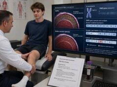
A Duchenne muscular dystrophy genetic test looks for disease-causing changes in the DMD gene, which provides instructions for dystrophin, a protein that protects muscle fibers during contraction. Testing usually begins with deletion and duplication analysis because missing or extra DMD exons cause most cases. If that result is negative, sequencing is used to find smaller variants. A confirmed molecular diagnosis can distinguish a dystrophinopathy from other causes of childhood weakness, determine whether a variant disrupts the gene’s reading frame, identify eligibility for mutation-specific treatment, and guide testing for relatives. The result must be interpreted with symptoms, creatine kinase level, examination, and sometimes dystrophin studies in muscle. Although Duchenne muscular dystrophy mainly affects boys, girls and women with a DMD variant can have muscle symptoms or cardiomyopathy. A negative test does not fully exclude a dystrophinopathy when the assay did not assess all variant types or when a complex or deep intronic change remains undetected.
- DMD deletion/duplication testing is usually the first molecular test because exon-level copy-number changes are the most common cause.
- Sequencing follows when no deletion or duplication is found and detects many nonsense, frameshift, splice, and missense variants.
- An out-of-frame variant usually predicts Duchenne muscular dystrophy, but the reading-frame rule has important exceptions.
- The exact exon pattern can affect eligibility for exon-skipping therapy, while other treatment decisions depend on age and clinical status.
- Mothers and other female relatives may need targeted testing and cardiac surveillance when a familial DMD variant is identified.
Table of Contents
- When DMD Genetic Testing Is Ordered
- How Dystrophin and the DMD Gene Relate to Disease
- Stepwise Testing for Deletions, Duplications, and Small Variants
- Reading the Laboratory Result
- Duchenne Versus Becker and the Reading-Frame Rule
- How the Variant Affects Treatment Options
- Inheritance, Carrier Testing, and Family Planning
- Negative, Complex, and Uncertain Results
When DMD Genetic Testing Is Ordered
Duchenne muscular dystrophy often becomes apparent between ages 2 and 5, although elevated muscle enzymes may be found earlier. Typical signs include delayed walking, frequent falls, difficulty running or climbing stairs, walking on the toes, enlarged calves, and using the hands to push up the thighs when rising from the floor, known as Gowers maneuver. Speech or learning differences may appear before obvious weakness.
Creatine kinase, or CK, is usually markedly elevated—often more than 10 times the upper limit of normal and commonly far higher. Aspartate aminotransferase and alanine aminotransferase may also be elevated because these enzymes are released from muscle as well as liver. A child with high “liver enzymes” but normal bilirubin and no clear liver disease should have CK measured before undergoing an extensive liver evaluation.
Testing may also be ordered after newborn screening, an incidental high CK result, a family history of Duchenne or Becker muscular dystrophy, unexplained cardiomyopathy, or a relative’s known DMD variant. An adult with longstanding weakness can still receive a useful molecular diagnosis, especially when the original diagnosis relied only on muscle biopsy.
The clinical spectrum is broader than a classic childhood presentation. DMD variants can cause Becker muscular dystrophy, isolated dilated cardiomyopathy, exertional cramps, elevated CK without obvious weakness, or symptoms in a female heterozygote. The test should therefore be selected for the suspected dystrophinopathy rather than limited by the label used at referral.
A prompt diagnosis matters because muscle, respiratory, cardiac, bone, endocrine, rehabilitation, and psychosocial care should begin before advanced weakness develops. Molecular confirmation is also required for many clinical trials and mutation-specific therapies. Testing should not delay baseline cardiac assessment or referral to a neuromuscular center when the clinical picture is strongly suggestive.
Muscle biopsy is no longer the usual first diagnostic step when high-quality genetic testing is available. It remains useful when DNA testing is nondiagnostic, the phenotype and variant do not agree, RNA analysis is needed, or another muscular dystrophy is being considered. Dystrophin immunostaining and protein analysis can show absent, reduced, or abnormal-sized protein but do not always identify the underlying DNA change.
How Dystrophin and the DMD Gene Relate to Disease
The DMD gene lies on the X chromosome and contains 79 exons. It is one of the largest human genes, which helps explain why deletions, duplications, and complex rearrangements are common. Muscle cells join the exons in order to produce messenger RNA, which is then translated into dystrophin protein.
Dystrophin connects the internal framework of a muscle fiber to a group of proteins at the cell membrane. Without enough functional dystrophin, repeated contraction damages the membrane. Muscle fibers degenerate and are gradually replaced by fat and connective tissue. Skeletal muscle, the diaphragm and other breathing muscles, and the heart can all be affected.
Classic Duchenne muscular dystrophy usually results from a variant that prevents production of functional full-length dystrophin. Many variants shift the reading frame, introduce an early stop signal, disrupt essential splicing, or remove critical portions of the gene. The resulting messenger RNA may be destroyed through nonsense-mediated decay, leaving little or no dystrophin.
Becker muscular dystrophy more often results from an in-frame variant that allows production of a shorter but partly functional protein. This distinction is useful but not absolute. Some in-frame deletions cause a severe Duchenne course because they remove an essential domain, and some out-of-frame changes permit alternative splicing or translation that preserves more function than expected.
DMD expression also occurs in the brain. This helps explain why some people have language delay, learning disability, attention-deficit/hyperactivity disorder, autism-related features, anxiety, or other neurobehavioral differences. Certain variants affect shorter dystrophin isoforms expressed in the brain and may be associated with greater cognitive risk, but the genotype cannot predict an individual child’s abilities.
In females, the second X chromosome often provides enough dystrophin to prevent the classic Duchenne phenotype. X-chromosome inactivation is uneven, however, and some women develop weakness, cramps, elevated CK, or cardiomyopathy. Rarely, a girl has severe Duchenne-like disease because of skewed X inactivation, an X-chromosome rearrangement, Turner syndrome, or two disease-causing variants.
Stepwise Testing for Deletions, Duplications, and Small Variants
A complete DMD evaluation must detect both copy-number changes and small sequence variants. The order can vary by laboratory, but each category should be addressed.
Deletion and duplication analysis
Multiplex ligation-dependent probe amplification, or MLPA, is widely used to measure the copy number of all 79 exons. Chromosomal microarray, comparative genomic hybridization, quantitative polymerase chain reaction, and validated next-generation sequencing algorithms can also detect exon-level changes.
A deletion result should list every missing exon, such as deletion of exons 45–50. A duplication result should list the duplicated exons, but copy number alone may not reveal where the extra segment sits, its orientation, or whether it disrupts the transcript. Some apparently simple duplications are complex rearrangements, so confirmation or further structural analysis may be needed when the predicted effect is uncertain.
Single-exon deletions or duplications deserve careful confirmation because a sequence variant under a probe can mimic a copy-number change. Laboratories often use an independent method or examine parental samples before reporting a definitive result.
Sequence analysis
When copy-number testing is negative, sequencing examines exons and nearby splice junctions for small variants. Next-generation sequencing can detect nonsense variants, frameshift insertions or deletions, canonical splice variants, and some missense changes. Sanger sequencing may confirm a finding or fill poorly covered regions.
Missense variants are relatively uncommon causes of Duchenne muscular dystrophy. A novel missense change should not be assumed pathogenic merely because it is in DMD. Evidence may include absence from population databases, location in an important protein domain, functional studies, segregation, muscle dystrophin findings, and RNA effects.
RNA analysis and expanded genomic testing
If routine testing is negative but suspicion remains high, RNA extracted from muscle or another informative tissue can reveal abnormal splicing. Deep intronic variants may create a new splice site and insert a pseudoexon into the messenger RNA. Genome sequencing, long-read sequencing, or optical genome mapping can identify structural rearrangements that standard assays miss.
RNA from blood is often uninformative because the muscle-specific DMD transcript is not expressed at the same level. Muscle biopsy may therefore be required for definitive transcript analysis. Some laboratories can use cultured cells or induced muscle models, but availability varies.
Testing after newborn screening
Newborn screening programs commonly begin with CK or a related muscle enzyme marker. An abnormal screen is not a diagnosis; birth stress, prematurity, and other muscle injury can elevate the marker. Follow-up uses repeat biochemical testing and molecular analysis. Early genetic confirmation should distinguish a pathogenic DMD variant from a carrier finding or an unrelated cause of high CK.
| Method | Variant types detected well | Important limitation |
|---|---|---|
| MLPA or equivalent copy-number assay | Whole-exon deletions and duplications | May not define complex duplication structure or small variants |
| DMD sequencing | Small substitutions and insertions/deletions | May miss deep intronic and complex structural variants |
| RNA analysis | Abnormal splicing and transcript effect | May require muscle or another informative tissue |
| Genome or long-read analysis | Complex, deep intronic, and structural changes | Interpretation and clinical availability vary |
Reading the Laboratory Result
A useful report names the variant using current DNA and protein notation, lists affected exons, classifies the finding, explains the predicted reading frame, and states the test’s limitations. The interpretation should also identify whether the change has been reported in people with a dystrophinopathy.
Pathogenic or likely pathogenic
A pathogenic or likely pathogenic DMD variant that fits the phenotype establishes a molecular diagnosis. “Likely pathogenic” means the evidence strongly supports disease causation and is generally sufficient for clinical management, although variant classification can evolve.
For a deletion or duplication, the report should indicate whether the predicted transcript is in frame or out of frame. Online reading-frame tools can help, but the clinical laboratory’s interpretation should take priority because transcript structure and exceptions can be complex.
A nonsense variant creates a premature stop codon. A frameshift changes the sequence of codons after the variant and usually creates an early stop. Canonical splice variants disrupt the normal removal of introns. These changes often cause Duchenne muscular dystrophy, but RNA studies may alter the predicted consequence.
Variant of uncertain significance
A variant of uncertain significance, or VUS, is not a confirmed diagnosis. This category is especially important for missense and noncanonical splice variants. The team may request maternal testing, analysis of other affected relatives, dystrophin studies, RNA analysis, or periodic reclassification.
A VUS should not be used alone to declare a female relative a carrier, make prenatal decisions, or establish eligibility for mutation-specific treatment. If the child has a classic phenotype and no other finding, the uncertainty warrants further study rather than assuming the variant is causal.
Benign or likely benign
Benign and likely benign variants do not explain disease. The DMD gene contains normal variation, and not every change in a very large gene is harmful.
Mosaic result
Mosaicism means the variant is present in only a proportion of cells. A boy with somatic mosaicism may have a milder or uneven phenotype, though severity is not predictable from a blood percentage alone. A mother with low-level mosaicism may have a higher recurrence risk than routine carrier estimates suggest.
Duchenne Versus Becker and the Reading-Frame Rule
The reading frame organizes messenger RNA bases into groups of three. If a deletion or duplication preserves those triplets, the transcript is in frame and can often produce a shorter dystrophin. If the variant shifts the triplets, the transcript is out of frame and usually produces little functional protein.
This rule explains many genotype–phenotype relationships, but it is a probability, not a guarantee. Exceptions occur because of alternative splicing, use of internal translation start sites, differences in deleted protein domains, complex rearrangements, and residual dystrophin production. A laboratory may predict Duchenne, Becker, or an intermediate phenotype, but the child’s actual progression remains essential.
The distinction can also change over time. A toddler with high CK and mild weakness may not yet show enough clinical progression to separate Duchenne from Becker. Genetic results provide an early estimate, while serial motor assessments, dystrophin testing, and cardiac or respiratory findings refine the diagnosis.
Some exon deletions that preserve the frame remove regions critical for dystrophin function and can cause severe disease. Conversely, selected out-of-frame deletions may trigger naturally occurring exon skipping that restores part of the frame. These exceptions explain why two people with apparently similar DNA changes can have different outcomes.
Clinical labels should not obscure care. A person with an intermediate dystrophinopathy still needs surveillance for cardiomyopathy, respiratory weakness, contractures, bone health, and learning or behavioral needs. Female heterozygotes also require risk-based cardiac follow-up even if they have no skeletal muscle symptoms.
How the Variant Affects Treatment Options
The molecular result can determine whether a person’s transcript is amenable to exon skipping. Exon-skipping medicines use short synthetic strands to alter splicing so the cell bypasses a specific exon and restores the reading frame. The goal is to convert an out-of-frame Duchenne transcript into an in-frame transcript that can produce a shorter dystrophin.
Eligibility depends on the exact deletion, not simply the presence of a DMD diagnosis. A person may be described as amenable to skipping exon 45, 51, or 53, for example, based on which additional exon would restore the frame. The laboratory report should state the exon boundaries precisely. Treatment availability and regulatory status vary by country and can change, so a neuromuscular specialist should confirm current eligibility.
Nonsense variants may be relevant to stop-codon read-through approaches in regions where such therapy is authorized. Again, the exact variant and jurisdiction matter. A generic “point mutation” description is not enough.
Gene-transfer therapy delivers a shortened micro-dystrophin because the full DMD gene is too large for commonly used viral vectors. Eligibility and safety assessment involve age, ambulatory status, antibodies to the vector, liver health, cardiac status, steroid use, and other factors—not only the DMD variant. Regulatory indications and safety restrictions have changed over time, so decisions require the current product label and an experienced center.
Corticosteroid or steroid-like treatment, cardiac therapy, respiratory care, physical therapy, contracture prevention, nutrition, bone protection, and psychosocial support are not limited to a particular mutation. A genetic result should not create the impression that mutation-specific treatment replaces comprehensive care.
The variant can also affect trial eligibility for gene editing, cell therapy, or other experimental strategies. Research participation involves uncertain benefit and potential risk. Families should distinguish a trial’s biological rationale from proven clinical effectiveness.
Treatment monitoring may use motor-function measures, pulmonary tests, cardiac imaging, CK trends, and imaging or biopsy biomarkers. A change in CK alone does not show whether meaningful muscle function has improved. Genetic confirmation establishes the target but does not serve as a treatment-response marker because the inherited variant remains present.
Inheritance, Carrier Testing, and Family Planning
Duchenne muscular dystrophy follows X-linked inheritance. A boy usually has one X chromosome, so a disease-causing DMD variant on that chromosome affects his only copy. A woman with one altered copy is heterozygous and may be called a carrier, although some carriers have clinical symptoms.
A substantial proportion of affected boys have a de novo variant with no detectable variant in the mother’s blood. When the mother carries the familial variant, each pregnancy has a 50% chance of inheriting that X chromosome. A child who inherits it and has one X chromosome is expected to be affected; a child with two X chromosomes may be a carrier and may develop muscle or cardiac manifestations.
When maternal blood testing is negative, recurrence risk is lower but not zero because of germline mosaicism. More than one egg cell can carry the variant even when the mother’s blood does not. The exact recurrence estimate depends on the variant and family history and should be discussed with a genetics professional.
Carrier testing should target the known familial variant. CK alone is not reliable because many carriers have a normal level and some noncarriers have elevations for other reasons. A negative limited deletion test also does not exclude a family’s sequence variant; the assay must match the proband’s exact finding.
Female relatives who carry a pathogenic DMD variant need cardiac evaluation even if they feel well. Dilated cardiomyopathy can occur without obvious skeletal muscle weakness. Follow-up intervals depend on age, symptoms, imaging, family history, and specialist guidance. Muscle pain, weakness, exercise intolerance, or elevated CK should prompt neuromuscular assessment.
Reproductive options may include prenatal diagnosis, preimplantation genetic testing, donor eggs, adoption, or natural conception without testing. Prenatal diagnosis requires direct testing of fetal DNA for the known familial variant; fetal sex alone cannot establish or exclude disease. Noninvasive screening based only on cell-free DNA is not equivalent to diagnostic testing unless a validated family-specific method is used.
Negative, Complex, and Uncertain Results
A negative DMD test should be interpreted according to what was actually performed. If only deletion analysis was completed, duplications and small variants remain possible. If sequencing was performed without copy-number analysis, exon deletions or duplications may be missed. A comprehensive negative result still leaves deep intronic variants, complex rearrangements, low-level mosaicism, and technical blind spots.
When CK is very high and the phenotype remains strongly suggestive, next steps may include review of raw coverage, RNA studies, genome sequencing, long-read sequencing, or muscle biopsy. Testing for other muscular dystrophy genes is appropriate when dystrophin studies are not typical or when features point to another condition.
A complex duplication should not be interpreted only by multiplying exon numbers. The duplicated segment can insert in tandem, invert, move elsewhere, or coexist with a deletion. Its effect on messenger RNA may differ from the simple copy-number prediction. Long-range methods or RNA analysis can resolve clinically important ambiguity.
Reclassification can alter a VUS or an older result. Families should keep the original report, laboratory name, and accession number and ask how updates are issued. Reanalysis is reasonable when new symptoms appear, a therapy depends on precise exon structure, or improved methods become available.
An older diagnosis based on a muscle biopsy should be revisited with modern molecular testing. Exact variant identification can clarify relatives’ risks and access to treatment even when the clinical diagnosis is certain. Conversely, a genetic result should be questioned when CK, dystrophin staining, or progression is inconsistent with the predicted effect.
Seek prompt medical assessment for new breathing difficulty, chest pain, fainting, palpitations, dark urine after exertion, marked loss of strength, or inability to clear respiratory secretions. These concerns require clinical care regardless of whether genetic testing is complete.
References
- The complex landscape of DMD mutations 2024 (Review).
- Clinical practice guidelines for the diagnosis and management of Duchenne muscular dystrophy: a scoping review 2024 (Review).
- Genetic strategies for therapy of Duchenne muscular dystrophy 2025 (Review).
- Consensus recommendations and considerations for the delivery and monitoring of gene therapy in patients with Duchenne muscular dystrophy 2025 (Consensus Statement).
- Genomic insights into Duchenne muscular dystrophy: Analysis of 1,250 patients with a focus on mutation patterns 2024 (Cohort Study).
- EMQN best practice guidelines for genetic testing in dystrophinopathies 2020 (Guideline).
Disclaimer
This article is general education and does not diagnose Duchenne muscular dystrophy or establish treatment eligibility. Genetic results, cardiac risk, and mutation-specific options should be reviewed by a neuromuscular specialist and genetics professional using the complete laboratory report. Seek prompt care for breathing difficulty, chest symptoms, fainting, severe weakness, or dark urine.