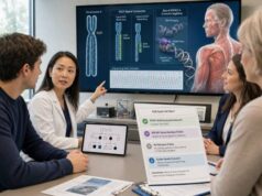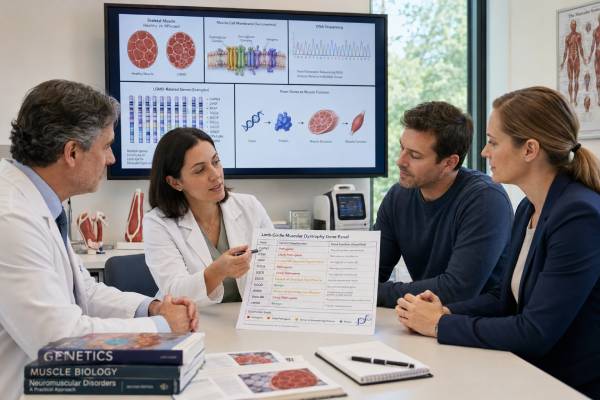
A limb-girdle muscular dystrophy genetic test looks for inherited causes of progressive weakness around the hips and shoulders. “Limb-girdle muscular dystrophy,” or LGMD, is a clinical pattern shared by many different diseases rather than one diagnosis. More than 30 genetic forms are recognized, and they can differ greatly in age of onset, speed of progression, heart and breathing risk, and inheritance. Testing usually begins with a broad neuromuscular gene panel that includes sequence and deletion/duplication analysis. Exome or genome sequencing may follow when the panel is negative, but some look-alike disorders require separate methods. A precise genetic diagnosis can reduce the need for muscle biopsy, identify relatives who may be at risk, guide cardiac and respiratory surveillance, and determine eligibility for gene-specific research or treatment. Results must be matched to the person’s examination, creatine kinase level, imaging, family history, and sometimes muscle protein studies because a single uncertain variant is often not enough to establish an LGMD subtype.
- LGMD is a group of genetic muscle diseases, not one gene or one uniform condition.
- Testing should include both small sequence variants and larger deletions or duplications.
- Two pathogenic variants are usually needed for a recessive subtype; one is usually needed for a dominant subtype.
- Some important mimics, including FSHD, myotonic dystrophy, and certain repeat disorders, need separate tests.
- A molecular diagnosis can change heart, lung, anesthesia, exercise, and family-planning recommendations.
- A negative panel does not rule out a genetic muscle disease.
Table of Contents
- When LGMD testing is considered
- Genes and subtypes included in testing
- Building an effective test strategy
- Reading positive, carrier, and uncertain results
- Why the genetic subtype matters
- Inheritance and testing relatives
- Why a result may be negative
- Care after genetic testing
When LGMD testing is considered
Clinicians consider LGMD when weakness is greatest in the muscles near the hips and shoulders. Early clues may include difficulty running, climbing stairs, rising from the floor, getting out of a chair, lifting objects overhead, or keeping up with peers. Some people develop a waddling gait, shoulder-blade winging, muscle cramps, calf enlargement, or frequent falls. Onset can occur in early childhood, adolescence, or adulthood.
The pattern must be evaluated carefully because proximal weakness has many causes. The examination looks at which muscles are weak and which are relatively spared, whether weakness is symmetric, whether facial or eye muscles are involved, and whether there are contractures, scapular winging, myotonia, neuropathy, sensory loss, or upper motor neuron signs. The tempo matters: inherited dystrophies usually progress over months to years, while rapid weakness may point toward inflammation, endocrine disease, toxin exposure, electrolyte disturbance, or another acquired cause.
Creatine kinase, or CK, is often elevated when muscle fibers are damaged. The degree of elevation can help narrow the differential, but it does not identify a subtype. Some LGMD forms produce very high CK before major weakness; others have modest elevation. Liver enzymes called AST and ALT can rise from muscle, so an unexplained “liver” abnormality should be interpreted with CK and clinical findings before invasive liver testing is pursued.
Electromyography can support a muscle process but is not specific. Muscle MRI or ultrasound may reveal a characteristic pattern of affected and spared muscles and can guide biopsy if one is needed. Heart testing, pulmonary function, and sleep-related breathing assessment may be urgent even before the genetic diagnosis because some subtypes affect the heart or respiratory muscles early.
Family history is useful but can be misleading. Recessive LGMD often appears in only one person or among siblings while both parents are healthy. Dominant disease may look absent because a parent has mild, late-onset, or unrecognized weakness. Consanguinity or shared ancestry can increase the likelihood of a recessive condition, but recessive LGMD occurs in families without known parental relatedness.
The term LGMD should be used only after considering better-defined alternatives. Duchenne or Becker muscular dystrophy, facioscapulohumeral muscular dystrophy, Emery-Dreifuss muscular dystrophy, congenital muscular dystrophy, Pompe disease, myotonic dystrophy, spinal muscular atrophy, inflammatory myopathy, and hereditary motor neuropathy can all resemble a limb-girdle pattern. The genetic plan should be broad enough to capture likely mimics or pair the panel with the right separate assays.
Genes and subtypes included in testing
Modern LGMD names indicate inheritance and the affected protein. “LGMDD” refers to dominant forms and “LGMDR” to recessive forms. Older labels such as LGMD1A or LGMD2B may still appear in medical records and publications. Because nomenclature has changed, the gene symbol is often the clearest way to communicate a diagnosis.
Common or clinically important recessive genes include CAPN3, DYSF, FKRP, ANO5, SGCA, SGCB, SGCG, SGCD, TCAP, TRIM32, GMPPB, POMT2, POMGNT2, ISPD/CRPPA, PLEC, and others. Dominant forms include conditions related to DNAJB6, TIA1, CAV3, and selected variants in genes such as CAPN3. Some genes produce more than one phenotype. For example, DYSF variants can cause a limb-girdle pattern or predominantly distal weakness, while FKRP variants can range from later-onset LGMD to severe congenital muscular dystrophy.
A useful panel also evaluates major muscular dystrophy and myopathy genes that are not always classified as LGMD. DMD can cause Becker muscular dystrophy or symptomatic disease in females. LMNA, EMD, FHL1, DES, FLNC, TTN, VCP, COL6A1, COL6A2, COL6A3, GAA, and mitochondrial or metabolic genes may be relevant depending on the phenotype. Including mimics can prevent an artificial diagnosis based solely on the panel label.
The laboratory must test the variant types that matter for each gene. Next-generation sequencing detects many single-letter changes and small insertions or deletions. Deletion/duplication analysis is needed for larger exon-level copy-number variants. Some genes have technically difficult regions, pseudogenes, repetitive sequence, or deep intronic disease-causing variants that standard panel sequencing may miss. The report’s methodology and coverage table are therefore as important as the gene list.
Protein studies can still add value. Immunostaining or western blot from muscle may show absent or reduced sarcoglycans, dysferlin, caveolin-3, dystrophin, or other proteins. However, secondary protein loss can occur; an abnormal stain does not always identify the primary gene. Genetic confirmation is preferred before assigning a subtype, predicting inheritance, or testing healthy relatives.
The number of genes on a panel is not a quality score. A carefully validated panel with strong coverage, copy-number analysis, transparent limitations, and expert interpretation can be more useful than a very large list with uneven performance. The clinician should confirm that the panel covers the suspected diseases and that important separate tests are not being assumed to be included.
Building an effective test strategy
The most efficient strategy begins with phenotype review and a well-designed panel or comprehensive sequencing assay. When the presentation is a typical progressive muscular dystrophy and no single subtype is obvious, a broad neuromuscular panel is often appropriate. It should include deletion/duplication analysis and genes for treatable or medically urgent mimics, especially GAA for Pompe disease and genes associated with serious cardiomyopathy or arrhythmia.
Targeted testing may come first when the phenotype and family finding are specific. If a relative has a confirmed pathogenic variant, testing for that exact familial variant is usually faster and easier to interpret than repeating a broad panel. If a boy has classic Duchenne features, DMD deletion/duplication analysis followed by sequencing may be prioritized. If facial weakness and asymmetric scapular winging suggest FSHD, a dedicated D4Z4 repeat and haplotype assay is required; routine sequencing panels do not diagnose most FSHD1.
Myotonic dystrophy also requires repeat-expansion testing. The DMPK and CNBP expansions are not reliably detected by ordinary exome sequencing. A myotonic dystrophy genetic test is especially relevant when proximal weakness occurs with myotonia, cataracts, early balding, cardiac conduction disease, or multisystem symptoms. Other separate assays may be needed for mitochondrial DNA, complex structural variants, or repeat expansions.
Exome sequencing can be used early or after an unrevealing panel. Trio analysis with both biological parents helps evaluate recessive inheritance and de novo dominant variants. Genome sequencing can improve detection of some structural and noncoding changes, but performance varies. Neither test should be ordered without checking whether it includes copy-number calling, mitochondrial analysis, repeat detection, and adequate coverage of relevant muscle genes.
A muscle biopsy is no longer an automatic first step. It is most useful when genetics is inconclusive, an inflammatory or metabolic myopathy remains possible, protein analysis could resolve competing variants, or tissue is needed for RNA studies. Biopsy should be planned by a neuromuscular specialist, often using imaging to select a muscle that is affected but not replaced by fat.
Pretest counseling should cover possible positive, carrier, uncertain, and negative results. Broad sequencing may also identify medically important findings unrelated to muscle disease if the person elects secondary-findings analysis. Families should understand whether samples and data will be retained, whether reanalysis is available, and how parental or relative testing will be handled.
Testing should not be delayed when the result could change immediate care. Severe CK elevation, rapid loss of function, breathing symptoms, fainting, palpitations, chest pain, or a family history of sudden death warrants prompt clinical evaluation while molecular testing is underway.
Reading positive, carrier, and uncertain results
A diagnostic result must fit the gene’s inheritance pattern. In most recessive LGMD forms, two pathogenic or likely pathogenic variants are needed, one on each copy of the gene. The variants should be in trans, meaning on opposite chromosome copies. Testing parents can often show that one variant came from each parent. Two variants found on the same copy are in cis and may leave the other gene copy unaffected.
In a dominant LGMD, one pathogenic variant can be sufficient. It may be inherited from an affected or mildly affected parent or arise de novo. A parent who appears healthy may still carry the variant because some dominant forms have age-dependent or variable expression. A careful examination and condition-specific testing are more informative than assuming inheritance from a healthy parent makes the variant benign.
A single pathogenic variant in a recessive gene usually means carrier status, not a complete diagnosis. The laboratory and clinician should search for a missed second variant using deletion/duplication analysis, coverage review, RNA testing, genome sequencing, or another method when the phenotype strongly fits. Some genes, including CAPN3, can cause either recessive or dominant disease through different mechanisms, so the exact variant and published evidence matter.
A variant of uncertain significance, or VUS, has insufficient evidence to call it disease-causing or benign. One VUS in a dominant gene or two VUSs in a recessive gene do not automatically establish LGMD. The team evaluates population frequency, predicted protein effect, functional data, segregation in relatives, muscle protein findings, and fit with the expected phenotype. Medical care should not be reorganized around a VUS alone unless independent clinical findings justify it.
A positive result can still be only a partial answer. Some people have two genetic conditions, and a muscle diagnosis may not explain neuropathy, intellectual disability, unusual organ disease, or a very different weakness pattern. Conversely, a pathogenic variant with poor phenotype fit may be an incidental carrier or secondary finding rather than the cause of symptoms.
Reports should use current gene and variant nomenclature, list transcript identifiers, and describe classification evidence. Families should keep the original report because names and classifications change. Online interpretations or consumer raw data should be clinically confirmed before they are used for diagnosis or family testing. A general pathogenic, benign, and VUS result guide can help with terminology, but neuromuscular interpretation still requires gene-specific expertise.
Why the genetic subtype matters
The subtype helps determine which organs need surveillance. Some sarcoglycan, FKRP, LMNA-related, and other muscular dystrophies carry substantial cardiomyopathy or rhythm risk. Dysferlinopathy typically has less primary cardiac involvement, although individual assessment is still appropriate. Certain forms can impair breathing before loss of walking, while others have respiratory involvement mainly in advanced disease. A generic “LGMD” plan can therefore miss an important complication.
Cardiac screening may include electrocardiography, echocardiography, ambulatory rhythm monitoring, or cardiac MRI. Frequency depends on the gene, age, symptoms, and baseline findings. Palpitations, fainting, chest pain, unexplained shortness of breath, or a family history of sudden death require timely evaluation. Relatives with a cardiac-risk variant may need surveillance even if muscle strength seems normal.
Respiratory monitoring can include seated and supine lung function, cough strength, sleep assessment, and blood-gas or carbon dioxide evaluation when indicated. Morning headaches, daytime sleepiness, poor sleep, weak cough, recurrent chest infections, or breathlessness when lying flat can signal respiratory muscle weakness. Noninvasive ventilation and cough-assist strategies may improve health and quality of life when started at the right time.
The diagnosis can affect exercise advice. Regular low-to-moderate aerobic activity and individualized strengthening may support function, but extreme eccentric exercise can worsen muscle injury in some dystrophies. A physical therapist familiar with neuromuscular disease can balance conditioning, contracture prevention, energy conservation, and fall safety. CK alone should not be used as the sole measure of exercise harm or benefit.
Some subtypes have additional concerns. Dysferlinopathy can be mistaken for inflammatory myositis, leading to ineffective immunosuppression. ANO5-related disease may present with exercise intolerance or high CK before clear weakness. LMNA-related disease can involve early contractures and dangerous arrhythmias. FKRP and sarcoglycanopathies can resemble dystrophinopathy. Recognizing the gene changes both diagnostic confidence and the emphasis of care.
A molecular diagnosis is increasingly important for clinical trials and gene-targeted therapies. Eligibility may require specific variants, preserved muscle function, age limits, antibody testing, or other criteria. A trial should not be presented as guaranteed treatment, and commercial claims should be checked against regulatory approval and peer-reviewed evidence. Standard multidisciplinary care remains essential regardless of research participation.
The result can also prevent unnecessary treatment. A confirmed inherited dystrophy may reduce repeated immune testing or prolonged steroid treatment given for presumed inflammatory myopathy, though some muscular dystrophies and inflammatory conditions can coexist. Decisions should be based on the full clinical picture.
Inheritance and testing relatives
Most LGMD subtypes are autosomal recessive. When both parents are carriers of pathogenic variants in the same gene, each pregnancy has a 25% chance of an affected child, a 50% chance of a carrier child, and a 25% chance of a child who inherited neither familial variant. These probabilities reset with each pregnancy. Siblings of an affected person may be affected, carriers, or neither, depending on their results.
Autosomal dominant LGMD usually gives each child of a carrier a 50% chance of inheriting the variant. Inheriting the variant does not always predict the same age of onset or severity as the parent. Some dominant variants arise newly, but low-level parental mosaicism can occasionally affect recurrence counseling.
X-linked muscle disorders can mimic LGMD. A DMD variant may cause severe disease in boys and variable weakness or cardiomyopathy in females. Other X-linked genes can produce scapuloperoneal, Emery-Dreifuss, or myofibrillar patterns. Family counseling must therefore use the confirmed gene rather than the limb-girdle label.
Testing healthy relatives should be targeted to the established familial pathogenic variants. A broad panel in an unaffected relative can create unrelated uncertain findings and may fail to answer the specific family question clearly. Before predictive testing for an adult-onset dominant form, counseling should cover medical actionability, emotional impact, privacy, and insurance rules in the relevant jurisdiction.
Carrier testing for recessive LGMD can support reproductive planning. The reproductive partner of a known carrier may be offered sequencing of the same gene, often with deletion/duplication analysis. A negative partner result lowers but does not eliminate risk because no assay detects every possible variant. Population ancestry and the laboratory’s detection rate should be included in residual-risk counseling.
Reproductive options may include natural conception with or without prenatal diagnosis, in vitro fertilization with preimplantation genetic testing, donor gametes, adoption, or choosing not to pursue pregnancy. Prenatal or embryo testing requires a clearly established familial variant and laboratory planning. These options are personal decisions, not medical obligations.
A family letter can help relatives understand which gene and variants were found and where to seek counseling. It should avoid predicting that every carrier will have the same course. For recessive disease, parents and many siblings may be healthy carriers; for dominant disease, apparently healthy relatives may need age-appropriate evaluation.
Why a result may be negative
A negative panel means the laboratory did not find a reportable explanation within the genes, regions, and variant types analyzed. It does not prove that the weakness is non-genetic. The causal change may be a deep intronic variant, repeat expansion, structural rearrangement, mitochondrial variant, low-level mosaic change, or alteration in a gene not yet associated with disease. Coverage gaps can also hide an exon or technically difficult region.
The first step is to audit the test rather than simply order a larger one. Was deletion/duplication analysis included? Were DMD, GAA, mitochondrial genes, and common mimics covered? Did the assay evaluate CNVs from sequencing data, and was that performance validated? Were repeat-expansion disorders tested separately? Did the report list poorly covered regions?
The phenotype should also be reconsidered. Sensory loss may point toward neuropathy. Myotonia suggests a channelopathy or myotonic dystrophy. Fluctuating weakness may suggest neuromuscular junction disease. Facial weakness, early contractures, rash, pain, endocrine findings, episodic rhabdomyolysis, or medication exposure can redirect the workup. A new diagnosis may emerge as the pattern evolves.
Exome or genome sequencing may identify a gene omitted from the original panel or a newly recognized disease association. Reanalysis after one to three years can be valuable because gene-disease knowledge and variant databases change. RNA sequencing from muscle or cultured cells may reveal abnormal splicing. Long-read sequencing, optical mapping, or research studies can detect complex changes not accessible to standard assays, although availability is limited.
Muscle biopsy can help when the genetic result is unresolved. Histology may distinguish dystrophic, inflammatory, metabolic, congenital, or neurogenic patterns. Protein staining can prioritize a pathway, and stored tissue may support RNA or biochemical studies. A normal or nonspecific biopsy does not exclude genetic disease, especially early in the course or when an unsuitable muscle was sampled.
An unresolved result should not stop management. Cardiac and respiratory screening should follow the clinical risk while testing continues. Physical therapy, mobility support, fall prevention, pain management, vaccination, nutrition, and anesthesia planning can be based on observed needs. The label “unsolved” describes the molecular investigation, not the legitimacy of the illness.
Care after genetic testing
After a confirmed diagnosis, the neuromuscular team should translate the gene result into a written care plan. This includes baseline motor function, CK and other relevant laboratory tests, cardiac assessment, pulmonary function, swallowing and nutrition review, bone health, contracture risk, and rehabilitation goals. The schedule should be gene-specific and adjusted as symptoms change.
People with muscular dystrophy may have increased anesthesia risks from respiratory weakness, cardiomyopathy, rhabdomyolysis, or medication sensitivity. The exact risk differs by disorder and is not identical to malignant hyperthermia susceptibility. The person should share the molecular diagnosis and current heart and lung status with the anesthesia team before procedures. Emergency information can be carried in a wallet card or medical record alert.
Therapy should preserve participation and independence rather than focus only on muscle scores. Orthoses, mobility devices, home modifications, adaptive equipment, and energy-conservation strategies can be introduced before a crisis. Early use of a device is not a failure; it can reduce falls and fatigue and protect access to school, work, family activities, and community life.
Cardiac symptoms, new breathing difficulty, choking, rapid functional decline, dark urine after exercise, or severe muscle pain require prompt assessment. Dark urine with muscle pain can indicate rhabdomyolysis and kidney risk. Sudden weakness should not automatically be attributed to LGMD because infection, medication effects, electrolyte problems, and other treatable conditions can occur.
A confirmed result should be revisited when new therapies, trials, or surveillance recommendations emerge. The DNA finding usually does not need to be repeated, but its classification and clinical implications may change. Maintaining contact with a neuromuscular clinic and genetic counselor helps keep the plan current.
Emotional and social support are part of medical care. Progressive weakness can affect education, employment, parenting, relationships, and identity. Counseling, peer support, social work, vocational services, and disability advocacy can address burdens that a molecular report cannot. The strongest use of genetic testing is not merely naming a subtype; it is converting that name into safer care, realistic planning, and useful choices for the person and family.
References
- Expert Consensus on Genetic Diagnostic Approaches for Patients With Limb-Girdle Muscular Dystrophy (2025)
- Limb-Girdle Muscular Dystrophies (2025)
- Genetic and Clinical Spectrum of Limb-Girdle Muscular Dystrophy (2025)
- Limb–Girdle Muscular Dystrophies: Classification and Therapies (2023)
- Molecular Diagnosis of Limb-Girdle Muscular Dystrophy Using Next-Generation Sequencing (2023)
- Genetic Testing Options for Limb-Girdle Muscular Dystrophy (2022, Muscular Dystrophy Association)
Disclaimer
This article is for general education and does not replace diagnosis or treatment from a neuromuscular specialist, geneticist, cardiologist, pulmonologist, or genetic counselor. Testing methods and surveillance needs vary by gene, variant, age, symptoms, and laboratory. Seek prompt medical care for fainting, chest pain, severe breathing difficulty, dark urine with muscle pain, or sudden loss of function.