An androstenedione blood test measures an androgen precursor made by both the ovaries and adrenal glands. In women, it is most useful when acne, excess facial or body hair, irregular periods, infertility, or virilization suggests androgen excess but testosterone and DHEA-S do not fully explain the pattern. A high result can occur with PCOS, nonclassic congenital adrenal hyperplasia, ovarian hyperthecosis, medication or supplement exposure, and rare ovarian or adrenal tumors. A normal result does not rule out PCOS, and a mildly elevated result rarely identifies the cause by itself. Time of day, menstrual-cycle phase, age, pregnancy, hormonal contraception, and the laboratory method can all change the value. Clinicians usually interpret androstenedione alongside total and free testosterone, SHBG, DHEA-S, and 17-hydroxyprogesterone. Rapidly worsening symptoms or a markedly elevated androgen pattern deserves prompt assessment, while a small isolated rise is often repeated under standardized conditions before imaging or treatment decisions are made.
- Androstenedione reflects both ovarian and adrenal androgen production, so it cannot localize the source by itself.
- High levels are common in PCOS but also occur in nonclassic congenital adrenal hyperplasia and rare androgen-secreting tumors.
- Morning testing, ideally in the early follicular phase, improves consistency when cycles are present.
- Reference ranges vary widely by age and assay; LC-MS/MS is generally more specific than immunoassay.
- Rapid virilization or very high testosterone, DHEA-S, or androstenedione warrants faster specialist evaluation.
Table of Contents
- What Androstenedione Is
- Why the Test Is Ordered
- Timing and Preparation
- Normal, High, and Low Results
- PCOS, Adrenal Causes, and Tumors
- Follow-Up Tests and Imaging
- Treatment and Monitoring
What Androstenedione Is
Androstenedione is a steroid hormone and precursor. The body can convert it into testosterone or estrone, depending on the tissue and available enzymes. In reproductive-age women, both the adrenal glands and ovaries contribute substantially to circulating androstenedione. That mixed origin distinguishes it from DHEA-S, which is produced mainly by the adrenal glands, and makes it useful as part of a broader androgen evaluation.
The ovaries produce androstenedione in the theca cells under stimulation from luteinizing hormone. The adrenal glands produce it through ACTH-regulated steroid pathways. Peripheral tissues can also convert related steroids into androstenedione and then into active androgens or estrogens. Because several organs and pathways contribute, one abnormal number cannot reveal exactly where the excess began.
Androstenedione follows both daily and menstrual rhythms. Values tend to be higher in the morning and can change across the cycle. Concentrations are generally higher during the reproductive years and decline with age. Pregnancy changes steroid production substantially, so nonpregnant reference intervals should not be used during pregnancy.
The test is usually performed on serum. Laboratories may use an immunoassay or LC-MS/MS. Immunoassays are convenient but can cross-react with structurally similar steroids, particularly at the lower concentrations common in women. LC-MS/MS separates compounds before measurement and is usually preferred when the result is unexpected, severe androgen excess is suspected, or a borderline value will determine whether further testing is needed.
Androstenedione is not a direct measure of androgen action at the skin or hair follicle. Two women with similar blood levels can have different acne or hair growth because of genetics, local enzyme activity, androgen-receptor sensitivity, age, and duration of exposure. Clinical symptoms and the full hormone pattern therefore remain essential.
Why the Test Is Ordered
Clinicians commonly order androstenedione when a woman has signs of hyperandrogenism or when another androgen result is abnormal. It may add information when total testosterone is normal but symptoms are convincing, when DHEA-S is high, or when nonclassic congenital adrenal hyperplasia is being considered.
Common reasons include:
- Gradual or new hirsutism on the face, chest, abdomen, back, or inner thighs
- Persistent acne, especially with irregular periods
- Scalp hair thinning with other androgen signs
- Long cycles, absent periods, or anovulation
- Infertility associated with suspected PCOS
- High testosterone, DHEA-S, or 17-hydroxyprogesterone
- Early pubic hair development or rapid growth in an adolescent
- Suspected ovarian hyperthecosis after menopause
- Rapid virilization that raises concern for an androgen-secreting tumor
- Monitoring selected adrenal disorders under specialist care
The test is not required in every woman with mild acne or patterned hair loss. In many cases, total testosterone measured by a reliable method and an assessment of free testosterone provide the most useful first-line biochemical evaluation. Androstenedione is often a second-line or complementary marker.
In PCOS, androstenedione may be elevated even when testosterone is not. Measuring it can identify additional biochemical hyperandrogenism, but it does not diagnose PCOS on its own. Diagnosis still depends on ovulatory dysfunction, androgen excess, and qualifying ovarian morphology or another accepted criterion, after excluding mimicking conditions. A PCOS blood test panel is designed to evaluate that broader context.
After menopause, new severe androgen symptoms deserve particular attention. Mild increases in facial hair can occur as estrogen and SHBG fall, but rapid progression, voice deepening, clitoral enlargement, or pronounced scalp recession suggests a stronger androgen source. Androstenedione contributes to the workup but should not delay imaging when the clinical picture is concerning.
Timing and Preparation
For consistent results, a morning blood draw is usually preferred. Many clinicians choose approximately 7–10 a.m. because adrenal steroid production is influenced by ACTH’s daily rhythm. A late-day sample may be lower and harder to compare with morning-based reference data.
When menstrual cycles are present, testing in the early follicular phase—often cycle days 3 through 5 or within the first week of bleeding—reduces cycle-related variation. If periods are very irregular or absent, the test can be drawn on any day and interpreted with progesterone, estradiol, menstrual history, or ultrasound when needed.
Fasting is not usually necessary for androstenedione alone. It may be required if the same order includes fasting glucose, insulin, or lipids. The laboratory instructions should take priority.
Tell the clinician about all prescription drugs, hormones, and supplements. Important examples include:
- Combined hormonal contraception, progestins, or estrogen therapy
- Testosterone, DHEA, anabolic steroids, and bodybuilding products
- Glucocorticoids such as prednisone or dexamethasone
- Fertility medications and recent ovarian stimulation
- Antiandrogens, including spironolactone
- Some antiseizure medicines and drugs that affect liver metabolism
Hormonal contraception suppresses ovarian androgen production and raises SHBG. It can make untreated androgen excess harder to document. A clinician may recommend waiting after discontinuation before testing, but stopping contraception is not always safe or necessary. The purpose of the test determines whether an on-treatment or off-treatment result is more useful.
Acute illness, severe stress, and sleep disruption can alter adrenal hormones. A mildly abnormal result obtained during illness is often repeated after recovery. High-dose biotin can interfere with some immunoassays, so disclose hair and nail supplements even when they seem unrelated.
Laboratory comparison requires the same unit. Androstenedione may be reported in ng/dL, ng/mL, or nmol/L. One ng/mL equals 100 ng/dL, and one ng/mL is approximately 3.49 nmol/L. Never compare a number with an online range until the units match.
Normal, High, and Low Results
There is no universal adult female range. Many laboratories report an approximate reproductive-age interval somewhere around 30–200 ng/dL, but age-specific intervals can be narrower or wider. Some use nmol/L and provide separate ranges for follicular, midcycle, luteal, postmenopausal, adolescent, and pregnant patients. The result printed by the performing laboratory should be the primary comparison.
A “high” result means the concentration exceeds that laboratory’s expected interval for the patient’s age and physiologic state. The degree of elevation matters, but there is no single androstenedione threshold that reliably separates PCOS from a tumor. Marked elevation combined with rapid symptoms is more concerning than a stable value slightly above the upper limit.
| Result pattern | Possible meaning | Usual response |
|---|---|---|
| Normal androstenedione with symptoms | PCOS still possible; testosterone, free androgen exposure, or follicle sensitivity may be more important | Review testosterone, SHBG, DHEA-S, cycle pattern, and clinical signs |
| Mild elevation | PCOS, cycle timing, assay variation, supplements, or mild adrenal contribution | Repeat under standardized conditions and evaluate related hormones |
| Persistent moderate elevation | PCOS, NCCAH, ovarian hyperthecosis, or another steroid disorder | Add 17-OHP, testosterone, DHEA-S, and targeted endocrine testing |
| Marked elevation with virilization | Possible ovarian or adrenal androgen-secreting lesion | Prompt specialist review and imaging |
| Low value | Often age-related or medication-related; may occur with adrenal or ovarian insufficiency | Interpret with symptoms, cortisol, ACTH, FSH, and estradiol when indicated |
A low androstenedione level is rarely the main diagnostic target in women. It may occur after menopause, during glucocorticoid treatment, after ovarian or adrenal surgery, or with severe adrenal insufficiency. Low androgen concentrations alone do not establish an androgen-deficiency syndrome, and nonspecific symptoms such as fatigue or low libido have many causes.
A result can be falsely high because of assay cross-reaction. This is especially relevant when androstenedione is high but testosterone, DHEA-S, symptoms, and imaging do not fit. Confirmation by LC-MS/MS can prevent unnecessary scans and anxiety.
Serial results should ideally come from the same laboratory and method. A 20% change may reflect assay differences rather than true biology. Treatment decisions should follow symptoms and diagnosis, not small fluctuations within or near a reference interval.
Clinical interpretation is strongest when the result is compared with related markers:
- High androstenedione with high testosterone and normal DHEA-S leans toward an ovarian contribution, although overlap is common.
- High androstenedione with high DHEA-S suggests meaningful adrenal participation but does not prove an adrenal tumor.
- High androstenedione with high 17-OHP raises suspicion for a steroid-enzyme disorder such as NCCAH.
- High androstenedione with low SHBG may produce more free androgen exposure than the total concentration alone suggests.
- A normal androstenedione with high free testosterone can still represent clinically important androgen excess.
Reference intervals also change with age. A value acceptable for a young reproductive-age woman may be unexpected after menopause. For this reason, an apparently modest postmenopausal elevation paired with new virilization can deserve more attention than a similar number in a 22-year-old with long-standing PCOS.
PCOS, Adrenal Causes, and Tumors
PCOS
PCOS is the most common cause of androgen excess in reproductive-age women. Ovarian theca cells may produce excess androstenedione and testosterone, and insulin can amplify androgen production while reducing SHBG. The pattern is often gradual: irregular cycles begin around adolescence, acne or hirsutism progresses over years, and virilization is absent.
Androstenedione may improve biochemical sensitivity for PCOS, but a high result does not distinguish PCOS from other disorders. Metabolic assessment is also important because glucose intolerance, dyslipidemia, sleep apnea, and endometrial risk can be clinically more important than the precise androgen marker that is elevated.
Nonclassic congenital adrenal hyperplasia
NCCAH due to partial 21-hydroxylase deficiency can closely resemble PCOS. The screening test is an early-morning 17-OHP test in women. Androstenedione may be elevated because accumulated adrenal precursors are diverted toward androgen production. A borderline or high 17-OHP may lead to ACTH stimulation and, when appropriate, genetic testing.
DHEA-S can be normal in NCCAH, so a normal adrenal-specific androgen does not exclude it. The combination of menstrual irregularity, hirsutism, ancestry, family history, and 17-OHP response is more informative.
Adrenal tumors and Cushing syndrome
Adrenal androgen-secreting tumors are rare. They often cause rapid symptom progression and may raise DHEA-S, testosterone, and androstenedione. Some adrenal cancers also produce cortisol or other steroids, causing easy bruising, purple stretch marks, muscle weakness, high blood pressure, or new diabetes. Imaging is indicated when the biochemical and clinical pattern is severe, not for every small elevation.
Ovarian tumors and ovarian hyperthecosis
Ovarian androgen-producing tumors can elevate testosterone and androstenedione. Ovarian hyperthecosis involves nests of androgen-producing cells and occurs more often after menopause. It can cause substantial testosterone elevation, insulin resistance, and virilization. DHEA-S may remain normal because the source is ovarian.
No single laboratory ratio reliably separates ovarian from adrenal production. Suppression tests and selective venous sampling are reserved for unusual specialist cases when imaging is negative or conflicting.
The pace of change is often more informative than the absolute number. PCOS commonly causes symptoms that evolve over years. Tumors usually produce a faster and more dramatic pattern, although exceptions occur. A woman who has had stable chin hair since her twenties and a small elevation at age 34 is different from a woman who develops coarse facial hair, voice change, and frontal recession within six months. Both deserve evaluation, but the second situation requires a faster search for a discrete source.
Postmenopausal women need a slightly different frame. Estrogen and SHBG fall after menopause, so the relative effect of existing androgens can become more visible. Mild facial hair alone may be physiologic. New severe hirsutism, acne, alopecia, or virilization is not expected and may reflect ovarian hyperthecosis or a tumor. In that setting, testosterone often drives the imaging decision, while androstenedione helps define the steroid profile.
Medication and supplement exposure
DHEA supplements can increase androstenedione and testosterone. Testosterone gels used by a partner can transfer through skin contact. Compounded products, performance-enhancing drugs, and mislabeled supplements may contain androgenic substances. Exposure history should be reviewed before assuming an internal tumor.
Follow-Up Tests and Imaging
A rational follow-up sequence starts by confirming the result and defining the severity.
- Check collection details and units. Confirm morning timing, cycle phase, pregnancy status, medications, supplements, and assay.
- Repeat an isolated mild elevation. Use the same or a more specific method, preferably LC-MS/MS when available.
- Measure the main companion androgens. Total testosterone, calculated or directly measured free testosterone, SHBG, and DHEA-S describe the pattern.
- Screen for NCCAH. Add early-morning 17-OHP when symptoms and results suggest androgen excess.
- Evaluate menstrual causes. Pregnancy testing, TSH, prolactin, FSH, LH, and estradiol may be needed for irregular or absent periods.
- Assess metabolic health. Blood pressure, A1C or glucose testing, lipids, and waist-related risk are relevant when PCOS or severe insulin resistance is possible.
A hirsutism hormone test panel should be tailored to the pace and severity of symptoms. Mild, stable hirsutism with regular cycles may need fewer tests than rapid virilization.
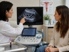
Pelvic ultrasound is useful for ovarian morphology, endometrial thickness, and some ovarian masses. It can miss small androgen-producing tumors, so normal ultrasound does not end a severe workup. MRI may provide better ovarian detail in selected cases. Adrenal CT or MRI is considered when DHEA-S is markedly elevated, several adrenal steroids are abnormal, or clinical findings suggest an adrenal lesion.
Urgent referral is appropriate when symptoms progress over a few months, the voice deepens, clitoral enlargement develops, muscle mass changes substantially, or androgen concentrations are several times the upper limit. The threshold for imaging is lower after menopause because new severe hyperandrogenism is less likely to be routine PCOS.
Treatment and Monitoring
Treatment depends on the cause, symptom burden, pregnancy plans, and health risks. Lowering one laboratory number is not the sole objective.
For PCOS, combined hormonal contraception can suppress ovarian androgen production and increase SHBG. Spironolactone or another antiandrogen may reduce hirsutism and acne when reliable contraception is used because antiandrogens can affect fetal sexual development. Hair-growth treatment takes time; visible improvement commonly requires six months or longer because hair follicles cycle slowly.
Lifestyle treatment should be individualized and weight-neutral when appropriate. Improvements in sleep, physical activity, nutrition quality, and insulin sensitivity can benefit cycles and metabolic health even without major weight change. Ovulation-induction therapy is used when pregnancy is desired rather than relying on antiandrogen treatment.
NCCAH management may include symptom-directed hormonal treatment or selective glucocorticoid therapy, especially for fertility. Glucocorticoids are not automatically required for every biochemical diagnosis and can cause harm when overused.
Androgen-secreting tumors and ovarian hyperthecosis require specialist treatment, often surgery. A falling androgen level after treatment can help document biochemical response, but imaging and clinical recovery remain important.
Routine serial androstenedione testing is not necessary for every woman with PCOS. Symptoms, cycle pattern, metabolic markers, blood pressure, and treatment safety often provide more useful follow-up. Repeat measurement is most valuable when the original result was markedly abnormal, when a specific adrenal disorder is being treated, or when symptoms change unexpectedly.
A small elevation should not be chased with repeated broad panels if the diagnosis and plan are already clear. Conversely, normalizing androstenedione does not guarantee that hirsutism will disappear immediately. Existing terminal hairs may need laser, electrolysis, or other cosmetic treatment even after androgen production improves.
Useful questions to ask after an abnormal result include whether the sample should be confirmed by LC-MS/MS, whether the elevation is isolated or part of a broader androgen pattern, whether 17-OHP has been checked, and whether symptoms are progressing quickly enough to justify imaging now. Asking what specific decision the repeat test will change can prevent months of low-value monitoring.
The absence of symptoms also changes the response. An incidental mild elevation in a woman with regular cycles, no hirsutism, no acne, and no virilization may reflect method variation. Confirmation is reasonable, but aggressive treatment is not. Laboratory abnormalities should be treated as evidence to interpret, not as diseases on their own.
Handling a result that does not match the symptoms
Discordant results deserve confirmation before imaging or treatment. If androstenedione is high but testosterone, DHEA-S, menstrual pattern, and examination are unremarkable, the clinician may repeat the test with LC-MS/MS and review the age-specific interval. Concentrations generally change across the life span, and a range designed for younger premenopausal women may be misleading after menopause. Biotin, steroid supplements, compounded hormones, and sample labeling or timing errors should also be considered.
The opposite mismatch matters too. A woman can have clinically important hirsutism or acne with a normal androstenedione because hair follicles differ in androgen sensitivity and because another androgen may be responsible. In that situation, repeating the same marker many times adds little. A broader but targeted assessment—reliable total testosterone, SHBG or free testosterone, DHEA-S, and 17-OHP when indicated—usually provides more information. The goal is to explain the clinical pattern, not force every symptom to match one laboratory value.
References
- Society for Endocrinology Clinical Practice Guideline for the Evaluation of Androgen Excess in Women 2025 (Guideline)
- Evaluating the diagnostic accuracy of androgen measurement in polycystic ovary syndrome: a systematic review and diagnostic meta-analysis to inform evidence-based guidelines 2025 (Systematic Review)
- Approach to androgen excess in women: Clinical and biochemical insights 2022 (Review)
- Practical Approach to Hyperandrogenism in Women 2021 (Review)
- International evidence-based guideline for the assessment and management of polycystic ovary syndrome 2023 – Summary 2023 (Guideline)
Disclaimer
This information cannot diagnose PCOS, congenital adrenal hyperplasia, or an ovarian or adrenal tumor. Androstenedione ranges and alert thresholds vary by age, assay, cycle phase, and laboratory. Seek prompt medical care for rapidly developing virilization, severe headache or weakness, or a markedly abnormal androgen pattern.