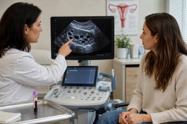
An anti-Müllerian hormone test estimates the number of small growing follicles in the ovaries and is used mainly to assess ovarian reserve and predict response to fertility medication. It is not a direct egg-count test, does not measure egg quality, and cannot tell whether a woman will conceive naturally in a particular month. Age remains the strongest predictor of egg quality and reproductive potential. AMH can be measured on most cycle days because it varies less across the menstrual cycle than FSH or estradiol, although results still change with age, hormonal contraception, pregnancy, ovarian surgery, chemotherapy, PCOS, and the assay used. Low AMH may predict a lower egg yield during ovarian stimulation, while high AMH may predict a stronger response and greater risk of ovarian hyperstimulation. Neither result is a diagnosis by itself. The most useful interpretation combines AMH with age, antral follicle count, menstrual history, medical treatment, and the reason the test was ordered.
- AMH estimates ovarian follicle quantity, not egg quality or the chance of natural pregnancy in one cycle.
- A low value often predicts fewer eggs during IVF stimulation but does not mean pregnancy is impossible.
- A high value is common in PCOS and can signal a stronger response to fertility medication.
- AMH can usually be tested on any cycle day, but hormonal contraception may temporarily lower it.
- Ranges are assay- and age-specific; common labels such as “low below 1 ng/mL” are only rough guides.
- AMH should not be used alone to predict the exact age of menopause or to screen healthy women for future fertility.
Table of Contents
- What AMH Measures
- Why AMH Is Tested
- How to Prepare
- Understanding AMH Results
- AMH and Fertility
- High AMH and PCOS
- Low AMH and Next Steps
What AMH Measures
Anti-Müllerian hormone is produced by granulosa cells surrounding preantral and small antral follicles. These are early-stage follicles that contain immature eggs. As the pool of remaining follicles declines with age, AMH generally declines as well. The test therefore acts as an indirect marker of ovarian reserve—the quantity of follicles available to respond to stimulation.
AMH does not count every egg in the ovaries. Most eggs are in microscopic resting follicles that do not directly release measurable AMH. The blood concentration reflects activity in a subset of growing follicles and is influenced by follicle biology as well as total reserve.
AMH also does not assess chromosome quality. A 40-year-old and a 28-year-old can have the same AMH level but different probabilities of embryo aneuploidy because egg quality is strongly age-related. Likewise, a woman with low AMH may release a healthy egg and conceive naturally, while a woman with high AMH may have ovulatory dysfunction or other infertility factors.
The hormone changes across life. It is low in early childhood, rises through adolescence, often peaks in the twenties, and declines toward menopause. The rate differs among individuals. Genetics, smoking, ovarian surgery, endometriosis, chemotherapy, radiation, and certain autoimmune or genetic conditions can affect ovarian reserve. PCOS often raises AMH because the ovaries contain more small follicles and granulosa-cell production may be increased.
Laboratories usually report AMH in ng/mL or pmol/L. One ng/mL is approximately 7.14 pmol/L. Results must be compared in the same units. Different commercial assays are not perfectly interchangeable, and a value may shift when testing moves between laboratories.
AMH is often interpreted with antral follicle count, or AFC, measured by transvaginal ultrasound. AFC counts visible small follicles, usually early in the cycle. AMH and AFC provide similar information about expected ovarian response, but each can be affected by technique and clinical context.
Why AMH Is Tested
The most established use of AMH is planning ovarian stimulation for IVF or egg freezing. It helps a fertility specialist estimate whether the ovaries are likely to produce a low, average, or high number of follicles in response to gonadotropin medication. That estimate supports dose selection and counseling about expected egg yield.
AMH may also be ordered for:
- Evaluation of diminished ovarian reserve in infertility
- Planning fertility preservation before chemotherapy, radiation, or ovarian surgery
- Assessing ovarian function after gonadotoxic treatment
- Supporting a diagnosis of PCOS in selected adults when other criteria are considered
- Clarifying possible primary ovarian insufficiency when FSH results are inconclusive
- Estimating risk of excessive response or ovarian hyperstimulation during treatment
- Monitoring ovarian reserve after endometrioma surgery or loss of ovarian tissue
The test has limitations outside these settings. In women without infertility, AMH has not reliably predicted the short-term chance of natural conception. A low result can create unnecessary fear, while a high result can create false reassurance and delay age-sensitive planning. Direct-to-consumer marketing often overstates what AMH can reveal about “fertility age” or future pregnancy.
AMH is not a standard test for confirming ovulation. Progesterone timed to the luteal phase or cycle tracking is more appropriate for that question. It is not a pregnancy test, and it does not measure estrogen, progesterone, or overall hormone balance.
In amenorrhea, FSH and estradiol usually provide the first distinction between ovarian insufficiency and central suppression. AMH may add context but is frequently unnecessary. A dedicated amenorrhea hormone panel addresses the broader causes of absent periods.
For PCOS, a high AMH can support evidence of polycystic ovarian morphology in some adult diagnostic pathways, but the cutoff depends on assay and population. AMH should not be used as the only diagnostic test, particularly in adolescents.
How to Prepare
AMH can usually be measured on any day of the menstrual cycle. It is more cycle-stable than FSH, LH, estradiol, or progesterone. Small fluctuations still occur, especially when values are near a decision threshold, so a repeat result may differ without indicating a true rapid loss of eggs.
Fasting is not required for AMH alone. Blood may be drawn at any time of day unless other tests on the order require morning or fasting collection.
Tell the clinician about:
- Combined birth-control pills, progestin-only contraception, implants, injections, or hormonal IUDs
- Pregnancy or recent delivery
- Recent ovarian stimulation, egg retrieval, or fertility medication
- Ovarian surgery, endometriosis, or ovarian cysts
- Chemotherapy, pelvic radiation, or medications that may affect ovarian function
- A history of one ovary, ovarian torsion, or genetic conditions linked with ovarian insufficiency
Hormonal contraception may lower measured AMH and AFC, often reversibly. The size of the effect varies. A low result while using contraception should not automatically be treated as permanent diminished reserve. If the result will change a major decision, a fertility specialist may recommend repeating it after a suitable interval off hormones when medically appropriate.
Pregnancy can alter AMH, and levels may remain changed for a period after delivery. Results obtained during pregnancy are generally not used for routine fertility prediction.
Recent ovarian stimulation can temporarily disturb the follicle population being measured. Clinics often schedule reserve testing after recovery rather than immediately after retrieval.
Biotin and common foods are not major established influences on AMH, but supplements should still be disclosed because assay platforms differ. Do not stop prescribed hormones solely to obtain a “truer” number without discussing contraceptive needs, symptoms, and the clinical purpose.
Using the same laboratory can improve comparability during follow-up. A change from 1.2 to 0.9 ng/mL across different assays may reflect laboratory variation, not a sudden 25% drop in ovarian reserve.
Understanding AMH Results
AMH interpretation should be age-specific and assay-specific. Many fertility clinics use rough categories similar to the following, but their treatment algorithms may differ.
| AMH result | Approximate interpretation | Important caution |
|---|---|---|
| Below 0.5 ng/mL | Very low ovarian reserve marker; low expected egg yield | Does not prove infertility or zero eggs |
| 0.5–1.0 ng/mL | Low range in many clinic systems | Age and AFC strongly affect meaning |
| 1.0–3.5 ng/mL | Often described as average | Not a guarantee of natural fertility or embryo quality |
| Above 3.5–5.0 ng/mL | High follicle count or PCOS pattern may be present | Can predict high stimulation response, not superior egg quality |
These are not universal diagnostic boundaries. A value of 1.0 ng/mL may be less expected at age 25 than at age 39. A woman with AMH 0.7 and a reassuring AFC may respond differently from someone with the same AMH and very few antral follicles. Clinics use age-adjusted centiles, assay-specific thresholds, and prior treatment response when available.
Results should also be viewed as estimates rather than exact inventories. AMH can vary between samples, and studies have documented enough inter-cycle variability to move some women from one descriptive category to another. That does not mean ovarian reserve changed dramatically within weeks. When a value sits close to a treatment cutoff, repeating it or adding AFC can be more informative than reacting to a single decimal point.
A useful report should identify the assay, unit, laboratory reference interval, and whether an age-specific interpretation is available. Online calculators that accept only age and AMH often omit diagnosis, contraception, ovarian surgery, and ultrasound findings. They may appear precise while producing only a rough population estimate.
Low AMH
Low AMH usually means fewer small follicles are producing hormone. It can occur with reproductive aging, diminished ovarian reserve, primary ovarian insufficiency, prior ovarian surgery, endometriosis, chemotherapy, radiation, smoking, or genetic factors. Temporary suppression from hormonal contraception can mimic a lower reserve.
Low AMH predicts egg quantity during stimulation better than it predicts pregnancy. Even extremely low values do not prove that no response will occur. They help set realistic expectations about the number of eggs and the possibility of needing more than one cycle.
High AMH
High AMH often reflects a high antral follicle count. It is common in PCOS and can signal a greater risk of excessive response to fertility medication. High AMH is not generally toxic and does not need treatment as a laboratory abnormality. Management focuses on the underlying cycle or metabolic condition and on safer stimulation dosing when fertility treatment is used.
Undetectable AMH
An undetectable result suggests very low measurable follicular activity, but assay detection limits differ. It does not by itself diagnose menopause or primary ovarian insufficiency. Menstrual history, repeated FSH, estradiol, symptoms, and age are required. Intermittent ovarian activity can occur in primary ovarian insufficiency even when AMH is very low.
AMH and Fertility
AMH answers “How might the ovaries respond to stimulation?” more reliably than “Can I become pregnant naturally?” Those are different questions.
Natural conception depends on ovulation, fallopian-tube function, sperm, uterine factors, intercourse timing, age-related egg quality, and chance. A woman usually ovulates one egg per cycle whether her reserve is average or low. Low reserve may shorten the remaining reproductive window, but a single AMH measurement cannot calculate that window precisely.
Prospective studies have found similar short-term natural pregnancy rates among some women with low and normal AMH. This does not mean reserve is irrelevant. It means AMH is not a strong stand-alone screening test for fecundability in women without diagnosed infertility.
In IVF, AMH is useful for predicting:
- The number of follicles likely to grow
- The expected number of retrieved eggs
- Risk of poor response or cycle cancellation
- Risk of excessive response and ovarian hyperstimulation
- Initial gonadotropin dose planning
AMH is much weaker for predicting:
- Whether an individual egg is chromosomally normal
- Embryo implantation
- Miscarriage independent of age
- Live birth without considering age and treatment details
- Natural conception in a specific month
A low AMH result can justify timely fertility consultation, especially when age is advancing or there are additional risk factors. It should not lead automatically to IVF, egg freezing, donor eggs, or the belief that spontaneous pregnancy is impossible. Those decisions depend on age, duration of trying, partner factors, reproductive goals, finances, and clinical findings.
Egg freezing outcomes depend heavily on age at freezing and the number of mature eggs stored. AMH helps estimate how many stimulation cycles may be needed to reach a target egg number. It does not guarantee that frozen eggs will produce a baby.
A full fertility evaluation may include semen analysis and tubal assessment as well as ovarian-reserve tests. Focusing only on AMH can miss the actual limiting factor.
For example, a 32-year-old with AMH 0.8 ng/mL, regular ovulation, open tubes, and normal semen may have a different prognosis from a 40-year-old with AMH 2.0 ng/mL. The second woman has a higher reserve marker but greater age-related egg-quality risk. In another case, a woman with AMH 5.5 ng/mL may have many follicles but rarely ovulate because of PCOS. These examples show why “higher” is not always “more fertile” and “lower” is not equivalent to infertility.
High AMH and PCOS
Women with PCOS often have more small follicles, and those follicles produce AMH. High levels can therefore fit a PCOS pattern. AMH may also participate in altered follicle development, although it remains primarily a marker in clinical practice.
PCOS is not diagnosed from AMH alone. The diagnosis requires a defined combination of irregular ovulation, clinical or biochemical androgen excess, and polycystic ovarian morphology or an accepted AMH-based equivalent in appropriate adults, after excluding thyroid disease, high prolactin, nonclassic congenital adrenal hyperplasia, and other mimics.
An AMH level cannot explain every symptom. Hirsutism relates more directly to androgen exposure and follicle sensitivity. Irregular periods require assessment of ovulation and endometrial protection. Metabolic risk requires blood pressure, glucose, lipid, sleep, and family-history review. A female hormone test panel may add context but cannot replace clinical criteria.
During IVF, high AMH allows clinics to choose lower starting doses, use antagonist protocols, modify the ovulation trigger, and freeze embryos when necessary to reduce ovarian hyperstimulation syndrome risk. The goal is a safe, controlled response—not the largest possible egg count.
High AMH without symptoms does not require treatment. A woman with regular cycles and no androgen excess should not be labeled with PCOS solely because an AMH value is above a generic cutoff.
Low AMH and Next Steps
The first response to low AMH is to confirm what question the test was intended to answer. A fertility-planning result, a post-chemotherapy result, and a test ordered during hormonal contraception require different interpretations.
Useful next steps include:
- Compare the result with age. Age-specific context is more informative than a single universal range.
- Review medications and timing. Hormonal contraception, pregnancy, and recent stimulation may suppress or distort the value.
- Obtain an antral follicle count. AMH and AFC together can improve confidence about expected ovarian response.
- Check FSH and estradiol when indicated. Early-follicular testing or random testing in amenorrhea can identify ovarian insufficiency patterns.
- Assess the complete fertility picture. Ovulation, tubes, uterus, semen, duration of trying, and prior pregnancy history matter.
- Discuss time-sensitive choices. Age, cancer treatment, surgery, and family goals may justify prompt consultation.
There is no proven supplement, diet, or procedure that reliably restores the depleted primordial follicle pool. AMH may fluctuate or rise slightly after lifestyle changes or stopping contraception, but such a change does not necessarily mean new eggs were created. Claims that a product can “rejuvenate” ovaries based only on a higher AMH result should be viewed cautiously.
Low AMH without infertility is not an emergency. It can be emotionally difficult, so results should be delivered with clear limits. The appropriate message is usually that expected response may be lower and planning may be time-sensitive—not that pregnancy cannot happen.
When periods are irregular or absent before age 40, evaluate for primary ovarian insufficiency rather than relying on AMH. Current guidelines use menstrual disturbance and elevated FSH for diagnosis, with AMH as an adjunct when FSH is inconclusive. If POI is confirmed, care includes bone, cardiovascular, symptom, fertility, genetic, and autoimmune considerations.
Repeat AMH testing is not needed every few months for most women. Biological and assay variation can create apparent trends that do not change management. Repeating after a meaningful clinical interval or medication change is more useful when the result will affect treatment.
Questions worth asking include: What decision will this result change? Is the value appropriate for my age? Could contraception or recent treatment have lowered it? Does AFC agree with the blood test? Is the concern natural conception, egg yield, or possible ovarian insufficiency? Clear answers to those questions are more valuable than assigning a generalized “ovarian age.”
A well-interpreted AMH result narrows expectations; it does not define a woman’s fertility, worth, or future. The number is most valuable when connected to a specific decision and explained alongside age and the complete reproductive picture.
AMH can be especially useful before time-sensitive treatment. A woman facing chemotherapy may use it with AFC and diagnosis to discuss embryo, egg, or ovarian-tissue preservation. A woman planning endometrioma surgery may use it to weigh the possible ovarian-reserve impact against pain, anatomy, and fertility needs. In both situations, the test supports a decision; it does not make the decision by itself.
When AMH and ultrasound disagree
AMH and antral follicle count usually move in the same direction, but disagreement is common enough that neither should automatically overrule the other. A low AMH with a reassuring AFC may reflect assay variation, temporary hormonal suppression, a recent pregnancy, or differences in how follicles were counted. A higher AMH with a lower AFC may occur when the ultrasound was performed under difficult technical conditions or when the ovaries were not fully visualized. Repeating the measurement is most useful only when the answer will change a treatment plan.
Context after ovarian surgery is particularly important. Endometrioma surgery can reduce AMH, yet the amount of change does not translate directly into a natural-pregnancy probability. Recent chemotherapy, ovarian torsion, or other gonadotoxic exposure also changes the meaning of a low value. A fertility specialist may combine age, AFC, prior response to stimulation, diagnosis, and AMH to estimate medication dose and expected egg yield. When those measures disagree, the safest interpretation is a range of likely response rather than a single precise forecast.
References
- Anti-Müllerian hormone for screening, diagnosis, evaluation, and prediction: A systematic review and expert opinions 2024 (Systematic Review)
- Anti-Müllerian hormone beyond an ovarian reserve marker 2023 (Review)
- Fertility evaluation of infertile women: a committee opinion (2021) 2021 (Committee Opinion)
- Evidence-based guideline: premature ovarian insufficiency 2024 (Guideline)
- Websites Selling Direct-to-Consumer Anti-Mullerian Hormone Tests: A Content Analysis 2023 (Research Study)
- Testing and interpreting measures of ovarian reserve: a committee opinion (2020) 2020 (Committee Opinion)
Disclaimer
This article provides general information and cannot predict an individual’s fertility, IVF outcome, or age at menopause. AMH ranges vary by assay, age, medication use, and clinical setting. Discuss low, high, or unexpected results with a fertility specialist or clinician who can interpret them with age, ultrasound, and the full reproductive history.