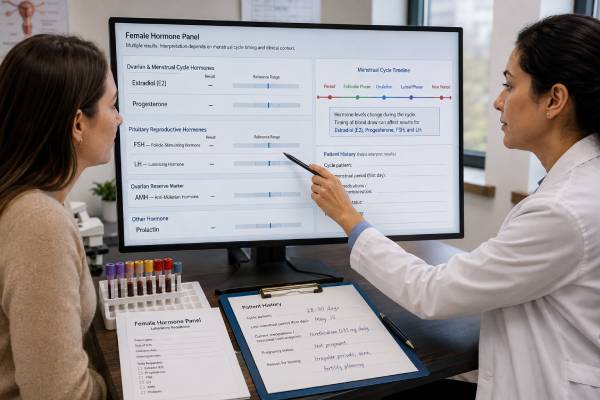
A female hormone test panel can help evaluate irregular or absent periods, infertility, suspected ovulation problems, ovarian reserve, early menopause, pituitary disorders, and selected symptoms of hormonal dysfunction. The panel commonly includes estradiol, progesterone, follicle-stimulating hormone, luteinizing hormone, anti-Müllerian hormone, and prolactin. These hormones do not have one fixed “balanced” range. Estradiol, progesterone, FSH, and LH change across the menstrual cycle; AMH depends strongly on age and ovarian follicle number; and prolactin can rise temporarily from stress, exercise, sleep, breast stimulation, or medications. Test timing and the clinical question are therefore as important as the numbers. A useful panel often requires more than one collection date, and pregnancy testing, thyroid tests, androgen tests, or pelvic ultrasound may be needed. Results should be read as patterns rather than isolated highs and lows. A normal panel does not guarantee fertility, while an abnormal result does not automatically mean permanent infertility or menopause.
- Cycle timing changes interpretation: early-cycle FSH and estradiol answer a different question from mid-luteal progesterone.
- AMH estimates ovarian response and follicle quantity, not egg quality or the chance of natural pregnancy by itself.
- Progesterone can confirm recent ovulation when measured about seven days before the expected period.
- High prolactin may be temporary: mild elevations often need a careful repeat before imaging or treatment.
- Hormone patterns guide diagnosis: pregnancy, thyroid disease, PCOS, hypothalamic amenorrhea, and ovarian insufficiency require different follow-up.
Table of Contents
- What the Female Hormone Panel Measures
- Why the Panel Is Ordered
- Test Timing and Preparation
- Estradiol and Progesterone Results
- FSH and LH Results
- AMH and Prolactin Results
- Interpreting Patterns and Planning Follow-Up
What the Female Hormone Panel Measures
The panel samples several parts of the hypothalamic-pituitary-ovarian system. The hypothalamus in the brain releases gonadotropin-releasing hormone in pulses. Those pulses stimulate the pituitary gland to release FSH and LH. The ovaries respond by developing follicles, producing estradiol, releasing an egg, and producing progesterone after ovulation. Prolactin comes from the pituitary and can suppress this pathway when elevated. AMH comes from small growing ovarian follicles and gives different information from the cycle hormones.
Estradiol
Estradiol, or E2, is the main active estrogen during the reproductive years. It rises as a dominant follicle develops, helps build the uterine lining, and contributes to the LH surge that triggers ovulation. Low or high estradiol has different meanings depending on cycle day, pregnancy, menopause status, and medication use. An estradiol test is commonly interpreted with FSH.
Progesterone
Progesterone rises after ovulation when the emptied follicle becomes the corpus luteum. A properly timed result can show that ovulation probably occurred. Progesterone is released in pulses, so one low value cannot reliably diagnose a defective luteal phase or predict whether a pregnancy will continue.
FSH and LH
FSH supports follicle growth. LH helps the follicle make androgens that are converted to estrogen and triggers ovulation through a midcycle surge. High levels can indicate reduced ovarian feedback, while low or inappropriately normal levels can indicate reduced hypothalamic or pituitary stimulation. Their meaning depends on estradiol and the clinical setting.
Anti-Müllerian hormone
AMH is produced by granulosa cells in preantral and small antral follicles. It changes less across the cycle than FSH or estradiol and is widely used to estimate likely ovarian response to fertility medication. AMH naturally declines with age, but values vary widely among women of the same age.
Prolactin
Prolactin stimulates milk production after childbirth. Outside pregnancy and breastfeeding, a sustained elevation can disrupt GnRH pulses, reduce FSH and LH activity, prevent ovulation, and cause irregular or absent periods. Mild elevations are common and often temporary.
The panel does not directly measure egg quality, fallopian-tube openness, uterine anatomy, embryo health, or sperm factors. It is one part of a reproductive or endocrine evaluation.
Why the Panel Is Ordered
Clinicians choose some or all of these tests when symptoms suggest disrupted ovulation, reduced ovarian function, or pituitary involvement. The exact combination should match the problem.
Common reasons include:
- periods that are very irregular, unusually frequent, or absent;
- difficulty becoming pregnant;
- suspected anovulation despite apparently regular bleeding;
- symptoms of early menopause or premature ovarian insufficiency;
- preparation for in vitro fertilization or egg freezing;
- evaluation of ovarian reserve after surgery, chemotherapy, or radiation;
- nipple discharge unrelated to breastfeeding;
- headaches or vision changes with suspected pituitary disease;
- monitoring selected fertility treatments;
- clarifying hormone status when medications or surgery make menstrual history unreliable.
Infertility evaluation
Infertility evaluation generally begins after 12 months of regular unprotected intercourse when the female partner is younger than 35, or after six months at age 35 or older. Evaluation may begin sooner with irregular periods, known endometriosis, prior pelvic treatment, suspected low ovarian reserve, sexual dysfunction, or a known male factor.
Hormones address ovulation and ovarian reserve, but a complete evaluation also considers semen analysis and the uterus and fallopian tubes. A normal AMH does not overcome blocked tubes, severe sperm abnormalities, or age-related changes in egg quality.
Irregular or absent periods
Pregnancy should be excluded first in anyone who could be pregnant. TSH is commonly checked because thyroid disease affects cycles, even though it is not listed in this panel. Androgen tests may be added for acne, hirsutism, or suspected PCOS. Pelvic ultrasound can reveal ovarian morphology, endometrial thickness, fibroids, or structural clues.
An amenorrhea hormone evaluation uses the FSH, LH, estradiol, and prolactin pattern to distinguish reduced ovarian function from hypothalamic-pituitary suppression and chronic anovulation.
Menopause questions
Routine blood testing is usually unnecessary to diagnose natural menopause in a woman older than 45 with typical symptoms and 12 months without periods. Hormones fluctuate widely during perimenopause. Testing is more important when symptoms occur before age 40, when hysterectomy prevents using periods as a guide, or when another condition may be causing the symptoms.
A panel should not be used as a general wellness screen in a person without a defined question. Random testing can find normal cycle variation and create an inaccurate diagnosis of “imbalance.”
Test Timing and Preparation
The panel may require two collection dates because the early follicular phase and luteal phase answer different questions. A laboratory can draw all six hormones together, but that does not make every result optimally timed.
Early-cycle testing
FSH, LH, and estradiol are often measured on cycle days 2 through 4, counting the first day of full menstrual flow as day 1. Early-cycle conditions reduce some variation and help assess ovarian feedback. Day 3 is traditional, but the exact accepted window varies by clinic.
If periods are absent, these tests can be drawn on a random day. The result is then interpreted with the length of amenorrhea, symptoms, pregnancy status, and medication use. A high estradiol can suppress FSH and make it look deceptively normal, which is why the two are read together.
Progesterone timing
A progesterone test for ovulation should be scheduled about seven days before the next expected period, not automatically on cycle day 21. Day 21 fits a 28-day cycle with ovulation near day 14. In a 35-day cycle, testing near day 28 is more appropriate. Ovulation predictor kits, basal temperature changes, ultrasound, or cycle history can guide timing.
A value above a laboratory or clinic threshold, often around 3 ng/mL, generally supports recent ovulation. Higher thresholds are sometimes used in fertility protocols, but a single value cannot grade luteal “quality” because progesterone can change several-fold within hours.
AMH and prolactin timing
AMH can usually be measured on any cycle day. It may vary modestly, and hormonal contraception can lower the measured concentration in some women. Fertility clinics interpret the result with age, antral follicle count, and treatment history.
Prolactin is often collected at least a few hours after waking, after the patient has rested quietly. A stressful venipuncture, strenuous exercise, sex, nipple stimulation, or a recent breast examination can cause a temporary rise. Fasting may be requested for a repeat mildly high result, although it is not always required.
Medicines and supplements
Report hormonal contraception, fertility drugs, menopausal hormone therapy, antipsychotics, antidepressants, anti-nausea medicines, opioids, thyroid medicine, glucocorticoids, and supplements containing biotin or hormones. Do not stop prescribed treatment without guidance. Hormonal contraception can suppress FSH and LH, alter estradiol, prevent ovulation, lower progesterone, and change AMH, making an untreated-cycle interpretation inappropriate.
High-dose biotin can interfere with some hormone immunoassays. The clinician or laboratory can specify whether and how long it should be withheld. Acute illness and major sleep disruption can also affect results.
Estradiol and Progesterone Results
Estradiol and progesterone are strongly phase-dependent. The laboratory report may provide follicular, ovulatory, luteal, postmenopausal, and pregnancy reference intervals. A result should be compared only with the relevant interval.
Estradiol patterns
Low estradiol with high FSH suggests reduced ovarian function because the pituitary is sending a stronger signal while the ovaries produce less estrogen. This pattern can occur in menopause and premature ovarian insufficiency.
Low estradiol with low or normal FSH and LH suggests reduced stimulation from the hypothalamus or pituitary. Functional hypothalamic amenorrhea can result from low energy availability, weight loss, intense exercise, stress, or an eating disorder. Pituitary disease, chronic illness, and medication effects are other possibilities.
Higher estradiol can reflect a developing follicle, the preovulatory phase, pregnancy, an ovarian cyst, fertility medication, exogenous estrogen, or rarely an estrogen-producing tumor. An early-cycle elevation can suppress FSH and complicate ovarian-reserve interpretation.
Estradiol values swing during perimenopause and do not reliably identify the exact stage from one sample. Symptoms can occur even when one result falls within a broad premenopausal range.
Progesterone patterns
Low progesterone before ovulation is normal. A low result late in the expected luteal phase may mean the test was mistimed, ovulation did not occur, or progesterone had dipped between pulses. Repeating the test with better timing or using ultrasound and LH tracking can clarify the issue.
A raised luteal progesterone supports ovulation but does not prove that the egg was normal, the fallopian tubes are open, implantation will occur, or pregnancy will continue. In early pregnancy, progesterone overlaps between viable and nonviable pregnancies. Serial hCG and ultrasound provide more direct information.
High progesterone is expected after ovulation and during pregnancy. It can also reflect supplementation, fertility medication, or some ovarian cysts. Treatment decisions should not be based on a reference range copied from a different cycle phase.
A progesterone test for ovulation is most useful when the date is tied to the expected period rather than a universal calendar day.
FSH and LH Results
FSH and LH are gonadotropins made by the pituitary. Their absolute numbers and their relationship with estradiol help locate dysfunction within the reproductive axis.
High FSH
FSH rises when the ovaries provide less estrogen and inhibin feedback. A persistent elevation with irregular or absent periods can support ovarian insufficiency. After natural menopause, FSH is typically high. A single elevated result in perimenopause is not enough to prove that periods have permanently ended because ovarian activity can resume temporarily.
For premature ovarian insufficiency, current guidelines emphasize menstrual disturbance for several months and an elevated FSH, with repeat testing when uncertainty remains. AMH should not replace FSH as the main diagnostic test, although it may support difficult cases.
Low or normal FSH
A low FSH may be appropriate at parts of the cycle. When estradiol is also low and periods are absent, low or inappropriately normal FSH suggests hypothalamic-pituitary suppression. Causes include energy deficiency, intense exercise, stress, pituitary lesions, postpartum pituitary injury, high prolactin, and medications.
A normal FSH does not guarantee normal ovarian reserve. Estradiol can suppress it, and ovarian reserve can decline before FSH becomes consistently abnormal.
LH patterns
LH rises sharply before ovulation. A random high result near midcycle may be normal. Persistently high LH and FSH after menopause reflect reduced ovarian feedback. Low LH with low estradiol supports central suppression.
PCOS may be associated with relatively higher LH than FSH, but the LH-to-FSH ratio is not required for diagnosis and has no universal cutoff. Some women with PCOS have a normal ratio, while a high ratio can appear for other reasons or simply because the blood was drawn near ovulation. PCOS assessment focuses on ovulatory dysfunction, clinical or biochemical hyperandrogenism, ovarian morphology or AMH under validated criteria, and exclusion of mimicking disorders.
The FSH test and LH result are therefore most informative when paired with estradiol, age, cycle day, and symptoms.
AMH and Prolactin Results
AMH and prolactin answer different questions and should not be grouped as general measures of “female hormone health.”
AMH interpretation
A lower AMH generally indicates fewer small recruitable follicles and may predict a lower egg yield during ovarian stimulation. A higher AMH often predicts a stronger response and is common in PCOS. Very high values can signal increased risk of ovarian hyperstimulation during fertility treatment, so clinics adjust medication doses accordingly.
AMH does not measure egg quality. Age remains a stronger predictor of chromosome-related egg quality and natural fertility than AMH. A low result does not mean pregnancy is impossible, and a high result does not guarantee pregnancy. AMH is also not a precise clock for the age of menopause.
Values differ by assay, age, contraception, ovarian surgery, chemotherapy exposure, and individual biology. An AMH test is best interpreted with antral follicle count and the treatment goal.
High prolactin
A persistently high prolactin can suppress ovulation, lower estrogen, cause irregular or absent periods, and produce milk-like nipple discharge. Causes include pregnancy, breastfeeding, hypothyroidism, medications, a prolactinoma, kidney or liver disease, chest-wall stimulation, and stress.
Mild elevations often need a repeat under calm conditions. The laboratory may test for macroprolactin, a larger form that can raise the measured number while causing fewer biological effects. If prolactin remains elevated after pregnancy, thyroid disease, medication effects, and macroprolactin are addressed, pituitary imaging may be appropriate.
Very high levels make a prolactinoma more likely, but assay limitations and medication history still matter. Large pituitary masses can cause headaches or peripheral vision loss.
Low prolactin
Low prolactin usually has limited significance outside the postpartum period. Failure to produce breast milk after severe postpartum bleeding can indicate pituitary injury and requires assessment with other pituitary hormones. A low result alone is not a common explanation for infertility.
A prolactin test should be repeated and investigated in proportion to the degree of elevation and the symptoms.
Interpreting Patterns and Planning Follow-Up
Hormone panels are most useful when results form a coherent pattern. The following examples are guides, not diagnostic formulas.
| Pattern | Possible interpretation | Common next steps |
|---|---|---|
| High FSH, low estradiol, irregular or absent periods | Menopause or ovarian insufficiency | Repeat when needed, pregnancy test, POI evaluation if under 40, bone and health planning |
| Low/normal FSH and LH, low estradiol | Hypothalamic-pituitary suppression | Nutrition, exercise and stress review, prolactin, TSH, pituitary assessment when indicated |
| Irregular periods, androgen signs, normal or higher AMH | Possible PCOS | Testosterone, DHEA-S, metabolic screening, ultrasound or validated AMH criteria |
| Low luteal progesterone | Mistimed test or anovulation | Recalculate timing, track LH, repeat progesterone, ultrasound if needed |
| High prolactin with low gonadotropin activity | Hyperprolactinemia affecting ovulation | Repeat prolactin, pregnancy and TSH tests, medication review, macroprolactin, possible MRI |
| Low AMH with otherwise regular cycles | Reduced ovarian reserve | Age-based counseling, antral follicle count, fertility planning; do not assume sterility |
Pregnancy testing should come first when periods are late or absent. TSH is commonly added. A pelvic ultrasound can assess ovarian follicles, the endometrium, fibroids, and structural causes. Infertility evaluation also includes semen analysis and tubal assessment when appropriate.
Results from at-home finger-prick tests may be useful for access or screening, but collection timing, assay performance, and lack of clinical context can limit interpretation. A surprising result should be confirmed through an accredited laboratory before major decisions.
Treatment targets the diagnosis, not a generic effort to “balance” all six hormones. PCOS management may address cycle protection, androgen symptoms, metabolic health, and fertility goals. Hypothalamic amenorrhea requires restoration of energy availability and attention to bone health. Hyperprolactinemia may improve after a medication change or dopamine-agonist treatment. Premature ovarian insufficiency often requires hormone replacement and fertility counseling. Reduced ovarian reserve may alter fertility-treatment strategy without requiring treatment in a person who is not seeking pregnancy.
Seek prompt assessment for severe headache with vision change, heavy bleeding with dizziness, sudden pelvic pain, possible pregnancy with pain or bleeding, or rapid virilization. Otherwise, review the panel with the ordering clinician using the exact cycle date, units, laboratory intervals, medications, and symptoms.
A practical way to avoid the one-draw panel trap
Before ordering all six hormones on one day, write down the decision each result is meant to support. For irregular or absent periods, pregnancy testing, prolactin, TSH, FSH, and estradiol may be obtained without waiting for a textbook cycle day. For ovarian reserve, AMH can usually be measured on any day, while AFC adds ultrasound context. For ovulation, progesterone should be timed about seven days before the expected next period rather than automatically on day 21. Baseline FSH, LH, and estradiol are often most interpretable early in a spontaneous cycle when that timing is feasible.
A staged plan also prevents conflicting snapshots. One visit may establish whether ovulation is suppressed; a later, correctly timed progesterone can answer whether ovulation occurred. A repeat prolactin under calm collection conditions can confirm a mild elevation before imaging. This approach may involve more than one date, but it produces fewer false conclusions than a convenient package collected at an arbitrary point in the cycle. The best panel is the smallest set of tests collected at the time that answers the clinical question.
The same planning prevents false reassurance. For example, a normal mid-cycle estradiol does not establish that ovulation occurred, and a low progesterone collected too early does not prove anovulation. AMH may be reassuring for expected egg yield while age-related egg quality remains the dominant fertility factor. Prolactin can be temporarily elevated by stress or difficult venipuncture. Each marker has a narrow job, and no combination removes the need to interpret symptoms, timing, and the rest of the reproductive evaluation.
References
- Current evaluation of amenorrhea: a committee opinion (2024) 2024 (Guideline)
- Fertility evaluation of infertile women: a committee opinion (2021) 2021 (Guideline)
- Evidence-based guideline: premature ovarian insufficiency 2024 (Guideline)
- Anti-Müllerian hormone for screening, diagnosis, evaluation, and prediction: A systematic review and expert opinions 2024 (Systematic Review)
- International evidence-based guideline for the assessment and management of polycystic ovary syndrome 2023 – Summary 2023 (Guideline)
- Testing and interpreting measures of ovarian reserve: a committee opinion (2020) 2020 (Guideline)
Disclaimer
This article provides general educational information and does not replace individualized medical evaluation. Female hormone results must be interpreted with cycle timing, age, pregnancy status, medicines, symptoms, and the laboratory’s method and ranges. Seek urgent care for severe pain, heavy bleeding, fainting, or headache with vision changes.