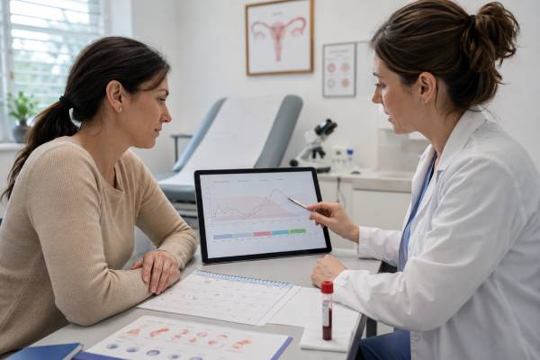
An estradiol blood test measures E2, the main estrogen during the reproductive years. It is used to evaluate menstrual changes, ovarian function, fertility treatment, menopause-related questions, primary ovarian insufficiency, and some pituitary or ovarian disorders. Estradiol rises and falls throughout the menstrual cycle, so the meaning of a result depends heavily on cycle day, age, pregnancy status, medications, symptoms, and the reason for testing. A low value may be expected early in a cycle or after menopause, but it can also reflect hypothalamic suppression, ovarian insufficiency, or medication effects. A high value may occur before ovulation, during pregnancy, with fertility medication, ovarian cysts, or exogenous estrogen. No single estradiol level diagnoses PCOS, confirms fertility, or proves that symptoms are caused by estrogen. Results are most useful when interpreted with FSH, LH, progesterone, hCG, ultrasound findings, and the laboratory’s method-specific reference range.
- Estradiol normally changes several-fold across a menstrual cycle, so cycle timing is essential.
- Low estradiol with high FSH suggests ovarian insufficiency; low estradiol with low or normal FSH suggests central suppression.
- High estradiol can be normal near ovulation or in pregnancy and is not automatically a disease signal.
- Early-follicular E2 is often measured with FSH on cycle days 2–4 for ovarian-reserve context.
- Very low concentrations are measured more accurately by sensitive LC-MS/MS methods than by many routine immunoassays.
- Unexpected bleeding, severe pelvic pain, fainting, or pregnancy-related pain requires prompt medical assessment.
Table of Contents
- What Estradiol Does
- Why E2 Is Tested
- Timing and Preparation
- Estradiol Ranges Across Life
- High Estradiol Results
- Low Estradiol Results
- Follow-Up and Next Steps
What Estradiol Does
Estradiol is one of the three main naturally occurring estrogens, along with estrone and estriol. During the reproductive years, it is the most biologically active circulating estrogen. Ovarian follicles produce most estradiol through a coordinated process involving theca cells, granulosa cells, LH, FSH, and aromatase enzymes.
Estradiol helps regulate the menstrual cycle and reproductive tract. It thickens the uterine lining during the follicular phase, supports the midcycle LH surge, influences cervical mucus, and contributes to vaginal and vulvar tissue health. Its effects extend well beyond reproduction. Estradiol supports bone remodeling, temperature regulation, skin and connective tissue, cardiovascular physiology, brain function, and sexual health.
The level is dynamic. It is usually relatively low during the first days of menstruation, rises as a dominant follicle develops, peaks before ovulation, falls briefly, and then reaches a smaller luteal-phase rise as the corpus luteum functions. If pregnancy occurs, placental and ovarian estrogen production increases substantially. After menopause, ovarian estradiol production falls, and estrone becomes the more prominent circulating estrogen.
Estradiol circulates partly bound to SHBG and albumin. Routine tests measure total estradiol, not only the free fraction. Changes in SHBG can affect total sex-steroid interpretation, although this issue is generally more prominent for testosterone.
The test may use an immunoassay or LC-MS/MS. Immunoassays work reasonably well across many reproductive-age concentrations but can be inaccurate at the very low levels seen after menopause, during aromatase-inhibitor therapy, or in some pituitary disorders. Sensitive LC-MS/MS is preferable when a small difference at a low concentration will change management.
Estradiol is not the same as ethinyl estradiol in many birth-control pills, and routine E2 assays may not accurately measure synthetic estrogens. A woman can have strong estrogen effects from medication while her measured serum estradiol appears low or unchanged.
Why E2 Is Tested
Estradiol is ordered for several different questions, and each uses the result differently.
Common reasons include:
- Investigating absent or irregular periods
- Distinguishing ovarian insufficiency from hypothalamic or pituitary suppression
- Evaluating delayed or early puberty
- Assessing ovarian-reserve context with FSH
- Monitoring follicle development during IVF or ovulation induction
- Evaluating menopausal symptoms in selected situations
- Monitoring estrogen replacement in specific clinical settings
- Investigating an ovarian cyst, estrogen-producing tumor, or unexplained uterine bleeding
- Assessing ovarian function after chemotherapy, radiation, or surgery
- Supporting evaluation of infertility or anovulation
In amenorrhea, E2 is especially useful with FSH and LH. Low estradiol with high FSH suggests that the pituitary is stimulating the ovaries but the ovaries are not responding adequately. Low estradiol with low or normal FSH and LH suggests that hypothalamic or pituitary signaling is reduced. A broader amenorrhea hormone panel includes pregnancy, prolactin, and thyroid evaluation because estradiol alone cannot identify the cause.
For ovarian reserve, estradiol is often drawn with FSH on cycle days 2–4. An early elevated E2 can suppress FSH into a deceptively normal range, so the pair is more informative than FSH alone. AMH and antral follicle count have largely replaced basal E2 as primary reserve markers, but E2 still adds context.
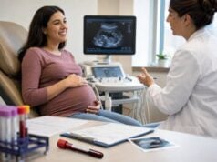
During IVF, serial estradiol results reflect growing follicle activity and help assess response. Clinics do not use one universal target because expected levels depend on the number of follicles, stimulation protocol, trigger plan, and assay. Ultrasound remains essential.
Routine estradiol testing is usually not needed to diagnose natural menopause in women over 45 with typical symptoms and cycle changes. Values fluctuate widely during perimenopause, and one normal or high result does not rule out the transition. Testing may be more useful at younger ages, after hysterectomy, or when another endocrine disorder is possible.
Estradiol is not a reliable standalone explanation for fatigue, weight change, mood symptoms, or low libido. These symptoms are real but have many potential causes. Testing should connect to a specific clinical question.
Timing and Preparation
Cycle timing is the most important practical factor. Record the first day of full menstrual bleeding as cycle day 1. Light spotting before full flow is usually not counted as day 1 unless a clinic gives different instructions.
Common timing choices include:
- Cycle days 2–4: basal E2 with FSH for ovarian-reserve or menstrual evaluation
- Late follicular phase: follicle monitoring before ovulation
- Midcycle: assessment of an expected estrogen peak, usually with ultrasound or LH
- Luteal phase: evaluation in a known cycle, though progesterone is usually more useful for ovulation
- Any day: amenorrhea, menopause, or urgent evaluation when there is no reliable cycle
Fasting is not required for estradiol alone. Other tests on the same order may require fasting or morning collection.
List all hormones and medications, including:
- Birth-control pills, patches, rings, injections, implants, and hormonal IUDs
- Oral, transdermal, vaginal, injectable, or compounded estrogen
- Fertility medication
- Aromatase inhibitors, selective estrogen-receptor modulators, and GnRH medicines
- Testosterone or DHEA
- Glucocorticoids and medications affecting the liver or pituitary gland
Do not stop hormone therapy for testing unless the prescriber directs it. The correct timing depends on whether the clinician wants to assess untreated ovarian function, absorption of a prescribed product, or suppression during therapy.
Transdermal estradiol can produce peaks and troughs depending on patch placement and replacement timing. Oral estradiol undergoes first-pass liver metabolism and raises estrone more than transdermal therapy. Blood drawn soon after a dose may not represent average exposure. A monitoring plan should specify when to draw relative to the dose.
High-dose biotin can interfere with some immunoassays. The laboratory may advise withholding it for a defined period. Pregnancy, recent fertility treatment, and ovarian cysts should also be reported because each can alter the expected result.
Use the same laboratory when following a trend. Estradiol assays can differ enough that small changes across platforms are not clinically meaningful.
Estradiol Ranges Across Life
Estradiol is commonly reported in pg/mL or pmol/L. To convert pg/mL to pmol/L, multiply by approximately 3.67. Reference intervals vary by laboratory and method.
| Physiologic stage | Approximate E2 range | Interpretive note |
|---|---|---|
| Early follicular phase | About 20–80 pg/mL | Often interpreted with FSH on cycle days 2–4 |
| Late follicular or preovulatory | About 150–750 pg/mL | Wide range based on follicle number and timing |
| Luteal phase | About 30–450 pg/mL | Varies across the luteal phase |
| Postmenopause without estrogen therapy | Often below 20–30 pg/mL | Low-level assay accuracy is important |
| Pregnancy | Progressively much higher | Must use gestational-age and laboratory-specific ranges |
These ranges are examples, not universal targets. A result of 180 pg/mL may be normal before ovulation, unexpectedly high on cycle day 3, or low during a stimulated IVF cycle with many follicles. The same number has different meanings in different settings.
The concept of an “optimal” estradiol level is misleading without context. Menstrual phase, bone protection, symptom treatment, fertility stimulation, and cancer therapy have different goals. A concentration should be judged against the intended physiologic or therapeutic state.
A single result also cannot show total estrogen exposure over weeks or months. Someone with irregular ovulation may alternate between low and high values. Someone using oral estrogen may have relatively high estrone but a modest E2 concentration. Someone using a vaginal product may have strong local tissue benefit with little change in serum E2. The route, timing, and target tissue all influence what the blood number represents.
When comparing results, note whether the laboratory changed its method. At low concentrations, a result of 12 pg/mL by LC-MS/MS and 28 pg/mL by immunoassay may reflect assay bias rather than a biologic rise. Trend interpretation is most reliable when the same platform is used.
Why values can look inconsistent
Estradiol can change rapidly as a follicle grows or regresses. An ovulatory peak may be missed by a single draw. Perimenopausal ovaries may produce intermittent high peaks followed by low periods. A result can therefore conflict with symptoms without either being false.
Assay limitations matter at low concentrations. An immunoassay may read 20 pg/mL when a more specific method reads substantially lower. This distinction can be important during aromatase-inhibitor treatment or when confirming profound suppression, but it may not change routine menopause care.
High Estradiol Results
High estradiol is often physiologic. The most common normal causes are the preovulatory surge and pregnancy. Fertility medication can produce much higher concentrations because several follicles develop at once.
Other possible causes include:
- A functional ovarian cyst
- Exogenous estrogen or a dose drawn near peak absorption
- Higher body-fat-related aromatization, especially after menopause
- An estrogen-producing ovarian tumor, which is rare
- Liver disease that alters hormone metabolism
- Some adrenal or ovarian steroid disorders
- Assay interference or cross-reactivity
Symptoms of high estrogen exposure may include breast tenderness, bloating, heavy or irregular bleeding, headaches, or nausea. These symptoms are nonspecific and can occur at normal levels.
In early-follicular testing, E2 above roughly 60–80 pg/mL may suppress FSH and reduce the usefulness of the pair for ovarian-reserve assessment. The cause may be an early recruited follicle or cyst rather than a general state of “estrogen dominance.” Ultrasound and repeat timing can clarify the result.
In fertility treatment, estradiol is interpreted in relation to follicle count and size. A rapid rise with many follicles can increase ovarian hyperstimulation risk, but clinics use the whole protocol rather than a single threshold. Symptoms such as increasing abdominal distension, shortness of breath, reduced urination, or severe pain after stimulation require urgent contact with the fertility clinic.
After menopause, persistent high E2 without prescribed estrogen deserves review of medication exposure, assay accuracy, body composition, liver function, and rare hormone-producing lesions. Vaginal estrogen often produces low systemic exposure, but the amount varies by product and dose.
High estradiol does not diagnose endometriosis, fibroids, breast cancer, or PCOS. Those conditions may be estrogen-responsive, but a routine blood level is not a diagnostic test for them.
The phrase “estrogen dominance” is often used online to describe a wide range of symptoms, but it is not a single laboratory diagnosis. Heavy bleeding can result from fibroids, adenomyosis, anovulation, pregnancy complications, medication, or a bleeding disorder even when E2 is within range. A proper evaluation may require blood count, pregnancy testing, pelvic examination, ultrasound, or endometrial assessment rather than repeated estradiol panels.
Low Estradiol Results
Low estradiol is expected after menopause and can be normal in the first days of a cycle. Outside those settings, the associated FSH and LH pattern helps identify the cause.
Possible causes include:
- Functional hypothalamic amenorrhea from low energy availability, weight loss, intensive exercise, illness, or stress
- Primary ovarian insufficiency before age 40
- Natural menopause or late perimenopause
- Pituitary or hypothalamic disease
- Ovarian surgery, chemotherapy, or pelvic radiation
- GnRH therapy, aromatase inhibitors, or other suppressive medication
- Severe chronic illness
- Pregnancy-related or postpartum hormonal transitions in specific contexts
Low-estrogen symptoms can include hot flashes, night sweats, sleep disruption, vaginal dryness, pain with sex, urinary symptoms, lower bone density, and reduced menstrual bleeding. Not everyone has symptoms, and symptoms can persist even when a single test is not very low because levels fluctuate.
Low E2 with high FSH suggests ovarian failure or menopause. In a woman under 40 with irregular or absent periods, the pattern may support primary ovarian insufficiency and requires confirmation and evaluation. AMH may add information when FSH is inconclusive, but it is not the primary diagnostic test.
Low E2 with low or normal FSH and LH suggests reduced central signaling. Functional hypothalamic amenorrhea is common, but pituitary disease must be considered when there are headaches, visual changes, high prolactin, excessive thirst, postpartum hemorrhage, head injury, or multiple low pituitary hormones.
Prolonged low estradiol can affect bone health. Women with many months of amenorrhea, stress fractures, restrictive eating, or premature ovarian insufficiency may need bone-density assessment and targeted treatment. Simply taking a birth-control pill may create withdrawal bleeding without correcting an underlying energy deficit.
Low estradiol is not a validated diagnosis of “estrogen deficiency” in every woman with fatigue or low libido. A careful history, examination, and companion tests remain necessary.
In adolescents, interpretation requires pubertal stage rather than adult cycle ranges. In older adults, a low result may be expected and treatment is based on symptoms and health risks, not on restoring a premenopausal laboratory number. Across ages, the clinical setting determines whether low E2 is physiologic, temporary, or evidence of a disorder.
Follow-Up and Next Steps
When a result is unexpected, first verify cycle day, medication use, pregnancy status, units, and assay. Repeating under standardized conditions may be more useful than immediately ordering imaging.
Common follow-up tests include:
- FSH and LH to locate ovarian versus central dysfunction
- Pregnancy testing when cycles are late or symptoms suggest pregnancy
- Progesterone when confirming ovulation in a timed luteal sample
- Prolactin and TSH for irregular or absent periods
- AMH and antral follicle count for ovarian-response planning
- Testosterone, DHEA-S, and 17-OHP when androgen symptoms are present
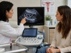
- Pelvic ultrasound for cysts, follicle development, endometrium, or ovarian masses
- Pituitary testing and MRI when central disease is suspected
A female hormone test panel can be helpful when each component is timed and selected for a reason. Drawing every hormone on a random day may create a confusing mixture of normal physiologic variation and unrelated findings.
Treatment depends on the cause. Functional hypothalamic amenorrhea requires adequate energy intake, exercise adjustment when needed, and support for stress or disordered eating. Primary ovarian insufficiency usually prompts hormone replacement until the average age of natural menopause unless contraindicated. Thyroid or prolactin disorders need specific therapy. Ovarian cysts may be observed or treated based on size, appearance, symptoms, and persistence.
For menopausal symptoms, treatment decisions are usually based on symptoms, age, time since menopause, medical history, and contraindications rather than reaching a specific serum E2 target. Routine monitoring is not required for most women using standard menopausal hormone therapy. Testing is more useful when absorption is uncertain, symptoms persist despite adherence, or unusually high exposure is suspected.
During fertility treatment, follow the clinic’s timing and do not compare results with natural-cycle online ranges. Ultrasound and protocol-specific trends matter more than a generic “normal.”
A high stimulation value may be expected when many follicles are growing, while the same concentration in an unstimulated early cycle would be unusual. Clinics may change medication, trigger choice, or embryo-transfer timing based on the combined estradiol trend, follicle measurements, progesterone, and symptoms. Patients should contact the clinic rather than adjusting doses from online charts.
Seek prompt care for severe pelvic or abdominal pain, fainting, heavy bleeding, a positive pregnancy test with pain, shortness of breath after ovarian stimulation, or neurologic symptoms. These findings require direct assessment rather than repeat hormone testing alone.
A useful estradiol result answers a defined question: whether ovarian estrogen production is low, whether a follicle is developing, whether stimulation is progressing, or whether a medication is producing expected exposure. Without that question, the number can be technically accurate but clinically unhelpful.
Questions to ask after an abnormal result include: Was this the expected cycle day? Could a cyst or recent medication explain it? Does FSH point toward an ovarian or central cause? Is the assay sensitive enough for the concentration being measured? Will repeating the test change treatment, or is ultrasound or another hormone more informative? These questions turn a confusing number into a specific next step.
Why symptoms and treatment route may not match one E2 value
Serum estradiol is a snapshot, while symptoms reflect tissue exposure over time. A woman using a transdermal patch may have a level influenced by when the patch was changed, where it was applied, skin absorption, and the timing of the blood draw. Oral estrogen also produces a different liver and metabolite pattern, so equal doses by different routes do not create interchangeable laboratory values. For these reasons, a single number should not be used to compare people or to declare that symptoms are “not hormonal.”
When menopausal symptoms continue during prescribed therapy, the clinician first reviews adherence, application technique, dose, route, drug interactions, and other causes such as thyroid disease, anemia, sleep disorders, or medication effects. Testing may help when absorption is uncertain or exposure appears unusually high, but treatment is usually adjusted according to symptom control and safety rather than a universal target E2. Unexpected bleeding still requires its own evaluation even when the estradiol result appears normal.
References
- Metabolism of endogenous and exogenous estrogens in women 2024 (Review)
- Estrogen deficiency in the menopause and the role of hormone therapy: integrating the findings of basic science research with clinical trials 2024 (Review)
- Current evaluation of amenorrhea: a committee opinion (2024) 2024 (Committee Opinion)
- Fertility evaluation of infertile women: a committee opinion (2021) 2021 (Committee Opinion)
- Monitoring of estradiol levels in premenopausal women receiving adjuvant aromatase inhibitors 2024 (Research Study)
- Evidence-based guideline: premature ovarian insufficiency 2024 (Guideline)
Disclaimer
This article provides general education and cannot diagnose an ovarian, pituitary, fertility, or menopausal condition. Estradiol ranges vary by cycle phase, age, pregnancy, treatment, and assay. Seek prompt medical care for severe pain, heavy bleeding, fainting, pregnancy-related symptoms, or complications after fertility treatment.