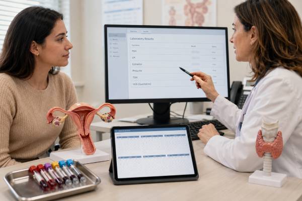
An amenorrhea hormone panel helps identify why menstrual periods have not started or have stopped. The first test is usually a pregnancy test, even when pregnancy seems unlikely. After pregnancy is excluded, FSH, LH, estradiol, prolactin, and TSH can show whether the problem is more likely to involve the ovaries, hypothalamus, pituitary gland, thyroid, or another cause. The pattern matters more than any single number. High FSH with low estradiol can suggest ovarian insufficiency, while low or normal FSH and LH with low estradiol can point toward hypothalamic or pituitary suppression. High prolactin may disrupt ovulation, and abnormal TSH may reveal thyroid disease. PCOS often produces a different pattern, but no LH-to-FSH ratio can diagnose it. Results must be interpreted with age, cycle history, medications, weight changes, exercise, stress, symptoms, and imaging. Some causes need prompt treatment, while others improve when an underlying energy, thyroid, medication, or pituitary problem is addressed.
- Pregnancy testing comes before interpreting FSH, LH, estradiol, prolactin, or TSH.
- High FSH with low estradiol suggests reduced ovarian function; low or normal gonadotropins with low estradiol suggests central suppression.
- Prolactin should be repeated under calm conditions when mildly elevated because stress and medications can raise it.
- TSH is a first-line screen, but free T4 is needed when central hypothyroidism is possible.
- Amenorrhea lasting more than 3 months after regular cycles or 6 months after irregular cycles generally needs evaluation.
- Severe headache, vision change, fainting, pregnancy pain or bleeding, or rapid virilization requires urgent assessment.
Table of Contents
- When Amenorrhea Needs Testing
- What Each Hormone Shows
- Preparing for the Panel
- Common Result Patterns
- Additional Tests and Imaging
- Treatment and Follow-Up
- Urgent Warning Signs
When Amenorrhea Needs Testing
Amenorrhea means absent menstruation. Primary amenorrhea refers to periods that have not begun. Evaluation is generally recommended when menstruation has not started by age 15 despite otherwise typical development, when more than about five years have passed since breast development began, or when breast development has not begun by age 13. Earlier assessment is appropriate when there is pelvic pain, a known genetic condition, abnormal development, or concern about an outflow obstruction.
Secondary amenorrhea means periods stopped after they had begun. A common clinical threshold is no bleeding for more than three months in someone whose cycles were previously regular, or six months in someone whose cycles were already irregular. A shorter delay may still require pregnancy testing. Menstrual suppression caused by certain contraceptives can be expected, but a clinician should confirm that the pattern fits the method and that no other symptoms are present.
The menstrual cycle depends on coordinated signals among the hypothalamus, pituitary gland, ovaries, uterus, and genital outflow tract. Amenorrhea can result from a disruption at any level. Common causes include:
- Pregnancy and lactation
- PCOS or another chronic anovulation disorder
- Functional hypothalamic amenorrhea from low energy availability, weight loss, intensive exercise, stress, or an eating disorder
- Hyperprolactinemia, including a prolactinoma or medication effect
- Thyroid disease
- Primary ovarian insufficiency before age 40 or the normal menopause transition later in life
- Pituitary injury, tumor, surgery, radiation, or postpartum infarction
- Intrauterine scarring or an anatomic obstruction
- Certain medications, chemotherapy, and exogenous hormones
A hormone panel narrows the possibilities, but history often determines which tests are truly needed. Important details include the age at first period, usual cycle length, pregnancy possibility, recent weight change, food intake, exercise load, stress, hot flashes, vaginal dryness, headaches, vision symptoms, milk discharge, acne, excess hair growth, pelvic procedures, chronic illness, and medication use.
The evaluation is not simply a search for one “imbalanced” hormone. Amenorrhea is a sign. The same FSH value can mean different things in a 16-year-old with delayed puberty, a 28-year-old endurance athlete, a 37-year-old after chemotherapy, and a 52-year-old in the menopause transition.
The duration of estrogen deficiency also matters. A brief interruption after illness may resolve without lasting harm, while many months of low estradiol can affect bone density, vaginal tissue, sleep, and mood. Chronic anovulation with normal estrogen creates a different risk: the uterine lining may remain exposed to estrogen without regular progesterone, increasing the chance of prolonged or unpredictable bleeding and endometrial overgrowth. The same symptom—no period—can therefore reflect opposite hormone environments and require different treatment.
What Each Hormone Shows
FSH
Follicle-stimulating hormone is released by the pituitary gland. It stimulates ovarian follicles and estradiol production. When the ovaries respond poorly, feedback falls and FSH rises. A repeatedly high FSH with low estradiol can support primary ovarian insufficiency or menopause, depending on age and context. A normal FSH does not guarantee normal ovulation or ovarian reserve because FSH fluctuates and may remain normal in PCOS or early ovarian decline.
For fertility and ovarian-reserve questions, FSH is often measured in the early follicular phase. With amenorrhea there may be no identifiable cycle day, so a random measurement is reasonable. A FSH test in women is most useful when interpreted together with estradiol rather than by itself.
LH
Luteinizing hormone also comes from the pituitary gland. It supports ovarian steroid production and triggers ovulation during a normal cycle. Low LH can accompany hypothalamic suppression. In PCOS, LH may be higher relative to FSH, but many women with PCOS have a normal ratio. The ratio varies with time, assay, body weight, and medications and is not a stand-alone diagnostic test.
Very low FSH and LH together suggest reduced hypothalamic or pituitary signaling, especially when estradiol is low. Normal-range values may still be inappropriately low when the ovaries are producing little estradiol; the physiologic response should be a rise in gonadotropins.
Estradiol
Estradiol, or E2, is the main circulating estrogen during the reproductive years. It reflects current ovarian follicular activity. Low estradiol can explain hot flashes, vaginal dryness, sleep changes, and bone loss risk. In functional hypothalamic amenorrhea, estradiol is often low while FSH and LH are low or normal. In ovarian insufficiency, estradiol is low while FSH is high.
Estradiol changes substantially across a normal cycle. Early-follicular values are often around 30–50 pg/mL, while preovulatory values can rise to 150–300 pg/mL or higher. A random result should therefore be interpreted as part of a pattern, not compared with a single universal target.
Prolactin
Prolactin is produced by the pituitary gland. High prolactin suppresses hypothalamic GnRH signaling and can reduce FSH and LH, causing irregular periods, amenorrhea, infertility, low estrogen symptoms, or milk discharge. Galactorrhea is helpful when present but is not required.
Pregnancy, breastfeeding, sleep, recent exercise, breast stimulation, stress from venipuncture, hypothyroidism, kidney disease, and many medications can raise prolactin. Antipsychotics, some antidepressants, metoclopramide, domperidone, opioids, and estrogen-containing drugs are important examples. A mild elevation is often repeated in the morning after quiet rest. Persistent elevation may require testing for macroprolactin and pituitary imaging.
TSH
Thyroid-stimulating hormone is a first-line screen because both hypothyroidism and hyperthyroidism can disrupt cycles. High TSH with low free T4 suggests primary hypothyroidism. Low TSH with high free T4 or T3 suggests hyperthyroidism. In central hypothyroidism, TSH may be low, normal, or only mildly abnormal despite low free T4, so free T4 is essential when pituitary disease is suspected.
An abnormal TSH may explain amenorrhea, but it should not automatically stop the rest of the evaluation. Mild thyroid abnormalities can coexist with PCOS, hyperprolactinemia, pregnancy, or ovarian insufficiency.
Preparing for the Panel
Most amenorrhea hormone tests do not require fasting, but fasting may be requested when glucose, insulin, or lipids are included. Follow the laboratory order rather than assuming every panel is the same.
Pregnancy testing should be done first or at the same visit. A urine test is often sufficient, but a blood hCG test may be preferred when pregnancy is very early, symptoms are concerning, or a urine result conflicts with the clinical picture.
Morning testing is helpful for prolactin and androgens. Before a prolactin draw, sitting quietly for 15–30 minutes can reduce a stress-related rise. Avoid vigorous exercise and breast stimulation shortly beforehand when practical. There is no need to create extreme restrictions; a markedly high value is unlikely to be explained by a few minutes of ordinary activity.
Cycle-day timing is less important when periods are absent because there may be no reliable day 2 or day 3. If bleeding occurs intermittently, the clinician may choose early-cycle testing for FSH and estradiol. Progesterone is not usually helpful for confirming ovulation when there has been no recent cycle, but it may be used when the timing of an apparent cycle is known.
Provide a full medication and supplement list. Do not stop antipsychotics, thyroid medicine, hormones, glucocorticoids, or other prescribed drugs without medical guidance. Hormonal contraception can suppress FSH and LH, alter estradiol, and produce amenorrhea by design. Results obtained while taking it answer a different question from results in an untreated natural cycle.
Biotin in high-dose hair or nail supplements can interfere with some thyroid and reproductive hormone immunoassays. The laboratory or clinician may recommend withholding it for a specified interval. The appropriate interval depends on dose, kidney function, and the platform used.
Record the dates and nature of any bleeding, even if spotting is light. Note weight trends, exercise hours, dietary restriction, recent illness, surgery, pregnancy loss, delivery complications, and symptoms such as headache or hot flashes. These details often explain the laboratory pattern more accurately than a broad commercial “female hormone panel.”
Common Result Patterns
No single table can diagnose amenorrhea, but recurring combinations help organize the differential diagnosis.
| Pattern | Possible explanation | Common next step |
|---|---|---|
| Positive hCG | Pregnancy, including possible ectopic pregnancy depending on symptoms and trend | Pregnancy assessment, serial hCG or ultrasound when indicated |
| High FSH, low estradiol | Primary ovarian insufficiency or menopause | Repeat FSH when appropriate, review age and medications, evaluate POI causes if under 40 |
| Low or normal FSH/LH, low estradiol | Functional hypothalamic amenorrhea or pituitary disease | Assess energy availability, stress, chronic illness, and pituitary symptoms |
| Normal estradiol with irregular cycles and androgen excess | PCOS or another hyperandrogenic disorder | Test testosterone, DHEA-S, 17-OHP, metabolic markers, and consider ultrasound |
| High prolactin | Medication effect, hypothyroidism, pregnancy, macroprolactin, or pituitary lesion | Confirm, review drugs, check TSH and macroprolactin, consider MRI |
| High TSH with low free T4 | Primary hypothyroidism | Treat thyroid disease and reassess cycles |
High FSH does not equal permanent sterility
In primary ovarian insufficiency, ovarian activity can be intermittent. FSH and estradiol may fluctuate, and occasional ovulation can occur. The diagnosis has important fertility implications, but it should not be translated into an absolute claim that spontaneous pregnancy is impossible. A repeat test and specialist counseling are appropriate, especially before decisions about contraception or fertility treatment.
Low hormones are not always a pituitary tumor
Functional hypothalamic amenorrhea is common in women with low energy availability, significant weight loss, restrictive eating, intensive exercise, or sustained psychological stress. A person does not need to be underweight. Energy deficiency can occur at any body size. The pattern often includes low estradiol, low or normal FSH, and LH that is lower than FSH. Because it is a diagnosis of exclusion, pregnancy, thyroid disease, prolactin disorders, PCOS, and structural disease still need consideration.
High prolactin needs context
A modest prolactin elevation can disappear on repeat testing. Macroprolactin is a large, less biologically active form that may register as high in an assay without causing symptoms. Marked or persistent elevation, especially with headaches or visual symptoms, deserves pituitary evaluation. Prolactin concentration alone does not reliably tell whether a tumor is present or how large it is.
PCOS is not diagnosed by LH/FSH ratio
PCOS is considered when ovulatory dysfunction is accompanied by clinical or biochemical androgen excess and/or qualifying ovarian morphology, after excluding alternatives. An amenorrhea hormone panel helps rule out other causes, but a ratio above 2 or 3 is neither required nor sufficient.
Additional Tests and Imaging
The initial panel often leads to more focused testing rather than a larger indiscriminate panel.
When androgen symptoms are present, common additions are total testosterone, free or calculated free testosterone, SHBG, DHEA-S, and 17-hydroxyprogesterone. Rapid virilization or very high values may require urgent pelvic and adrenal imaging. Gradual acne and hirsutism with long-standing irregular cycles are more consistent with PCOS, although other causes still need exclusion.
Pelvic ultrasound can assess the uterus, endometrial thickness, ovaries, follicles, masses, and some anatomic causes. A thin endometrium may support a low-estrogen state. Polycystic ovarian morphology can support PCOS in appropriate adults but is not diagnostic by itself. Ultrasound is also useful when amenorrhea follows uterine instrumentation and scarring is suspected, though saline sonography or hysteroscopy may be needed.
Pituitary MRI is considered when prolactin remains elevated, when multiple pituitary hormones are abnormal, or when headache, visual change, excessive thirst, or other neurologic symptoms are present. A history of severe postpartum hemorrhage raises concern for pituitary injury. Morning cortisol, ACTH, IGF-1, and other pituitary tests may then be appropriate.
AMH can reflect the pool of small ovarian follicles, but it is not required in most amenorrhea evaluations. Very low AMH can support reduced ovarian reserve, while high AMH is common in PCOS. It should not replace FSH and estradiol when diagnosing ovarian insufficiency, and it cannot reliably predict natural fertility or the exact age of menopause. A dedicated AMH test in women answers a narrower ovarian-reserve question.
Other possible tests include free T4, thyroid antibodies, A1C, fasting glucose, lipids, cortisol testing, a karyotype, FMR1 premutation testing, autoimmune evaluation, or pregnancy-related imaging. The choices should follow the most likely diagnosis, age, family history, and treatment goals.
Treatment and Follow-Up
Treatment targets the cause and the consequences of low estrogen or chronic anovulation.
Functional hypothalamic amenorrhea is treated by restoring adequate energy availability, reducing excessive exercise when needed, and addressing stress or disordered eating. Care may involve a clinician, registered dietitian, and mental-health professional. Recovery can take months, and weight alone is not the only marker. Bone health matters because prolonged low estradiol increases bone loss and stress-fracture risk.
PCOS management depends on whether the priorities are cycle protection, acne or hirsutism, metabolic health, or pregnancy. Regular progestin exposure is important when prolonged anovulation leaves the endometrium unopposed. Combined hormonal contraception, cyclic progestin, insulin-sensitizing strategies, or ovulation-induction treatment may be used in different circumstances.
Hyperprolactinemia may improve after changing a causative medication, correcting hypothyroidism, or treating a prolactinoma. Medication changes involving psychiatric drugs require coordination with the prescribing clinician. Dopamine agonists can restore ovulation in many women with prolactinomas, but dosing and monitoring are individualized.
Primary ovarian insufficiency usually requires discussion of hormone therapy until the average age of natural menopause unless contraindicated. The purpose is not only symptom relief; estrogen replacement also supports bone and cardiovascular health. Fertility counseling, genetic testing, and autoimmune evaluation may be appropriate. A repeat FSH confirms the biochemical pattern when results are uncertain, but treatment should not be delayed when symptoms and risk are clear.
Thyroid-related amenorrhea often improves when thyroid function normalizes. Cycle recovery may lag behind laboratory improvement. Persistent amenorrhea after treatment deserves reevaluation rather than repeated dose changes based solely on menstruation.
Follow-up commonly includes cycle tracking, symptom review, repeat targeted laboratories, bone-density assessment in prolonged hypoestrogenism, and monitoring of metabolic risk when PCOS is present. The aim is not to force every hormone into a narrow “optimal” range. It is to restore safe physiology where possible, protect bone and endometrium, treat the underlying disorder, and align care with fertility goals.
A practical follow-up plan should state what result or symptom would change the next step. For example, a mildly high prolactin may be repeated after medication review; persistent elevation may trigger MRI. A single high FSH may be repeated to confirm ovarian insufficiency; a consistent pattern may lead to genetic and autoimmune evaluation. Low estradiol with an energy deficit may prompt nutrition and bone-health care rather than repeated hormone panels. Clear decision points prevent endless testing without treatment.
Commercial panels sometimes add progesterone, testosterone, DHEA-S, AMH, cortisol, or insulin to every order. Those markers can be useful, but only when the history supports them. More numbers do not automatically produce a more accurate diagnosis, and poorly timed results can create false alarms.
Urgent Warning Signs
Amenorrhea is usually evaluated in an outpatient setting, but some associated symptoms need faster care.
Seek urgent assessment for:
- A positive pregnancy test with one-sided pelvic pain, shoulder pain, dizziness, fainting, or heavy bleeding
- A sudden severe headache, new vision loss, double vision, weakness, confusion, or vomiting
- Rapid deepening of the voice, clitoral enlargement, sudden severe hair growth, or major muscle change
- Severe weakness, low blood pressure, dehydration, or persistent vomiting
- Pelvic pain with fever or heavy bleeding
- Symptoms of a serious eating disorder, chest pain, fainting, or an unusually slow heart rate
Primary amenorrhea with cyclic pelvic pain may indicate an obstruction that traps menstrual blood and should not be dismissed as late development. Amenorrhea after severe postpartum bleeding, head injury, brain surgery, or radiation also deserves prompt endocrine evaluation.
A missed period can be benign, medication-related, or temporary, but repeated absence is a clinical signal. Testing FSH, LH, estradiol, prolactin, and TSH in the right sequence turns that signal into a manageable differential diagnosis rather than a collection of disconnected numbers.
When the hormone panel is normal
Normal FSH, LH, estradiol, prolactin, and TSH do not prove that the reproductive system is normal. The next question is whether the uterus and outflow tract are present and functioning, whether ovulation is occurring intermittently, and whether a medication or chronic illness is suppressing bleeding without producing a dramatic laboratory abnormality. Pelvic ultrasound may show a thin or thick endometrium, polycystic ovarian morphology, fibroids, or a structural difference. In selected cases, a progestin challenge, saline ultrasound, hysteroscopy, or evaluation for intrauterine adhesions may be considered.
History is especially important after pregnancy. Breastfeeding can suppress cycles, but severe postpartum bleeding followed by failure to lactate, fatigue, low blood pressure, or persistent amenorrhea raises concern for pituitary injury. Uterine procedures after delivery or miscarriage can also lead to adhesions. A “normal panel” should therefore redirect the evaluation rather than close it.
References
- Current evaluation of amenorrhea: a committee opinion (2024) 2024 (Committee Opinion)
- Evidence-based guideline: premature ovarian insufficiency 2024 (Guideline)
- Functional Hypothalamic Amenorrhea: Recognition and Management of a Challenging Diagnosis 2023 (Review)
- Hyperprolactinemia in Women: General Approach and Reproductive Health Implications 2024 (Review)
- Psychological factors in functional hypothalamic amenorrhea: A systematic review and meta-analysis 2023 (Systematic Review)
Disclaimer
This article provides general information about amenorrhea testing and cannot determine the cause of absent periods for an individual. Hormone ranges vary by age, assay, medication use, and clinical setting. Seek urgent care for pregnancy-related pain or bleeding, severe headache or vision change, fainting, or rapidly developing virilization.