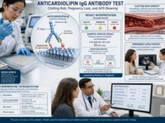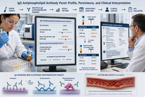
An antiphospholipid IgG antibody panel checks for IgG autoantibodies that recognize phospholipid-binding proteins or related targets. The most important components are anticardiolipin IgG and anti-beta-2 glycoprotein I IgG. These antibodies are central to laboratory assessment for antiphospholipid syndrome (APS), an autoimmune condition associated with blood clots and certain pregnancy complications.
A positive panel is not enough to diagnose APS. The result must fit a qualifying clinical event, and clinically relevant antibodies generally need to remain positive on repeat testing at least 12 weeks later. The exact antibody, level, test method, and presence of lupus anticoagulant or other antiphospholipid antibodies all affect interpretation. IgG results often carry more weight than isolated low-level IgM or non-criteria antibodies, particularly when the level is moderate or high. Even so, treatment should never be based on the panel name alone. A person with no clot history, someone with a recent pulmonary embolism, and someone planning pregnancy require very different decisions despite having the same laboratory label.
- Core IgG markers are anticardiolipin IgG and anti-beta-2 glycoprotein I IgG; some laboratories add non-criteria antibodies.
- Moderate or high persistent IgG positivity is generally more meaningful than a single weak positive result.
- APS requires clinical correlation; an antibody result alone does not establish the syndrome.
- A repeat sample is usually needed after at least 12 weeks to confirm persistence.
- Sudden chest pain, shortness of breath, one-sided weakness, or a swollen painful leg requires urgent care, not routine follow-up.
Table of Contents
- Tests in an IgG Panel
- How IgG Antibodies Relate to Clotting
- Interpreting Levels and Patterns
- Role in APS Diagnosis
- Risk After a Positive Result
- Testing Process and Repeat Timing
- Clinical Follow-Up and Treatment Questions
Tests in an IgG Panel
An antiphospholipid IgG panel is a laboratory grouping rather than a single universal test. Its value depends on the specific markers included. The report should list each antibody separately.
The two established solid-phase IgG tests are:
- Anticardiolipin IgG (aCL IgG): measures IgG binding in an assay built around cardiolipin and associated proteins. Clinically important aCL reactivity is often dependent on beta-2 glycoprotein I.
- Anti-beta-2 glycoprotein I IgG (anti-β2GPI IgG): measures IgG directed more specifically against beta-2 glycoprotein I, a circulating protein that attaches to phospholipid surfaces.
Some panels also report IgG antibodies to phosphatidylserine, phosphatidylserine/prothrombin complexes, prothrombin, phosphatidylethanolamine, or other targets. Those are often called non-criteria antibodies because they are not among the standard laboratory markers used in current APS classification. Their evidence and assay standardization vary, so they should not be combined into a single score without explanation.
Lupus anticoagulant is not an IgG concentration test and may not be included in a product labeled “IgG panel.” It is detected through a sequence of phospholipid-dependent clotting assays. Yet lupus anticoagulant is one of the three established laboratory marker groups for APS and may carry substantial thrombotic significance. A complete investigation commonly pairs solid-phase antibody tests with lupus anticoagulant testing.
The term IgG describes the antibody class. IgG circulates throughout the bloodstream, can persist for long periods, and participates in immune responses through several mechanisms. In APS, pathogenic IgG antibodies may interact with beta-2 glycoprotein I and cell surfaces, promoting endothelial activation, platelet activation, complement signaling, and coagulation. Not every detected IgG antibody has these effects, which is why level, persistence, target, and clinical context matter.
A detailed explanation of the individual components is available in the anticardiolipin IgG antibody test and beta-2 glycoprotein 1 IgG antibody test articles.
How IgG Antibodies Relate to Clotting
IgG antiphospholipid antibodies do not simply make blood “thicker.” They can disturb several systems that normally keep clot formation limited to the right place and time. Proposed mechanisms include activating the lining of blood vessels, increasing tissue factor activity, stimulating platelets, interfering with protective anticoagulant pathways, and amplifying complement-mediated inflammation.
These antibodies are better viewed as part of a prothrombotic tendency than as a guarantee that a clot will occur. Many people need an additional trigger—the “second hit”—before thrombosis develops. Surgery, prolonged immobility, pregnancy, estrogen exposure, infection, smoking, cancer, or severe inflammation can add to the underlying risk.
The clinical events linked with APS include:
- Deep vein thrombosis and pulmonary embolism
- Ischemic stroke or transient ischemic attack
- Arterial thrombosis in the limbs or other organs
- Small-vessel disease affecting organs such as the kidneys, skin, brain, or lungs
- Certain placental and pregnancy complications
- Less commonly, rapidly progressive multiorgan thrombosis in catastrophic APS
The target of the IgG antibody influences how strongly it is associated with these outcomes. Anti-beta-2 glycoprotein I IgG and anticardiolipin IgG are established markers, but their predictive value is not identical in every population or assay. The strongest laboratory concern usually comes from a persistent profile that includes lupus anticoagulant, high-level IgG antibodies, or positivity across multiple established marker groups.
One positive result cannot provide an exact percentage risk for an individual. Study populations differ in age, autoimmune disease, prior thrombosis, treatments, antibody methods, and follow-up. A laboratory value must therefore be placed into a clinical risk model rather than treated as a standalone forecast.
Interpreting Levels and Patterns
The word positive covers a wide range of findings. A result barely above the laboratory cutoff is not equivalent to a repeatedly high value, and isolated anticardiolipin IgG is not equivalent to combined anticardiolipin, anti-beta-2 glycoprotein I, and lupus anticoagulant positivity.
Negative, low, moderate, and high results
Laboratories may report numerical units, percentile-based cutoffs, or categories. Under the 2023 ACR/EULAR classification framework, moderate and high anticardiolipin or anti-beta-2 glycoprotein I levels are defined for specified enzyme-linked immunosorbent assays as 40–79 units and at least 80 units, respectively. Those research thresholds should not be transferred blindly to every automated assay or manufacturer platform.
A laboratory may use GPL units for anticardiolipin IgG, units per milliliter, chemiluminescent units, or an index value. The correct first step is to read that report’s reference interval. The second is to identify the assay method. A value cannot be compared safely with a different laboratory unless the methods are known to be comparable.
| Finding | Possible interpretation | Important caution |
|---|---|---|
| Negative aCL IgG and anti-β2GPI IgG | No IgG antibody detected above the assay cutoffs | Does not exclude lupus anticoagulant, IgM antibodies, or every APS-like condition |
| Single low positive | May be transient, nonspecific, or of limited clinical weight | Needs event history and often repeat testing before conclusions |
| Moderate or high IgG positive | More compatible with clinically important antiphospholipid autoimmunity | Still requires persistence and a relevant clinical picture |
| Both aCL IgG and anti-β2GPI IgG positive | Broader established antibody profile | Risk also depends on lupus anticoagulant, level, and prior events |
| IgG positivity plus lupus anticoagulant | Potentially higher-risk profile, especially if persistent | Anticoagulant drugs can complicate lupus anticoagulant testing |
Single, double, and triple positivity
These terms describe established marker groups, not the number of positive lines printed on a commercial panel:
- Single positive: one established group is positive, such as anticardiolipin alone.
- Double positive: two established groups are positive.
- Triple positive: lupus anticoagulant, anticardiolipin, and anti-beta-2 glycoprotein I are all positive.
Triple positivity is associated with a higher-risk APS phenotype in many studies. It should be confirmed carefully, particularly when a patient is taking an anticoagulant that can distort lupus anticoagulant assays. Non-criteria antibody results should be described separately rather than used to manufacture a “triple-positive” label.
Persistent versus transient results
Persistence adds weight because antiphospholipid antibodies can appear temporarily during infection, inflammatory illness, or other immune stimulation. A repeat result at least 12 weeks later helps reduce the chance that a short-lived antibody is being mistaken for APS-related autoimmunity. Testing sooner may document change, but it does not satisfy the usual persistence interval.
Role in APS Diagnosis
APS is a clinical-laboratory syndrome. It is not diagnosed from antibodies alone and not diagnosed from thrombosis alone. The clinician must connect a sufficiently credible clinical manifestation with a persistent, relevant antiphospholipid antibody profile.
The 2023 ACR/EULAR criteria use an entry requirement followed by weighted clinical and laboratory domains. The clinical domains include venous thrombosis, arterial thrombosis, microvascular disease, obstetric events, heart valve findings, and thrombocytopenia. The laboratory domains include lupus anticoagulant and solid-phase anticardiolipin or anti-beta-2 glycoprotein I IgG/IgM results. These criteria were developed for classification in studies and are highly specific; they are not a substitute for individualized clinical diagnosis.
For routine care, specialists generally ask four questions:
- Was there a documented APS-type event? Imaging, pathology, obstetric records, and timing matter.
- Is an established antibody present at a meaningful level? IgG results often contribute strongly when moderate or high.
- Did the antibody persist? A second positive sample at least 12 weeks later is usually needed.
- Is another explanation more convincing? Major provoking factors or alternative diseases can change attribution and treatment.
A person may meet classification criteria and still need careful diagnostic review. The reverse can also occur: someone with a compelling clinical picture may fall outside a research classification boundary. The medical record should distinguish “positive antiphospholipid antibodies,” “possible APS,” and “established APS” instead of using them interchangeably.
Three contrasting examples show why the distinction matters. A patient with a high anticardiolipin IgG result during pneumonia, no thrombosis history, and a negative repeat sample does not have the same evidence as someone with high anticardiolipin IgG on two samples and an imaging-confirmed unprovoked deep vein thrombosis. A third patient may have persistent anti-beta-2 glycoprotein I IgG but only a superficial vein problem that does not meet an accepted APS clinical event. The antibody can still be relevant to future risk assessment, but it does not automatically turn every vascular symptom into APS.
Clinical timing also matters. The antibody test does not need to be drawn on the day of the clot to be informative, but the 2023 classification framework requires a positive test within three years of the clinical criterion for entry. In practice, clinicians may evaluate older events, especially when records are strong, yet uncertainty grows when neither the event nor the original laboratory data can be verified. A copied diagnosis in a chart is weaker evidence than the original imaging report and laboratory values.
The complete APS blood test panel is important because an IgG-only order can leave major gaps. Missing lupus anticoagulant testing, failing to test both targets, or omitting the repeat sample can lead to underdiagnosis or overdiagnosis.
Risk After a Positive Result
Risk depends more on the profile and the person than on the presence of any IgG antibody. Two patients with the same anticardiolipin IgG value may have very different outlooks if one has never had a clot and the other has recurrent unprovoked thrombosis plus lupus anticoagulant.
Factors that increase concern include:
- A previous unprovoked or recurrent clot
- Arterial thrombosis, especially at a young age without a better cause
- Persistent lupus anticoagulant
- Double or triple established-marker positivity
- High-level anticardiolipin IgG or anti-beta-2 glycoprotein I IgG
- Systemic lupus erythematosus
- Traditional vascular risks such as smoking, hypertension, high cholesterol, and diabetes
- Temporary high-risk situations such as surgery, immobilization, postpartum recovery, or severe infection
A weak antibody found during broad screening in a person with no symptoms usually has a lower immediate significance. It may require verification rather than treatment. Long-term anticoagulation is not routinely started simply because an asymptomatic person has one positive IgG result; bleeding risk must be weighed against a clearly defined thrombotic benefit.
For a person with a prior clot, the antibody profile can affect recurrence assessment and the choice or duration of anticoagulation. Vitamin K antagonists such as warfarin remain important in thrombotic APS, especially in high-risk profiles. Direct oral anticoagulants are not considered interchangeable with warfarin for every APS patient, and they may be unsuitable in some people with arterial events or triple positivity. Medication selection belongs with a clinician who can assess the complete history.
Pregnancy creates a separate clinical setting. Persistent criteria IgG antibodies may support a diagnosis of obstetric APS when the pregnancy history meets defined patterns. Management can include low-dose aspirin and heparin in established cases, but a positive blood test without qualifying pregnancy morbidity does not automatically require the same regimen. Preconception consultation helps separate antibody carriage from treated obstetric APS.
The pregnancy record should identify gestational age, fetal findings, placental disease, maternal blood pressure, and other possible causes. Recurrent very early losses, an unexplained fetal death, and delivery before 34 weeks because of severe preeclampsia or placental insufficiency are clinically different patterns. They should not be reduced to the single phrase “pregnancy loss.” In someone with persistent IgG antibodies, those details determine whether the history resembles criteria obstetric APS, another placental disorder, or a problem requiring a different evaluation.
People with known antibodies should receive individualized plans for major surgery, hospitalization, long-distance immobility, estrogen-containing medication, and pregnancy. Routine daily life does not require constant fear, but preventable risk factors deserve attention.
Testing Process and Repeat Timing
The IgG solid-phase tests use a blood sample and normally do not require fasting. Eating, drinking water, and ordinary activity do not meaningfully change anticardiolipin IgG or anti-beta-2 glycoprotein I IgG results. The clinician should still know about recent infection, autoimmune flares, pregnancy, and current medication.
Acute thrombosis does not automatically invalidate solid-phase IgG assays, but it can create a difficult testing environment. Hospitalized patients may have inflammation, receive heparin or a direct oral anticoagulant, and undergo several blood draws on different platforms. The safest interpretation separates the antibody assays from clot-based lupus anticoagulant testing and records which medicines were present at collection. A negative or positive lupus anticoagulant result obtained under interference may need later confirmation even when the IgG antibody measurements are technically sound.
The timing plan should be deliberate:
- Initial testing: obtain the full relevant antibody profile when APS is clinically suspected.
- Clinical verification: confirm the clot or pregnancy event with records rather than waiting for the repeat result to reconstruct the history.
- Repeat testing: if the initial established antibody is positive and the result may affect diagnosis or treatment, repeat it no sooner than 12 weeks later for persistence.
- Method comparison: use the same laboratory and platform when possible, and retain the original units and reference interval.
A repeat is not needed merely to watch a number fluctuate when it will not change care. Once APS is firmly established, repeated antibody titers are not a simple measure of treatment success. Anticoagulation is monitored with medication-appropriate tests, not by trying to make the antibody disappear.
Solid-phase IgG assays are generally less affected by anticoagulant drugs than lupus anticoagulant testing. However, warfarin, heparin, and direct oral anticoagulants can interfere with clot-based lupus anticoagulant methods. Patients should never stop anticoagulation on their own for testing. The laboratory and treating clinician can choose safer timing, interpretive comments, alternative assays, or specialized sample handling.
Analytical variation remains a major limitation. Antigen source, coating conditions, calibration, instrument type, and cutoff selection can all influence results. This explains why a positive sample may not reproduce identically on another platform. It also explains why classification thresholds designed around one method require caution when applied to another.
The separate antiphospholipid antibody repeat testing guide covers timing and confirmation in greater detail.
Clinical Follow-Up and Treatment Questions
The next step after receiving the report is to translate the laboratory pattern into a clear clinical statement. A useful summary might read: “Persistent high anti-beta-2 glycoprotein I IgG with lupus anticoagulant after an unprovoked pulmonary embolism,” rather than simply “positive IgG panel.”
Bring the following information to the follow-up visit:
- The complete report from both the initial and repeat samples
- Imaging reports or discharge records for any thrombosis
- Dates and details of pregnancy losses or placental complications
- A medication list, including estrogen, anticoagulants, aspirin, and autoimmune therapies
- History of lupus, kidney disease, low platelets, migraine, livedo, or heart valve disease
- Family history of thrombosis and major personal risk factors
Questions for the clinician may include:
- Which antibody is positive, and is the level low, moderate, or high on this exact assay?
- Does the result meet a recognized laboratory threshold?
- Is lupus anticoagulant testing valid while I am taking my current medication?
- Do I have antibody positivity alone, possible APS, or established APS?
- Does my prior event require long-term anticoagulation for reasons independent of the antibody?
- Should pregnancy, surgery, travel, or estrogen exposure be managed differently?
- Who should coordinate care: hematology, rheumatology, maternal-fetal medicine, neurology, or another specialty?
Do not use a falling IgG value as permission to stop prescribed anticoagulation. Do not begin aspirin because a panel is positive without reviewing bleeding risk and the reason it was ordered. Do not assume a negative repeat erases a previous clot or proves the first test was meaningless. Each decision depends on the whole record.
Emergency symptoms override outpatient interpretation. Call emergency services for sudden neurologic deficits, severe unexplained shortness of breath, chest pain, coughing blood, or signs of threatened limb circulation. A known antibody result can support the history, but urgent diagnosis depends on clinical examination and imaging.
When the result is approached systematically—marker, level, persistence, profile, event, and competing causes—the IgG panel becomes useful evidence rather than a confusing label. That is the level of detail needed for accurate APS diagnosis and sensible risk management.
References
- An update on laboratory detection and interpretation of antiphospholipid antibodies for diagnosis of antiphospholipid syndrome: guidance from the ISTH-SSC Subcommittee on Lupus Anticoagulant/Antiphospholipid Antibodies 2025 (Guideline)
- 2023 ACR/EULAR antiphospholipid syndrome classification criteria 2023 (Classification Criteria)
- Guidelines on the investigation and management of antiphospholipid syndrome 2024 (Guideline)
- Diagnosis and management of antiphospholipid syndrome 2024 (Review)
- New definitions for antiphospholipid syndrome 2024 (Review)
- Laboratory Diagnosis of Antiphospholipid Syndrome: Insights and Hindrances 2022 (Review)
Disclaimer
This article provides general education and cannot determine whether a positive IgG result represents APS or whether anticoagulation is appropriate. Interpretation requires the complete antibody profile, repeat timing, documented clinical events, medications, and bleeding risk. Seek emergency care immediately for symptoms of a possible clot or stroke.