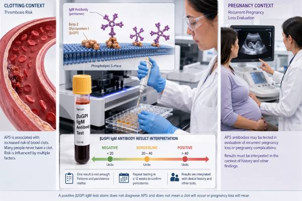
The beta-2 glycoprotein 1 IgM antibody test looks for an autoimmune antibody linked to antiphospholipid syndrome, or APS. APS can raise the risk of blood clots and certain pregnancy complications, but a positive IgM result does not prove that a person has the condition. Doctors interpret the result alongside symptoms, clotting history, pregnancy history, other antiphospholipid antibodies, and a repeat sample collected at least 12 weeks later. The strength and pattern of positivity also matter. An isolated, low-level IgM result is often less concerning than persistent moderate or high levels combined with lupus anticoagulant or anticardiolipin antibodies. Because laboratories use different assay systems and units, the numerical value must be compared with the reference range printed on the same report. The test is most useful as one part of a complete APS evaluation rather than as a stand-alone measure of clotting or miscarriage risk.
- The test measures IgM antibodies against beta-2 glycoprotein I, a blood protein that binds to negatively charged phospholipid surfaces.
- A positive result alone does not diagnose APS; a related clinical event and persistent antibody positivity are usually needed.
- Repeat testing should be at least 12 weeks after the first positive sample to distinguish persistent antibodies from a temporary response.
- Low or borderline isolated IgM results often carry less risk than lupus anticoagulant, IgG antibodies, or triple-positive profiles.
- No fasting is usually required, and anticoagulant medicines generally interfere less with this solid-phase antibody test than with lupus anticoagulant assays.
- New chest pain, sudden shortness of breath, one-sided weakness, or a swollen painful leg requires urgent medical assessment, regardless of the antibody result.
Table of Contents
- What the Test Measures
- Why the Test Is Ordered
- How to Read Negative, Borderline, and Positive Results
- Clotting Risk and APS Diagnosis
- Pregnancy Loss and Obstetric Risk
- Testing Process, Timing, and Interference
- Next Steps After a Result
What the Test Measures
Beta-2 glycoprotein I, also written as β2GPI or apolipoprotein H, is a protein made mainly by the liver and found in the bloodstream. It can attach to phospholipids, which are fat-based components of cell membranes and activated platelet surfaces. In some people, the immune system produces antibodies that recognize beta-2 glycoprotein I, especially when the protein changes shape after binding to a surface.
The test measures the IgM class of these antibodies. IgM is one of several antibody types. It often appears early in an immune response and has a different structure from IgG, the other routinely measured beta-2 glycoprotein I antibody class. Laboratories may use enzyme-linked immunosorbent assays, chemiluminescent assays, or related solid-phase methods. The report may list the marker as anti-β2GPI IgM, aβ2GPI IgM, B2GP1 IgM, or beta-2 glycoprotein I antibody IgM.
A positive test indicates that the laboratory detected antibody binding above its established cutoff. It does not show whether a clot is present, how quickly blood is clotting, or whether a pregnancy complication will occur. It is an immune marker, not a direct measurement of blood flow or coagulation speed.
The test belongs to the three-part group of established antiphospholipid antibody tests:
- Lupus anticoagulant, measured with phospholipid-dependent clotting assays
- Anticardiolipin IgG and IgM antibodies
- Anti-beta-2 glycoprotein I IgG and IgM antibodies
These tests detect related but nonidentical antibody effects. A person may be positive on one, two, or all three. The overall pattern usually gives more information than any single value. A broader antiphospholipid syndrome blood test panel can show whether the IgM finding is isolated or part of a higher-risk profile.
Beta-2 glycoprotein I antibodies may promote clotting through several pathways. They can activate endothelial cells lining blood vessels, stimulate platelets and monocytes, amplify complement activity, and interfere with natural anticoagulant surfaces. These mechanisms help explain why APS behaves as a thrombo-inflammatory disease rather than a simple excess of one clotting factor. However, not every detected antibody has the same biological activity. Assay design, antibody concentration, target region on the protein, and coexistence of other antibodies all influence clinical significance.
Why the Test Is Ordered
Doctors usually order anti-beta-2 glycoprotein I IgM when a person has a clinical event that could fit APS or when another autoimmune condition raises suspicion. Testing healthy people without a suggestive history is generally less useful because low-level antibodies can occur temporarily and may create anxiety or lead to unnecessary repeat testing.
Common reasons for testing include:
- An unprovoked deep vein thrombosis or pulmonary embolism
- A blood clot at an unusual site, such as a cerebral, abdominal, or retinal vein
- Ischemic stroke or transient ischemic attack in a younger person without a clear cause
- Recurrent pregnancy loss or a later fetal death
- Early delivery related to severe preeclampsia, placental insufficiency, or fetal growth restriction
- Unexplained low platelets, livedo racemosa, heart valve abnormalities, or other features that can accompany APS
- Systemic lupus erythematosus or another autoimmune disease with a relevant clinical history
- A prolonged clotting test that suggests lupus anticoagulant
Testing may also follow a positive anticardiolipin IgM antibody test. Because anticardiolipin antibodies can partly recognize beta-2 glycoprotein I bound to cardiolipin, the two markers may appear together, but they are not interchangeable.
APS can be primary, meaning it occurs without another defined systemic autoimmune disease, or associated with conditions such as lupus. Infection, inflammation, and some medications can also coincide with transient antiphospholipid antibody positivity. The test is therefore most informative when ordered for a specific clinical reason and interpreted after the acute situation has been reviewed.
A clinician may avoid ordering the test after every ordinary miscarriage or in routine preconception screening. Early pregnancy loss is common and has many causes, especially chromosomal abnormalities. APS testing is more likely to help after recurrent losses, fetal death at a later gestation, severe placenta-mediated complications, or thrombosis. Exact thresholds for evaluation vary by clinical guideline and individual circumstances, so obstetric history should be reviewed in detail rather than reduced to a single number of losses.
The test is not a general screen for “thick blood,” inherited thrombophilia, bleeding disorders, or all causes of infertility. Factor V Leiden, prothrombin gene variants, protein C or S deficiency, and antithrombin deficiency are different conditions. Likewise, a normal anti-beta-2 glycoprotein I IgM result does not exclude every acquired or inherited cause of thrombosis.
How to Read Negative, Borderline, and Positive Results
The laboratory’s own reference interval should guide the first reading. Results may be reported in U/mL, SMU, MPL-equivalent units, arbitrary units, or another assay-specific format. One laboratory’s value cannot safely be converted into another laboratory’s value without validated standardization.
| Report category | Usual meaning | Typical follow-up |
|---|---|---|
| Negative | No IgM antibody detected above the assay cutoff | Review the other APS markers and the clinical history |
| Borderline or low positive | Weak antibody binding that may be transient or assay dependent | Repeat after at least 12 weeks if APS remains clinically plausible |
| Moderate or high positive | Stronger antibody signal with greater potential relevance, especially if persistent | Confirm persistence and assess the complete antibody profile |
| Persistent positive | Positive on two appropriately spaced samples | Interpret with thrombosis or obstetric criteria and specialist review |
Older APS classification frameworks often used a result above the 99th percentile as the laboratory threshold for anti-beta-2 glycoprotein I antibodies. The 2023 ACR/EULAR research classification criteria distinguish moderate and high antibody levels for solid-phase assays and give different weight to antibody types and profiles. These criteria were designed to create specific research groups, not to replace individualized clinical diagnosis. A person can need clinical care even when a research classification score is not met, and a laboratory result should not be self-scored without the full clinical record.
A negative IgM result means only that this particular isotype was not detected above the cutoff. IgG may still be positive, and lupus anticoagulant may still be present. Conversely, an isolated low IgM result may have limited significance when all other tests are negative and there has been no qualifying clinical event.
The word “positive” can sound absolute, but several features alter its weight:
- Persistence: A result present at least 12 weeks apart is more convincing than a single result during infection or inflammation.
- Level: Moderate or high values are generally more relevant than values just above the cutoff.
- Isotype: IgG anti-beta-2 glycoprotein I has shown a more consistent relationship with thrombosis in many studies, while isolated IgM associations are less uniform.
- Other antibodies: Lupus anticoagulant and triple positivity often indicate greater risk than isolated IgM positivity.
- Clinical history: A documented unprovoked clot or characteristic pregnancy complication changes the interpretation substantially.
- Assay consistency: Repeating the test at the same laboratory can reduce variation caused by different platforms and cutoffs.
A falling number does not necessarily mean that risk has disappeared, and a rising number does not prove that a clot is forming. Antibody levels are not used like an INR or blood glucose value for day-to-day dose adjustment. Routine serial measurement outside a defined clinical plan may produce noise rather than useful guidance.
Clotting Risk and APS Diagnosis
A positive anti-beta-2 glycoprotein I IgM test can support APS, but APS requires more than an antibody. Clinicians look for a compatible clinical manifestation, most often objectively confirmed thrombosis or specific pregnancy morbidity, together with persistent antiphospholipid antibodies.
Thrombosis may occur in veins, arteries, or small vessels. Venous events include deep vein thrombosis and pulmonary embolism. Arterial events often involve the brain, causing stroke or transient ischemic attack, although other organs may be affected. Microvascular APS can involve small vessels in the kidneys, lungs, skin, heart, or other tissues.
Risk is not identical for every positive person. The strongest laboratory warning pattern is usually persistent lupus anticoagulant combined with anticardiolipin and anti-beta-2 glycoprotein I antibodies, known as triple positivity. A person with this pattern has a different risk profile from someone with one low, isolated IgM result. The triple-positive antiphospholipid antibody profile also affects medication choices after a clot, because some oral anticoagulants perform less reliably in high-risk APS than vitamin K antagonist therapy.
Isolated IgM deserves balanced interpretation. It should not be dismissed automatically, especially when the level is clearly elevated, persistent, and paired with a compelling clinical event. At the same time, evidence linking isolated IgM anti-beta-2 glycoprotein I to thrombosis is weaker and more variable than evidence for lupus anticoagulant or high-risk multiple positivity. This uncertainty is one reason a hematologist or rheumatologist may review original imaging, the timing of the event, provoking factors, and the complete antibody panel before labeling a person with APS.
Factors unrelated to the antibody can further raise clotting risk:
- Smoking or nicotine exposure
- Estrogen-containing contraception or hormone therapy
- Pregnancy and the postpartum period
- Major surgery, trauma, or prolonged immobility
- Active cancer
- Obesity and metabolic disease
- Severe infection or systemic inflammation
- Poorly controlled lupus activity
- Prior thrombosis
A positive antibody result does not automatically mean lifelong anticoagulation. People with no previous thrombosis may need risk-factor control and situation-specific prevention rather than full-dose blood thinners. People with confirmed thrombotic APS often need long-term anticoagulation, but the drug and intensity depend on whether the event was venous or arterial, whether it recurred, the antibody profile, bleeding risk, and other medical conditions. Treatment should never be started or stopped from a laboratory result alone.
Catastrophic APS is a rare emergency in which thrombosis develops rapidly in multiple organs, often with severe inflammation and organ failure. It cannot be diagnosed from an IgM value. Rapidly worsening shortness of breath, confusion, kidney problems, skin discoloration, or several organ symptoms require emergency care, particularly in a person with known APS.
Pregnancy Loss and Obstetric Risk
Persistent antiphospholipid antibodies can contribute to pregnancy complications through placental inflammation, complement activation, impaired trophoblast function, and clotting in placental vessels. The result can be early loss, fetal death, restricted fetal growth, preeclampsia, or medically necessary preterm delivery.
Classic obstetric APS patterns include recurrent early miscarriages, otherwise unexplained fetal death at or beyond 10 weeks, and delivery before 34 weeks because of severe preeclampsia, eclampsia, or recognized placental insufficiency. Clinical practice is more nuanced than a checklist. A maternal-fetal medicine specialist may consider gestational age, fetal testing, placental findings, blood pressure, growth measurements, and alternative explanations.
An isolated IgM result does not establish that APS caused a loss. Many early miscarriages result from chromosome errors, and later complications can arise from hypertension, diabetes, uterine abnormalities, infection, genetic conditions, or other placental disorders. The antibody result becomes more persuasive when it is persistent, moderate or high, accompanied by another antiphospholipid antibody, and matched to a characteristic pregnancy history.
Lupus anticoagulant is often the strongest laboratory predictor of adverse pregnancy outcome among the established tests. That does not make anti-beta-2 glycoprotein I IgM irrelevant, but it means the recurrent pregnancy loss antiphospholipid panel should be read as a pattern rather than as separate yes-or-no answers.
When obstetric APS is confirmed, treatment commonly involves low-dose aspirin and prophylactic or therapeutic low-molecular-weight heparin, depending on prior thrombosis and pregnancy history. Warfarin is usually avoided during pregnancy because it can harm fetal development, although it may be appropriate outside pregnancy and is often compatible with breastfeeding. Exact treatment should be planned before conception when possible.
Pregnancy care may include:
- Preconception review of the antibody profile and previous records
- A medication plan for the weeks before and after conception
- Early confirmation of pregnancy location and viability
- Blood pressure and urine protein monitoring
- Serial ultrasound assessment of fetal growth and placental function
- A delivery and postpartum anticoagulation plan
The postpartum period deserves special attention because clotting risk remains elevated after delivery. A person with APS or a high-risk antibody profile may need anticoagulant prevention for a defined period after birth. The plan depends on delivery method, bleeding, anesthesia timing, and whether there is a history of thrombosis.
Most people with treated obstetric APS can still have a successful pregnancy. A positive result should prompt coordinated care, not a prediction that loss is inevitable.
Testing Process, Timing, and Interference
The test uses a routine blood sample from a vein. Fasting is usually unnecessary. Normal food intake, hydration, and ordinary daily activity do not meaningfully change the antibody concentration in the way they can affect some metabolic tests.
Before testing, give the clinician and laboratory a complete medication list. Anticoagulants such as heparin, warfarin, apixaban, rivaroxaban, edoxaban, and dabigatran can complicate functional lupus anticoagulant tests. They generally have much less direct effect on solid-phase anti-beta-2 glycoprotein I antibody assays. This difference is useful when a person cannot safely pause anticoagulation, although the whole panel may still require careful interpretation.
Acute infection can trigger temporary antiphospholipid antibodies. Testing during or soon after a viral or bacterial illness may therefore produce a low positive result that later disappears. Surgery, major inflammation, and pregnancy can also change the clinical context. Clinicians often document these circumstances and rely on repeat testing rather than assuming that the first value is permanent.
The minimum interval for confirming persistence is 12 weeks, not 12 days and not simply “three months” if that results in a shorter interval. The repeat sample should ideally use the same laboratory and method. A dedicated antiphospholipid antibody repeat test helps prevent a transient immune response from being mistaken for APS.
Several practical issues can affect comparability:
- Laboratories use different antigen preparations, calibrators, instruments, and cutoff calculations.
- Some reports label a narrow zone as equivocal or indeterminate.
- A result near the cutoff can switch categories because of normal analytical variation.
- IgM assays may be affected by nonspecific binding or other immunoglobulin-related interference in some samples.
- A unit value from one manufacturer may not represent the same antibody activity on another platform.
For these reasons, the numerical trend should not be overinterpreted when tests come from different laboratories. The report’s category, percentile or cutoff, method, and date all belong in the clinical record.
A clinician may postpone initial testing when the result will not change immediate care or when the acute setting makes interpretation difficult. After a new clot, treatment should not be delayed while waiting for APS antibodies. The clot itself is managed first, and testing is integrated safely around anticoagulation and follow-up.
Next Steps After a Result
A negative result, a borderline value, and a persistent high positive each call for a different response. The next step is to connect the laboratory finding to the reason the test was ordered.
After a negative result
Confirm that the complete APS panel was performed. A negative IgM result does not replace lupus anticoagulant, anticardiolipin IgG and IgM, or anti-beta-2 glycoprotein I IgG testing. If all established markers are negative, APS becomes less likely, but the clinician may still investigate other causes of thrombosis or pregnancy loss.
After a borderline or low positive result
Ask whether the sample was collected during infection, inflammation, or another temporary stress. Review the exact cutoff and whether other antibodies were positive. If the clinical history fits APS, repeat the same test at least 12 weeks later. Avoid starting aspirin or anticoagulation without medical advice, because these drugs can cause serious bleeding and may not help a person with an incidental antibody.
After a moderate or high positive result
Arrange review with the ordering clinician, especially after a clot, stroke-like event, fetal death, recurrent loss, or severe placenta-related complication. The clinician may confirm persistence, obtain missing panel components, and refer to hematology, rheumatology, neurology, or maternal-fetal medicine. A persistent result should be documented clearly because it can affect pregnancy planning, surgery, estrogen use, and thrombosis prevention.
Questions worth asking at the appointment
- Was the result low, moderate, or high by this laboratory’s method?
- Were IgG anti-beta-2 glycoprotein I, anticardiolipin antibodies, and lupus anticoagulant also tested?
- Does my medical history meet a clinical APS pattern, or is this an isolated laboratory finding?
- When exactly should the repeat sample be collected?
- Could infection, medication, or a different assay explain the result?
- Do I need changes before surgery, pregnancy, long travel, or estrogen treatment?
- Which symptoms should send me to urgent care?
Seek emergency help for sudden shortness of breath, coughing blood, severe chest pain, a new one-sided swollen leg, facial droop, speech difficulty, one-sided weakness, sudden vision loss, or an abrupt severe headache. During pregnancy, urgent assessment is also appropriate for heavy bleeding, severe persistent headache, new visual changes, marked upper abdominal pain, sudden swelling with high blood pressure, reduced fetal movement, or breathing difficulty.
A beta-2 glycoprotein I IgM result is most useful when treated as one piece of a clinical pattern. Persistence, antibody profile, event history, and competing explanations determine whether it represents APS, a possible risk marker, or a temporary laboratory finding.
References
- 2023 ACR/EULAR antiphospholipid syndrome classification criteria 2023 (Classification Criteria)
- An update on laboratory detection and interpretation of antiphospholipid antibodies in antiphospholipid syndrome 2025 (Review)
- Laboratory Diagnosis of Antiphospholipid Syndrome: Insights and Hindrances 2022 (Review)
- A systematic review of the association between anti-β-2 glycoprotein I antibodies and clinical manifestations of antiphospholipid syndrome 2021 (Systematic Review)
- IgM Antiphospholipid Antibodies in Antiphospholipid Syndrome 2025 (Review)
- Antiphospholipid syndrome in pregnancy 2025 (Review)
Disclaimer
This article provides general information about beta-2 glycoprotein I IgM testing and does not diagnose antiphospholipid syndrome or determine an individual treatment plan. Do not start, stop, or change aspirin, anticoagulants, hormone therapy, or pregnancy medication without guidance from a qualified clinician. Symptoms of a possible blood clot, stroke, or serious pregnancy complication require urgent medical care.