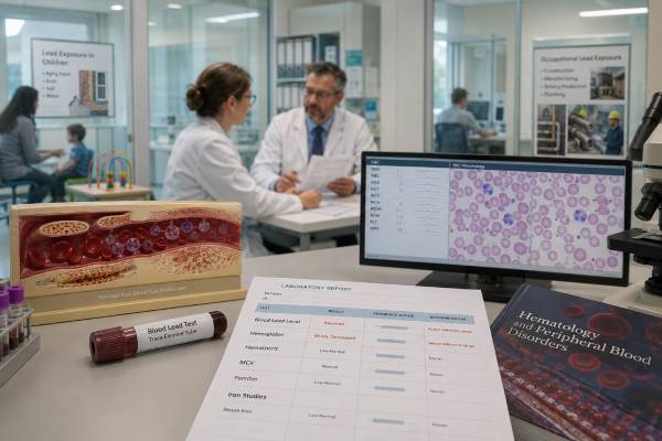
Blood lead and hemoglobin answer two different questions, but they often belong in the same conversation. A blood lead test shows how much lead is circulating in the blood at the time of testing. Hemoglobin shows how much oxygen-carrying protein is inside red blood cells. Lead can interfere with hemoglobin production, but low hemoglobin does not automatically mean lead poisoning, and a high blood lead level does not always cause anemia. The pattern depends on the lead level, the length of exposure, age, nutrition, iron status, kidney function, and whether another anemia cause is present. The most useful interpretation comes from reading blood lead alongside the complete blood count, MCV, RDW, reticulocyte count, ferritin or iron studies, and the exposure history. A child with a modest blood lead level may need environmental action before anemia appears, while an adult with anemia may need lead testing only when the history or blood smear pattern fits.
- Blood lead is measured in µg/dL and reflects recent or ongoing lead exposure, not the body’s full lifetime lead burden.
- Hemoglobin is measured in g/dL and is low when anemia is present, but it does not identify the cause by itself.
- Lead can cause anemia by disrupting heme production and shortening red blood cell survival, especially at higher or prolonged exposures.
- Iron deficiency and lead exposure can overlap, and iron deficiency may increase lead absorption, especially in children.
- Children with blood lead at or above 3.5 µg/dL need follow-up, because this level is used to identify children with higher blood lead than most U.S. children.
- Urgent medical guidance is needed for very high blood lead levels, neurologic symptoms, severe abdominal symptoms, pregnancy, or suspected ingestion of lead-containing objects.
Table of Contents
- How the Two Tests Fit Together
- How Lead Can Lower Hemoglobin
- Blood Count Patterns That Can Point Toward Lead
- Blood Lead Ranges and Next Steps
- Checking Iron and Other Causes of Anemia
- Children, Pregnancy, and Workplace Exposure
- What to Do After Abnormal Results
- Common Mistakes When Reading These Results
How the Two Tests Fit Together
A blood lead level and hemoglobin result should not be treated as two versions of the same test. They measure different things.
A blood lead level measures lead in the bloodstream. In the United States, it is usually reported as micrograms of lead per deciliter of blood, written as µg/dL. A detectable result means lead entered the body. The result is especially useful for recent exposure, ongoing exposure, or lead that has moved from body stores back into the blood. It does not fully measure lead stored in bone, where much of the body’s lead can remain for years.
Hemoglobin is the protein inside red blood cells that carries oxygen. It is reported in grams per deciliter, or g/dL. Low hemoglobin means anemia is present, but anemia is a description, not a diagnosis. The cause may be iron deficiency, chronic inflammation, kidney disease, blood loss, vitamin B12 or folate deficiency, hemolysis, inherited blood disorders, bone marrow problems, lead exposure, or more than one cause at the same time.
Lead becomes relevant to hemoglobin because the body needs an intact heme pathway to make hemoglobin. Heme is the iron-containing part of hemoglobin. Lead interferes with enzymes used in heme production, so red blood cells may become smaller, paler, and less efficient. This is why lead exposure can sometimes look like iron deficiency on a blood count.
The two tests are most helpful together in three situations:
- A blood lead level is elevated, and the clinician needs to know whether it has affected red blood cell production.
- Hemoglobin is low, especially with a microcytic pattern, and lead exposure is possible from housing, work, hobbies, imported products, or contaminated water or soil.
- A child, pregnant person, or lead-exposed worker needs monitoring because lead exposure and anemia risk can reinforce each other.
For a broader view of the red blood cell side of the result, hemoglobin is usually interpreted with hematocrit, RBC count, MCV, MCH, MCHC, RDW, and sometimes a blood smear. A separate hemoglobin and hematocrit comparison can help explain why these numbers often move together but are not identical.
Normal hemoglobin ranges vary by age, sex, pregnancy status, altitude, and the lab method. Many adult reference ranges are roughly 13.5–17.5 g/dL for men and 12.0–15.5 g/dL for women, but a local lab range and clinical context matter more than a single universal cutoff. Children have age-specific ranges, and infants normally pass through a physiologic hemoglobin drop in early life.
A normal hemoglobin result does not make lead exposure safe. Lead can harm the nervous system, kidneys, blood pressure regulation, pregnancy outcomes, and child development before it causes anemia. That is especially important in children, where prevention and exposure removal matter even at low levels.
How Lead Can Lower Hemoglobin
Lead can lower hemoglobin through several connected effects. The most familiar one is impaired heme synthesis.
Heme production happens through a sequence of enzyme-driven steps. Lead inhibits key enzymes in that pathway, especially delta-aminolevulinic acid dehydratase and ferrochelatase. Ferrochelatase helps insert iron into protoporphyrin to form heme. When that step is blocked, less heme is made, and zinc may be inserted instead of iron, forming zinc protoporphyrin. This is why zinc protoporphyrin, or ZPP, can rise in both lead exposure and iron deficiency.
Lead can also shorten red blood cell survival. Red blood cells normally circulate for about 120 days in adults. Lead-related oxidative stress and membrane injury can make them more fragile, which can contribute to anemia when exposure is significant. In severe cases, hemolysis can occur, although this is less common than impaired production.
A third issue is nutritional overlap. Iron, calcium, and other nutrients influence how much lead the body absorbs. Iron deficiency is especially important because the body may absorb more lead when iron stores are low. A child with low iron intake, pica, and old housing can develop both iron deficiency anemia and an elevated blood lead level. In that case, treating iron deficiency helps the anemia and may also reduce ongoing lead absorption, but it does not replace finding and removing the lead source.
Lead-related anemia is often described as microcytic, meaning the red blood cells are smaller than usual. The MCV is low. It may also be hypochromic, meaning red cells contain less hemoglobin and look paler on a smear. These features overlap with iron deficiency, thalassemia trait, and some chronic inflammatory patterns. That overlap is the reason blood lead is not interpreted from hemoglobin alone.
Lead can also produce basophilic stippling on a peripheral blood smear. This means small blue granules are visible inside red blood cells under the microscope. Basophilic stippling can support the suspicion of lead toxicity, but it is not specific enough to diagnose lead exposure by itself. It can appear in other conditions, including some inherited and acquired blood disorders.
The relationship between blood lead and hemoglobin is not perfectly linear. A mild blood lead elevation may not change hemoglobin at all. A person with severe iron deficiency may have very low hemoglobin with only modest lead exposure. Someone with ongoing occupational exposure may have symptoms or kidney and blood pressure concerns before the hemoglobin becomes clearly abnormal. The lab pattern only becomes meaningful when it is matched to the exposure history.
Blood Count Patterns That Can Point Toward Lead
The complete blood count gives the first clues about whether lead has affected red blood cell production. Hemoglobin tells whether anemia is present. MCV, RDW, reticulocyte count, and the smear help narrow the pattern.
A lead-related anemia often resembles other microcytic anemias. The MCV may be low, MCH may be low, and hemoglobin may fall slowly over time. RDW may rise if red blood cell size becomes more variable, especially when iron deficiency is also present. A MCV and RDW pattern can help separate common anemia patterns before more specific testing is added.
The following table shows how common patterns can differ.
| Pattern | Common lab clues | How lead fits |
|---|---|---|
| Lead exposure without anemia | Hemoglobin may be normal; CBC may look normal | Possible at lower or early exposures; still needs exposure control when blood lead is elevated |
| Lead-related microcytic anemia | Low hemoglobin, low MCV, sometimes basophilic stippling | More likely with higher or prolonged exposure, especially when the history fits |
| Iron deficiency anemia | Low hemoglobin, low MCV, often high RDW, low ferritin or low iron stores | Can mimic lead effects and can increase lead absorption |
| Thalassemia trait | Low MCV, often normal or high RBC count, RDW may be normal | Can be mistaken for lead or iron deficiency if iron studies and history are skipped |
| Anemia of inflammation | Low or normal MCV, low serum iron, normal or high ferritin | May coexist with lead exposure but needs separate evaluation |
The reticulocyte count adds another layer. Reticulocytes are young red blood cells. A low or inappropriately normal reticulocyte count means the marrow is not increasing production enough. This can happen when heme production is impaired, iron is unavailable, inflammation suppresses red cell production, or bone marrow function is reduced. A high reticulocyte count points more toward blood loss or hemolysis, although severe toxic injury can sometimes involve red cell destruction.
The blood smear may show small pale cells, target cells, basophilic stippling, or mixed changes. Smear findings rarely settle the diagnosis alone, but they can push the evaluation in the right direction. A peripheral blood smear is most useful when the CBC pattern is unusual, anemia is more than mild, or the diagnosis remains unclear after iron studies.
A low MCV should not automatically be called iron deficiency. Iron deficiency is common, but lead exposure, thalassemia trait, chronic inflammation, and sideroblastic processes can also create microcytic patterns. When MCV is low and RDW is high, iron deficiency becomes more likely, but blood lead testing may still be appropriate if the person has a plausible source of exposure. A focused low MCV and high RDW pattern can be a useful starting point, not the final answer.
Blood Lead Ranges and Next Steps
Blood lead levels are interpreted differently in children, adults, pregnancy, and workplace monitoring. The same number can trigger different actions depending on local rules and clinical risk.
For children in the United States, 3.5 µg/dL is used as the blood lead reference value. This is not a “safe” level or a toxicity threshold. It identifies children whose levels are higher than most children’s levels and should prompt follow-up actions. CDC guidance recommends confirmatory venous testing when a screening capillary result is at or above the reference value, because finger-prick samples can be contaminated by lead dust on the skin.
The general pattern is simple: as blood lead rises, confirmation and follow-up become more urgent.
| Blood lead level | Typical interpretation | Usual next step |
|---|---|---|
| <3.5 µg/dL in a child | Below the current U.S. child reference value | Continue prevention, age-appropriate screening, nutrition review, and risk-based follow-up |
| 3.5–9 µg/dL | Above the reference value | Confirm if needed, identify sources, review iron and calcium intake, repeat testing on schedule |
| 10–19 µg/dL | Higher exposure requiring more active follow-up | Report as required, investigate exposure, check for iron deficiency, repeat testing sooner |
| 20–44 µg/dL | Significant exposure | Medical exam, environmental investigation, possible abdominal X-ray in children with ingestion risk, specialist guidance |
| ≥45 µg/dL | High level with possible need for urgent treatment | Prompt specialist or poison center guidance; chelation may be considered depending on age, symptoms, and circumstances |
For adults, interpretation often includes workplace standards and occupational medicine guidance. A blood lead level that would trigger follow-up in a child may also matter in an adult, but the response depends on whether exposure is occupational, whether the person is pregnant or may become pregnant, and whether symptoms or organ effects are present. Occupational guidance often uses repeated testing, exposure reduction, and sometimes medical removal from lead work at higher levels.
A single blood lead result should answer three questions:
- Is the result confirmed? Capillary screening results at or above action levels usually need venous confirmation.
- Is exposure still happening? A falling level after removal from exposure is different from a rising level despite precautions.
- Is there evidence of harm? Hemoglobin, kidney function, blood pressure, neurologic symptoms, abdominal symptoms, pregnancy status, and child development all affect urgency.
A dedicated blood lead test discussion can help with units, testing method, and common causes of high results.
Checking Iron and Other Causes of Anemia
When blood lead is elevated and hemoglobin is low, iron status needs attention. Iron deficiency is common, treatable, and closely tied to lead absorption. It can also make the CBC look very similar to lead-related anemia.
The usual iron evaluation may include ferritin, serum iron, total iron-binding capacity, transferrin saturation, and sometimes C-reactive protein if inflammation may be affecting ferritin. Ferritin reflects iron stores, but it can rise during inflammation, liver disease, infection, and some chronic illnesses. That means a “normal” ferritin does not always rule out iron deficiency when inflammation is present.
An iron panel helps distinguish low iron stores from inflammation-related iron restriction, but it should be interpreted with the CBC pattern and the clinical story. In a child with pica, poor dietary iron intake, and older housing, iron deficiency and lead exposure may both be present. Treating one while ignoring the other can leave the child at risk.
Zinc protoporphyrin can be useful in selected settings because it rises when heme formation is disrupted. It may increase in lead exposure and iron deficiency, so it is not a stand-alone lead test. A normal ZPP also does not rule out a low-level or recent lead exposure. Its best role is as an additional clue when the timing, CBC pattern, and iron studies are considered together. A ZPP test is especially relevant when the question is whether heme production is being disrupted.
Other anemia causes still need to stay on the list. Heavy menstrual bleeding, gastrointestinal blood loss, celiac disease, chronic kidney disease, inflammatory disease, inherited hemoglobin disorders, B12 or folate deficiency, and medication effects can all lower hemoglobin. Lead exposure may be incidental in one person and central in another.
The age of the patient also changes the evaluation. In toddlers, nutritional iron deficiency and environmental exposure are common concerns. In menstruating adults, menstrual blood loss and iron intake are common. In older adults, new iron deficiency often raises concern for gastrointestinal blood loss until proven otherwise. In people with kidney disease, anemia may reflect reduced erythropoietin production, inflammation, or multiple overlapping causes.
A low hemoglobin result deserves a cause-based workup, not just an iron prescription. A low hemoglobin result becomes more useful when it is read with MCV, RDW, reticulocytes, ferritin, kidney markers, inflammation markers, and the exposure history.
Children, Pregnancy, and Workplace Exposure
Lead exposure has different stakes in different groups.
Children absorb more lead than adults and are more vulnerable to nervous system effects. They also explore with their hands and mouths, so lead dust, soil, peeling paint, and contaminated objects become direct exposure routes. A child may have developmental risk from lead exposure even when hemoglobin is normal. That is why pediatric follow-up focuses on source control, nutrition, developmental surveillance, and repeat blood lead testing instead of waiting for anemia.
Common child exposure sources include older housing with lead-based paint, renovation dust, contaminated soil, lead in drinking water from plumbing, imported spices or candies, traditional remedies, cosmetics, ceramics with lead glaze, toys or jewelry, fishing sinkers, ammunition, and take-home exposure from an adult’s job or hobby.
Pregnancy adds another concern because lead stored in bone can move into the bloodstream during pregnancy and lactation. Lead can cross to the fetus. A pregnant person with possible exposure, anemia, pica, older housing, lead-related work, or use of imported products should discuss testing with a clinician. Iron and calcium intake matter because deficiencies may increase absorption or mobilization risk.
Adults with workplace exposure need a different style of monitoring. Battery manufacturing, smelting, construction, demolition, bridge work, firing ranges, stained glass, ceramics, radiator repair, ammunition work, and some recycling jobs can expose workers to lead. Hobbies can do the same. Work clothes, shoes, tools, and vehicles can bring lead dust home, exposing children and other household members.
For an adult worker, a mildly elevated blood lead level may not lower hemoglobin, but it can still signal inadequate exposure control. Repeat testing, workplace hygiene, respirator fit and use, ventilation, housekeeping, medical surveillance, and temporary removal from exposure may be needed depending on the level and applicable rules.
Workplace and household prevention steps include:
- Change clothes and shoes before leaving a lead-exposed work area.
- Shower after lead work when possible.
- Wash work clothes separately.
- Avoid dry sweeping lead dust.
- Use wet methods or HEPA-filtered cleanup when appropriate.
- Keep children away from workspaces, tools, ammunition supplies, stained glass materials, and renovation dust.
- Use certified lead-safe contractors for work that disturbs old paint.
In children and pregnancy, the safest plan is primary prevention: remove the source before the level rises. In workers, the safest plan is exposure control before blood lead becomes a medical removal issue.
What to Do After Abnormal Results
The right response depends on which result is abnormal.
If blood lead is elevated but hemoglobin is normal, the priority is to confirm the result if needed, identify the source, stop exposure, and schedule follow-up testing. Normal hemoglobin should not delay action. Lead’s most important effects, especially in children, may occur without anemia.
If hemoglobin is low but blood lead is not elevated, the anemia workup should continue. Iron deficiency, blood loss, chronic inflammation, kidney disease, thalassemia trait, and vitamin deficiencies are usually more common than lead toxicity. The blood lead result may still need repeating if exposure is ongoing or the first sample was drawn soon after a suspected exposure.
If both blood lead and anemia are present, the evaluation should move in parallel. The clinician may check iron studies, reticulocyte count, kidney function, liver tests, inflammatory markers, and a smear. In children, developmental screening and environmental investigation may be needed. In adults, occupational exposure and take-home contamination should be addressed.
A practical sequence is:
- Confirm the blood lead result when appropriate. Venous testing is preferred for confirmation.
- Classify the anemia. Use hemoglobin, MCV, RDW, reticulocyte count, and smear findings.
- Check iron status. Ferritin and transferrin saturation often clarify whether iron deficiency is present.
- Find the lead source. Ask about housing, plumbing, renovations, work, hobbies, imported products, cosmetics, remedies, ceramics, bullets, fishing weights, and contaminated soil.
- Stop ongoing exposure. Treatment works poorly if exposure continues.
- Repeat testing on schedule. Falling levels suggest exposure control is working; rising levels mean the source is still active or another source was missed.
- Use specialist help for higher-risk cases. Poison control, occupational medicine, pediatric environmental health specialists, or medical toxicologists may be needed.
Symptoms change urgency. Severe abdominal pain, vomiting, confusion, weakness, seizures, coma, severe headache, wrist or foot drop, or suspected ingestion of a lead object requires urgent medical advice. A very high blood lead level can require hospital care, gastrointestinal decontamination, or chelation therapy. Chelation should not be started casually; it is used in specific circumstances and should be guided by clinicians experienced in lead poisoning.
For lower levels, the treatment is often environmental, nutritional, and preventive rather than medication-based. Removing lead hazards, improving iron and calcium intake, treating iron deficiency, and monitoring development can be more important than any single prescription.
Common Mistakes When Reading These Results
The first mistake is assuming anemia must be present for lead exposure to matter. Lead can cause harm before hemoglobin falls. This is especially true for young children, where learning, attention, and developmental effects are the main concern.
The second mistake is assuming low hemoglobin means lead poisoning. Lead is only one possible cause of anemia. Iron deficiency, blood loss, inherited hemoglobin disorders, inflammation, and kidney disease are often more likely. Blood lead testing is most useful when the exposure history, CBC pattern, or public health screening rules support it.
The third mistake is relying on a capillary result without confirmation. Finger-prick testing is useful for screening, but lead dust on the skin can contaminate the sample. An elevated capillary result usually needs a venous sample before major decisions are made.
The fourth mistake is ignoring iron deficiency when lead is elevated. Iron deficiency can worsen anemia and may increase lead absorption. A child with both problems needs source removal and nutrition-focused treatment, not one or the other.
The fifth mistake is treating ZPP as a direct replacement for blood lead. ZPP reflects disrupted heme production, not the amount of lead in the blood. It can rise from iron deficiency and other conditions. Blood lead remains the main test for lead exposure.
The sixth mistake is overlooking the household. A lead-exposed worker may bring dust home on clothes, shoes, skin, tools, or a vehicle. A child with elevated blood lead may be the first sign of an adult’s workplace or hobby exposure.
The seventh mistake is using one “normal range” for everyone. Children, adults, pregnant people, and lead-exposed workers have different risks and follow-up needs. A blood lead level that seems modest in an adult may be more concerning in a toddler or pregnancy.
The final mistake is waiting for symptoms. Many people with elevated blood lead levels have no obvious symptoms. Lab testing, exposure history, and prevention steps are often the only way to catch the problem early.
References
- Recommended Actions Based on Blood Lead Level 2025 (Guideline). ([CDC][1])
- CDC Updates Blood Lead Reference Value 2024 (Guideline). ([CDC][2])
- Blood Lead Level Guidance 2024 (Guideline). ([CDC][3])
- Guideline for clinical management of exposure to lead 2021 (Guideline). ([World Health Organization][4])
- Update of the Blood Lead Reference Value — United States, 2021 2021 (Report). ([CDC][5])
- Toxicological Profile for Lead 2020 (Review). ([wwwn.cdc.gov][6])
Disclaimer
Blood lead and hemoglobin results should be interpreted by a qualified healthcare professional who can consider age, pregnancy status, symptoms, exposure sources, and local reporting requirements. Very high blood lead levels, neurologic symptoms, severe abdominal symptoms, or suspected ingestion of lead-containing objects need urgent medical guidance. Do not start chelation or supplements for lead exposure without medical supervision.