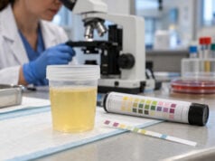

A complete urinalysis examines urine in three connected ways: physical appearance, chemical dipstick testing, and microscopic analysis of the sediment. Together, these measurements can provide quick clues about hydration, urinary tract infection, kidney disease, diabetes, bleeding, stones, liver or bile duct problems, and other conditions. The test is common because it is inexpensive, fast, and information-rich, but no single finding is diagnostic by itself. Color and clarity can be influenced by foods, medicines, concentration, and contamination. Dipstick pads are screening tools that can produce false-positive or false-negative results. Microscopy can confirm cells and reveal casts, crystals, bacteria, yeast, and other particles that the strip cannot identify directly. The most useful interpretation comes from the pattern of findings plus symptoms, collection quality, blood tests, and whether abnormalities persist when the urine is retested.
- A complete urinalysis has three parts: physical examination, chemical dipstick testing, and microscopic examination of cells and sediment.
- Normal results are usually negative for protein, glucose, ketones, bilirubin, nitrite, and significant blood or leukocyte esterase, but laboratory reference ranges vary.
- Microscopy adds detail: it can identify red and white blood cells, epithelial cells, casts, crystals, bacteria, yeast, and other formed elements.
- Collection quality matters: a clean-catch midstream sample and prompt laboratory analysis reduce contamination and deterioration.
- Abnormal findings often need confirmation: protein may need uACR, blood may need repeat microscopy, and suspected UTI may need urine culture depending on symptoms and risk.
Table of Contents
- What a Complete Urinalysis Includes
- Physical Urine Findings
- Chemical Dipstick Findings
- Microscopic Urine Findings
- Normal and Abnormal Result Patterns
- How Laboratories Report Urinalysis Results
- Collection and Testing Quality
- What Happens After the Test
What a Complete Urinalysis Includes
A complete urinalysis generally combines visual or instrument-based physical assessment, reagent-strip chemistry, and microscopic particle analysis. It is broader than a simple dipstick. The exact panel differs by laboratory, and some labs perform microscopy only when the dipstick or clinical request meets certain criteria.
The three parts answer different questions:
- Physical examination describes concentration and appearance, usually including color, clarity, and specific gravity. Some laboratories also report odor or other descriptive features, although odor is not a reliable stand-alone diagnostic marker.
- Chemical testing uses reagent pads to screen for pH, protein, blood, glucose, ketones, bilirubin, urobilinogen, nitrite, and leukocyte esterase. Specific gravity may also be measured by strip or other instrumentation.
- Microscopy looks for red blood cells, white blood cells, epithelial cells, casts, crystals, bacteria, yeast, sperm, mucus, and less common particles.
A related urinalysis test with dipstick and microscopy is often ordered for urinary symptoms, kidney evaluation, diabetes monitoring, pregnancy-related assessment, preoperative testing, or investigation of unexplained abnormal blood work. Routine screening in people without a clinical reason is less useful because minor abnormalities are common and can lead to unnecessary follow-up.
A complete urinalysis is best viewed as a pattern-recognition test. Protein plus red blood cell casts tells a different story from protein alone; leukocyte esterase plus nitrite plus dysuria has a different meaning from leukocytes in an asymptomatic contaminated sample.
Physical Urine Findings
Physical examination is the first layer of urinalysis. It gives immediate information about concentration and visible characteristics, but many changes are nonspecific.
Color
Normal urine ranges from nearly colorless to deep yellow depending mainly on concentration. Dark yellow urine often reflects reduced fluid intake. Red or pink urine can result from blood, but also from foods such as beets, medicines, or pigments. Brown or tea-colored urine may occur with concentrated urine, bilirubin, hemoglobin, myoglobin, or glomerular bleeding. Orange urine can appear with some medicines or concentrated bilirubin-containing urine.
Color alone cannot establish the cause. A urine color finding is most useful when paired with the dipstick and microscopy.
Clarity
Fresh urine is often clear, but slight cloudiness can be benign. Turbidity may come from white blood cells, red blood cells, epithelial cells, bacteria, yeast, crystals, mucus, semen, or contamination. Refrigerated urine can become cloudy because crystals precipitate as the sample cools.
A cloudy specimen therefore does not automatically mean infection. Microscopy and symptoms are needed to distinguish infection from crystalluria or contamination.
Specific gravity
Specific gravity estimates urine concentration relative to water. A common reference interval is roughly 1.005 to 1.030, but ranges vary by laboratory and method. Very dilute urine can occur after high fluid intake, while concentrated urine may occur with dehydration. Glucose, protein, contrast material, and some other solutes can influence measurement.
Persistent inability to concentrate or dilute urine appropriately may point to kidney concentrating problems or endocrine disorders, but interpretation requires serum sodium, fluid status, kidney function, and often urine osmolality. For a focused explanation, the urine specific gravity test is more informative than reading a single number in isolation.
Chemical Dipstick Findings
Dipstick chemistry uses multiple reagent pads that change color after contact with urine. The strip is read at specified times, often by an automated reader. Results are usually qualitative or semiquantitative rather than exact concentrations.
| Dipstick marker | Typical normal finding | What an abnormal result may suggest |
|---|---|---|
| pH | Often about 4.5–8.0 | Diet, metabolic state, stone risk, or some bacterial infections |
| Protein | Negative or trace | Transient proteinuria or kidney disease; dipsticks mainly detect albumin |
| Blood | Negative | Red blood cells, hemoglobin, or myoglobin |
| Glucose | Negative | Hyperglycemia, SGLT2 inhibitor effect, or renal glucosuria |
| Ketones | Negative | Fasting, vomiting, low-carbohydrate intake, illness, or diabetic ketoacidosis |
| Bilirubin | Negative | Conjugated bilirubin from hepatobiliary disease or obstruction |
| Urobilinogen | Low amount may be present | Altered bilirubin metabolism, liver disease, or hemolysis |
| Nitrite | Negative | Nitrate-reducing bacteria; a negative result does not rule out UTI |
| Leukocyte esterase | Negative | White blood cells from infection or inflammation |
Protein: A positive protein pad is most sensitive to albumin. Persistent protein should usually be quantified rather than followed by dipstick alone. The urine protein dipstick result can be affected by urine concentration and pH.
Blood: The reagent detects heme activity, so a positive result can occur with intact red cells, free hemoglobin, or myoglobin. Microscopy helps determine whether red cells are actually present.
Glucose and ketones: Urine glucose can reflect elevated blood glucose but does not replace blood testing. Ketones can appear during fasting or illness, and moderate to large ketones in a person with diabetes may require urgent evaluation for ketoacidosis.
Nitrite and leukocyte esterase: These are common UTI screening markers. Nitrite is relatively specific when positive but can be falsely negative when urine has not remained in the bladder long enough or when the organism does not reduce nitrate. Leukocyte esterase is sensitive to white blood cells but can also be positive in noninfectious inflammation.
Microscopic Urine Findings
Microscopy adds information that chemical testing cannot provide. Traditional analysis uses centrifuged urine sediment viewed under a microscope, although many laboratories now use automated digital microscopy or flow-based particle analyzers with manual review when needed.
Red and white blood cells
Red blood cells support true hematuria when the dipstick is positive for blood. Their shape can matter: markedly dysmorphic red cells may support a glomerular source. White blood cells suggest pyuria and are common with UTI, but they can also occur with stones, interstitial nephritis, sexually transmitted infections, and other inflammation.
Epithelial cells
Squamous epithelial cells often come from the skin or external genital tract and can indicate sample contamination when numerous. Transitional epithelial cells may originate from the bladder or urinary tract. Renal tubular epithelial cells are less common and, when increased in the right clinical setting, may indicate tubular injury.
Casts
Casts form inside renal tubules and therefore can localize a process to the kidney. A few hyaline casts may occur after exercise or dehydration. Red blood cell casts strongly suggest glomerular inflammation or bleeding; white blood cell casts may occur with pyelonephritis or interstitial nephritis; granular and epithelial casts can accompany tubular injury; fatty casts may occur with heavy proteinuria.
The urine microscopy test is especially valuable when kidney disease is suspected because some important casts are not reliably characterized by dipstick alone.
Crystals and microorganisms
Crystals can form from normal urinary solutes and may be harmless, particularly after cooling. Their meaning depends on type, quantity, urine pH, medication exposure, and symptoms. Calcium oxalate, uric acid, struvite, cystine, and drug-related crystals have different clinical implications.
Bacteria or yeast may reflect infection, colonization, or contamination. The number of organisms on microscopy does not replace a properly collected urine culture when culture is clinically indicated.
Normal and Abnormal Result Patterns
A “normal urinalysis” is not a single universal template. Laboratories use different instruments and reporting thresholds, and small numbers of cells or casts may fall within a normal reference interval. Still, several broad patterns are useful.
A largely normal sample is typically clear to slightly hazy, has a concentration appropriate to hydration, and is negative for significant protein, blood, glucose, ketones, bilirubin, nitrite, and leukocyte esterase. Microscopy generally shows few cells and no significant pathologic casts or heavy microbial burden.
Abnormal combinations can be more informative than isolated values:
- Protein plus dysmorphic red cells or red blood cell casts raises concern for glomerular kidney disease.
- Leukocyte esterase, pyuria, nitrite, and urinary symptoms make bacterial UTI more likely.
- Blood-positive dipstick with few red cells can suggest hemoglobinuria or myoglobinuria.
- Glucose plus ketones in a symptomatic person with diabetes can signal major metabolic decompensation.
- Very high specific gravity plus glucose may occur with marked glucosuria.
- Alkaline urine plus struvite crystals and infection findings can support infection with a urease-producing organism.
A useful principle is that discordant results deserve attention. For example, a positive dipstick blood result with no microscopic red cells is not simply “wrong”; it may be pointing to a different heme pigment. Similarly, a negative nitrite result does not cancel pyuria and classic UTI symptoms.
How Laboratories Report Urinalysis Results
Urinalysis reports mix several measurement styles, which can make them look more complicated than they are. Understanding the format helps prevent overinterpreting small differences.
Dipstick chemistry is usually categorical. A laboratory may report “negative,” “trace,” “small,” “moderate,” or “large,” or use grades such as 1+ through 4+. These grades are tied to the particular strip manufacturer and should not be treated as perfectly interchangeable across laboratories. For example, a 1+ protein result often corresponds to roughly 30 mg/dL of protein, but the actual threshold and color blocks vary by system. The result is also a concentration, so the same amount of protein excretion can appear stronger in concentrated urine and weaker in dilute urine.
Microscopy may use counts or categories. Red and white blood cells may be reported as cells per high-power field, cells per microliter, or ranges such as 0–2, 3–5, or 6–10 per field. Casts can be reported per low-power field, while bacteria and crystals may be described as rare, few, moderate, or many. Automated analyzers may report numerical particle counts that do not map perfectly to traditional manual microscopy categories.
Reference intervals are method-specific. A value flagged “H” or “abnormal” by one laboratory may fall near the boundary used by another. The laboratory’s own reference range therefore takes priority over a generic range from the internet. The same principle applies to pH and specific gravity, where normal variation is broad and strongly influenced by hydration, diet, and timing.
Reflex testing can change what appears on the report. Some laboratories automatically perform microscopy when dipstick blood, protein, leukocyte esterase, or other markers cross a threshold. Others perform microscopy only when specifically ordered. A report that lacks microscopic data does not necessarily mean the sediment was normal; it may mean microscopy was not triggered or requested.
When comparing results over time, use the same laboratory when practical and focus on meaningful trends rather than tiny shifts between categories. Persistent changes, a worsening pattern, or a combination of abnormalities usually matters more than a one-step change such as trace to 1+ on a single specimen.
For home or point-of-care strips, the same caution applies even more strongly. Lighting, timing, storage conditions, and visual color matching can affect interpretation. A home strip can be useful for a specific monitoring plan, but unexpected or clinically important results should be confirmed with a properly collected laboratory specimen rather than managed from the strip alone.
Collection and Testing Quality
Urine changes after it leaves the body, so preanalytical quality is a major part of urinalysis accuracy. Bacteria can multiply, cells can break down, casts can deteriorate, pH can shift, and light-sensitive compounds can degrade.
For many outpatient tests, a midstream clean-catch sample is appropriate. The usual sequence is to clean the collection area as instructed, begin urinating into the toilet, collect the midstream portion in a sterile container, and finish voiding into the toilet. The exact technique depends on the patient and test.
Fresh urine is preferred. Laboratories generally analyze specimens promptly or preserve/refrigerate them according to validated procedures. A sample that sits unpreserved at room temperature for several hours may no longer represent the original urine accurately.
Other common sources of misleading results include:
- menstrual or vaginal contamination;
- recent vigorous exercise;
- dehydration or unusually high fluid intake;
- medications that discolor urine or alter chemistry;
- vitamin C interference with some reagent pads;
- disinfectant or cleaning-agent contamination;
- reading a manual strip at the wrong time.
Modern automated analyzers improve consistency and throughput, but they do not eliminate the need for judgment. Recent studies show good performance for common elements such as red and white blood cells, while certain casts, renal tubular cells, dysmorphic red cells, crystals, and unusual particles may still require expert manual review. This is why laboratories often use reflex rules that trigger microscopic confirmation for selected abnormalities.
What Happens After the Test
Follow-up depends on the reason the test was ordered and which findings are abnormal. Many minor abnormalities disappear when the test is repeated under better collection conditions or after an acute illness resolves.
Typical next steps include:
- Persistent protein: quantify albumin with uACR or total protein with a protein-to-creatinine ratio, then assess serum creatinine and eGFR.
- Confirmed hematuria: repeat testing and risk-based evaluation for urinary, kidney, or urologic causes.
- Pyuria, nitrite, or bacteria: interpret with urinary symptoms and obtain culture when severity, recurrence, pregnancy, treatment failure, or complicated infection makes culture important.
- Glucose: check blood glucose and A1C unless glucosuria is expected from an SGLT2 inhibitor.
- Ketones: consider blood beta-hydroxybutyrate and urgent metabolic evaluation when diabetes or concerning symptoms are present.
- Casts or renal tubular cells: assess kidney function and the clinical setting; nephrology input may be appropriate when an intrinsic kidney process is suspected.
Seek prompt medical care when abnormal urine findings occur with high fever and flank pain, inability to urinate, visible blood with clots, severe muscle pain with dark urine, persistent vomiting, confusion, rapid or deep breathing, pregnancy with significant urinary symptoms, or signs of dehydration or serious illness.
The main value of a complete urinalysis is not that it gives one final answer. It efficiently narrows the possibilities, shows which findings need confirmation, and helps determine whether the next test should focus on infection, kidney function, metabolic disease, bleeding, stones, or another cause.
References
- The EFLM European Urinalysis Guideline 2023 2024 (Guideline)
- Urinalysis: Interpretation and Clinical Correlations 2023 (Review)
- Office-Based Urinalysis: A Comprehensive Review 2022 (Review)
- Urinary Biomarkers and Point-of-Care Urinalysis Devices for Early Diagnosis and Management of Disease: A Review 2023 (Review)
- The Clinical Impact of Urinalysis Screened by Automated Microscopy Compared to Reference Manual Analysis 2025
Disclaimer
A urinalysis is a screening and diagnostic aid, not a diagnosis by itself. Results should be interpreted with symptoms, medical history, medications, collection quality, and any needed confirmatory tests. Seek urgent care for severe symptoms such as high fever with flank pain, heavy visible bleeding, breathing changes, confusion, or persistent vomiting.