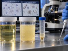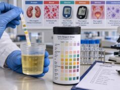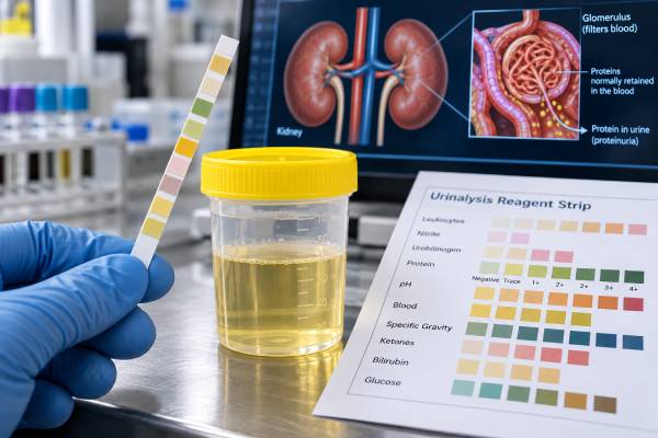
A urine protein dipstick test is a quick screening test for protein in urine, especially albumin. A positive result can be a sign of kidney damage, but one abnormal dipstick does not automatically mean chronic kidney disease. Temporary proteinuria can occur after strenuous exercise, fever, dehydration, acute illness, or prolonged standing. Concentrated or alkaline urine can also make the dipstick read higher than the true protein loss. When protein is detected, the usual next step is to confirm and quantify it with a laboratory urine albumin-to-creatinine ratio (uACR) or protein-to-creatinine ratio (UPCR), often using a first-morning sample. Persistent albuminuria is important because it can signal kidney disease even when blood creatinine and eGFR are still normal. The dipstick also has limits: it is much more sensitive to albumin than to some other proteins, so a negative result can miss certain forms of proteinuria.
- A normal urine protein dipstick is usually negative or sometimes trace; 1+ or greater generally deserves follow-up when it persists.
- Typical dipstick estimates are about 30 mg/dL for 1+, 100 mg/dL for 2+, 300 mg/dL for 3+, and 1,000 mg/dL for 4+, but brands vary and the result is only semiquantitative.
- A positive dipstick should usually be confirmed with a urine albumin-to-creatinine ratio or protein-to-creatinine ratio rather than judged from the color grade alone.
- Temporary proteinuria can follow exercise, fever, dehydration, or acute illness, so repeat testing after the trigger resolves is often important.
- Swelling, shortness of breath, very high blood pressure, blood in the urine, or reduced urine output with proteinuria needs prompt medical evaluation.
Table of Contents
- What the Protein Dipstick Measures
- What a Positive Protein Result Means
- Protein Dipstick Levels and Their Limits
- Common Causes of Proteinuria
- False-Positive and False-Negative Results
- Follow-Up Tests for Proteinuria
- When Proteinuria Needs Medical Attention
What the Protein Dipstick Measures
The protein pad on a routine urine dipstick changes color when protein is present. Standard dipsticks are designed to react most strongly with albumin, the main blood protein that leaks through the kidney filters in many glomerular diseases.
A healthy glomerulus keeps most albumin in the bloodstream. Small amounts may still appear in urine, and routine dipsticks are not sensitive enough to measure low-level albuminuria accurately. This is why a person can have an important increase in urine albumin while the standard protein dipstick remains negative.
Most dipsticks report protein as:
- negative;
- trace;
- 1+;
- 2+;
- 3+;
- 4+.
These categories estimate concentration in the urine sample, not the total amount lost over a day. A concentrated sample can therefore produce a stronger dipstick result than a dilute sample even if the person’s daily protein loss is unchanged.
The dipstick also does not detect all proteins equally. Albumin produces the strongest reaction. Low-molecular-weight proteins, free light chains, and some tubular proteins may be missed or underestimated. If a clinician suspects nonalbumin proteinuria, a negative routine dipstick does not end the evaluation.
A more sensitive urine albumin dipstick or, preferably, a quantitative uACR can detect smaller amounts of albumin that matter in diabetes, hypertension, and early chronic kidney disease.
What a Positive Protein Result Means
A positive urine protein dipstick means the sample contains enough protein—usually albumin—to trigger the reagent pad. It is a screening signal, not a diagnosis.
The first distinction is whether the proteinuria is temporary or persistent. Transient proteinuria can occur during conditions that briefly change kidney blood flow or glomerular permeability. If the dipstick normalizes after the trigger resolves, chronic kidney disease becomes less likely.
Persistent proteinuria is more concerning. It can reflect damage to the glomeruli, the kidney’s microscopic filtering units, or less commonly a tubular or overflow process. Repeated albuminuria can also indicate increased cardiovascular and kidney risk even before eGFR declines.
A positive result matters more when it appears with other findings such as:
- elevated blood pressure;
- reduced eGFR or rising creatinine;
- blood in urine;
- red blood cell casts or other abnormal urine sediment;
- swelling around the eyes, legs, or ankles;
- diabetes or long-standing hypertension;
- systemic autoimmune disease;
- a family history of kidney disease.
Protein can also appear during UTI, fever, or other acute illness. In those situations, repeating the test after recovery can prevent overdiagnosis of chronic proteinuria.
A single abnormal result does not establish CKD. Chronic kidney disease generally requires evidence of kidney damage or reduced filtration persisting for at least 3 months, unless chronicity is already clear from prior tests, imaging, pathology, or another established condition.
The amount and pattern of protein matter as well. Mild albuminuria can be an early marker of vascular and glomerular injury, while very heavy protein loss may point toward nephrotic syndrome. Proteinuria accompanied by blood, red blood cell casts, or a rapid decline in kidney function raises a different concern, including inflammatory glomerular disease. Dipstick protein alone cannot separate these patterns, which is why urine microscopy and blood tests often accompany quantitative protein measurements.
Pregnancy is another setting where context is essential. Proteinuria may predate pregnancy because of chronic kidney disease, appear transiently with infection, or develop with hypertensive disorders such as preeclampsia. A dipstick can be used as a screen when quantitative testing is not immediately available, but diagnosis and management should rely on blood pressure, symptoms, gestational timing, and quantitative urine protein assessment rather than the dipstick grade alone.
Protein Dipstick Levels and Their Limits
Dipstick protein grades provide a rough estimate of concentration. A commonly used approximation is shown below, but exact cutoffs vary by manufacturer.
| Dipstick result | Approximate protein concentration | Typical next step |
|---|---|---|
| Negative | Below routine dipstick detection | Consider uACR if early albuminuria screening is indicated |
| Trace | About 5–10 mg/dL | Repeat if unexpected or risk factors are present |
| 1+ | About 30 mg/dL | Confirm quantitatively if persistent |
| 2+ | About 100 mg/dL | Quantify with uACR or UPCR and assess kidney function |
| 3+ | About 300 mg/dL | Prompt quantitative evaluation is usually appropriate |
| 4+ | About 1,000 mg/dL | Heavy proteinuria is possible; evaluate without delay |
These values are not equivalent to grams of protein lost per day. A dipstick measures concentration at one moment. Daily protein excretion depends on both concentration and urine volume.
This concentration effect is easy to underestimate. For example, a person who is dehydrated may produce a small volume of concentrated urine and show 1+ protein, while the same person after normal fluid intake might test trace or negative. That does not necessarily mean kidney protein loss changed dramatically within hours. Ratios such as uACR and UPCR divide the protein or albumin concentration by urine creatinine, which helps correct for how concentrated or dilute the sample is.
Even ratios have biological variability. Exercise, fever, uncontrolled blood glucose, menstruation, urinary infection, and recent vigorous activity can raise albumin excretion temporarily. For borderline abnormalities, repeating a first-morning sample under stable conditions often provides a better estimate of the person’s usual level.
The grade also does not reliably distinguish moderate from nephrotic-range proteinuria. Nephrotic-range protein loss is generally defined as about 3.5 g/day or more of total protein in adults, or an equivalent spot protein-to-creatinine ratio. A 3+ or 4+ dipstick can raise concern for heavy proteinuria, but only quantitative testing can determine the amount.
Likewise, a negative routine dipstick does not exclude clinically important albuminuria below its detection threshold. KDIGO recommends quantitative urine albumin measurement for people at risk for CKD because albumin-to-creatinine ratio is more sensitive and adjusts for urine concentration.
Common Causes of Proteinuria
Proteinuria can be grouped by mechanism: transient, glomerular, tubular, or overflow.
Transient or functional proteinuria
Temporary protein leakage can occur with:
- fever;
- strenuous exercise;
- dehydration;
- acute illness;
- emotional or physical stress;
- seizures;
- prolonged standing in some younger people, called orthostatic proteinuria.
Orthostatic proteinuria usually disappears in a first-morning urine sample after lying down overnight. It is most often seen in adolescents and young adults and is generally benign after appropriate evaluation.
A typical evaluation compares a first-morning sample with a daytime sample. If protein is absent after overnight recumbency but appears later while upright, orthostatic proteinuria becomes more likely. Persistent first-morning proteinuria is less consistent with this benign pattern and deserves further assessment. Because orthostatic proteinuria becomes uncommon with increasing age, new proteinuria in an older adult should not be assumed to have this explanation.
Glomerular proteinuria
Glomerular disease is the major cause of significant albuminuria. Conditions include diabetic kidney disease, hypertensive kidney damage, glomerulonephritis, IgA nephropathy, lupus nephritis, focal segmental glomerulosclerosis, membranous nephropathy, and other diseases affecting the filtration barrier.
Diabetes is one of the most common settings in which albuminuria is monitored. A diabetic kidney disease urine panel may include uACR along with serum creatinine and eGFR rather than relying on a standard protein dipstick.
Tubular proteinuria
Tubular disorders can cause loss of smaller proteins that are normally filtered and then reabsorbed by kidney tubules. Examples include some forms of tubulointerstitial kidney disease, toxic injury, or inherited tubular disorders. Routine dipsticks may underestimate this type because they preferentially detect albumin.
Overflow proteinuria
Overflow occurs when the blood contains so much of a small protein that the filtered load exceeds tubular reabsorption. Monoclonal free light chains in plasma cell disorders are a classic example. A routine albumin-sensitive dipstick may be weakly positive or negative even when clinically important light-chain proteinuria is present.
This mismatch is one reason a clinician may order total protein measurement, serum and urine electrophoresis, or free light-chain testing when the clinical picture suggests a plasma cell disorder.
False-Positive and False-Negative Results
Dipstick protein testing is convenient but vulnerable to sample conditions.
False-positive or exaggerated results
Protein can appear higher than it truly is with:
- very concentrated urine from dehydration;
- strongly alkaline urine;
- some disinfectants or quaternary ammonium compounds contaminating the sample;
- phenazopyridine or other strongly colored substances that interfere with visual reading;
- blood, pus, or genital contamination in some situations.
A positive result in a highly concentrated specimen may shrink or disappear on a repeat first-morning or properly hydrated sample. Specific gravity can help show how concentrated the urine was at the time of testing.
False-negative or underestimated results
Protein may be missed when:
- the urine is very dilute;
- urine is strongly acidic;
- the protein is mainly nonalbumin, such as light chains;
- the amount of albumin is above normal but still below the routine dipstick detection threshold.
These limits explain why CKD screening increasingly relies on the urine albumin-to-creatinine ratio. The ratio corrects for urine concentration and detects lower albumin levels than a standard protein dipstick.
If a home or office dipstick is unexpectedly positive, repeating the result under controlled conditions is often reasonable. Persistent or high-grade protein should not be followed only with more dipsticks; it should be quantified. A quantitative baseline also makes future monitoring more meaningful because a clinician can compare actual ratios over time rather than subjective color grades from different urine concentrations.
Follow-Up Tests for Proteinuria
The most useful follow-up test depends on whether the concern is mainly albumin or total protein.
Urine albumin-to-creatinine ratio
For most adults being evaluated for CKD, uACR is the preferred initial quantitative test. It is usually reported in mg/g or mg/mmol. KDIGO albuminuria categories are commonly defined as:
- A1: less than 30 mg/g, or less than 3 mg/mmol;
- A2: 30–300 mg/g, or 3–30 mg/mmol;
- A3: greater than 300 mg/g, or greater than 30 mg/mmol.
An ACR of 30 mg/g or higher on a random sample is usually confirmed with a first-morning specimen because exercise, infection, menstruation, hyperglycemia, and other temporary factors can increase albumin excretion.
A spot urine albumin test is particularly useful for detecting early glomerular injury.
Urine protein-to-creatinine ratio
UPCR measures total urine protein relative to creatinine and can be useful when heavier proteinuria or nonalbumin protein is suspected. A urine protein-to-creatinine ratio provides an estimate of daily protein loss without requiring a full 24-hour collection in many cases.
24-hour urine protein
A timed collection can provide a direct total protein excretion estimate but is more cumbersome and prone to collection errors. It may be used when precise quantification is important, when spot ratios appear unreliable, or in selected clinical situations. A 24-hour urine protein test also allows evaluation of collection adequacy when paired with urine creatinine.
Blood tests typically include creatinine and eGFR. Depending on the pattern, clinicians may also order serum albumin, electrolytes, glucose or A1C, autoimmune tests, complement levels, hepatitis testing, electrophoresis, kidney ultrasound, or nephrology evaluation. Referral becomes more likely when proteinuria is heavy, kidney function is reduced, blood and protein appear together without a clear benign cause, or the diagnosis remains uncertain. In selected cases, kidney biopsy is needed to identify the exact glomerular disease and guide treatment.
When Proteinuria Needs Medical Attention
An isolated trace result after exercise or illness may simply need repeat testing. Persistent 1+ or greater protein, repeated elevated uACR, or a strong dipstick result without an obvious temporary trigger deserves a more complete evaluation.
Prompt medical assessment is especially important when proteinuria occurs with:
- swelling of the legs, face, or around the eyes;
- shortness of breath or rapid weight gain from fluid retention;
- visible blood in the urine;
- markedly reduced urine output;
- severe or newly uncontrolled high blood pressure;
- a rapid rise in creatinine or fall in eGFR;
- pregnancy with high blood pressure or concerning symptoms;
- systemic symptoms suggesting autoimmune or inflammatory disease.
Heavy proteinuria can lower blood albumin and cause edema, while inflammatory kidney diseases can progress quickly if they also produce hematuria, casts, hypertension, or acute loss of kidney function.
Foamy urine is a common reason people become concerned about protein. Persistent foam can occur with proteinuria, but bubbles are not diagnostic: forceful urination, concentrated urine, cleaning products in the toilet, and other factors can also create foam. If the change persists, a urinalysis and quantitative albumin or protein measurement are more useful than judging appearance alone.
People with diabetes, hypertension, cardiovascular disease, or a family history of kidney disease may need periodic quantitative albumin testing even when a routine protein dipstick is negative. Early albuminuria can precede a decline in filtration and can affect treatment decisions.
The key next step after a positive dipstick is not to guess the diagnosis from the number of plus signs. Confirm whether protein loss is real, quantify it, determine whether albumin is the main protein, and assess whether the finding persists. That sequence separates common temporary proteinuria from kidney disease that needs ongoing monitoring or treatment, and it reduces the chance of both unnecessary alarm and missed clinically important kidney injury altogether.
References
- Diagnostic accuracy of urine dipstick testing for albumin-to-creatinine ratio and albuminuria: A systematic review and meta-analysis 2021 (Systematic Review)
- Office-Based Urinalysis: A Comprehensive Review 2022 (Review)
- Measuring albuminuria or proteinuria: does one answer fit all? 2023 (Review)
- KDIGO 2024 Clinical Practice Guideline for the Evaluation and Management of Chronic Kidney Disease 2024 (Guideline)
- Executive summary of the KDIGO 2024 Clinical Practice Guideline for the Evaluation and Management of Chronic Kidney Disease: known knowns and known unknowns 2024 (Guideline)
Disclaimer
This article is for general educational purposes and does not diagnose kidney disease. A positive urine protein dipstick should be interpreted with urine concentration, repeat testing, uACR or UPCR, kidney function, and the clinical context. Seek prompt medical care for proteinuria with swelling, shortness of breath, visible blood in urine, very high blood pressure, or reduced urine output.