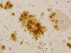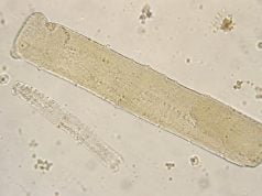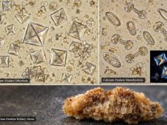

A urine microscopy test examines the solid particles in urine after a sample is collected and prepared for microscopic analysis. It can show red and white blood cells, epithelial cells, casts, crystals, bacteria, yeast, and other sediment that a chemical dipstick cannot identify directly. The test is commonly used when a routine urinalysis is abnormal, when urinary infection is suspected, or when clinicians are evaluating blood in the urine, kidney injury, stones, proteinuria, or unexplained urinary symptoms.
Microscopy is most useful as part of a complete urinalysis rather than as a stand-alone result. The meaning of “5–10 white blood cells,” “few bacteria,” “hyaline casts,” or “calcium oxalate crystals” changes with symptoms, sample quality, urine concentration, pH, kidney function, and culture results. Some findings are common and harmless in small amounts; others, such as red blood cell casts or abundant renal tubular epithelial cells with granular casts, can point toward important kidney disease. Accurate collection and prompt processing make the results more reliable.
- Urine microscopy identifies formed elements that dipsticks cannot fully characterize, including cells, casts, crystals, bacteria, and yeast.
- Small numbers of some particles can be normal, especially squamous epithelial cells, hyaline casts, or common crystals; the pattern and clinical context determine significance.
- White blood cells and bacteria can support a urinary infection, but culture and symptoms are often needed to confirm it.
- Red blood cell casts, white blood cell casts, renal tubular epithelial cells, and “muddy brown” granular casts are important kidney-localizing clues.
- A fresh, clean-catch specimen reduces misleading results from contamination, cell breakdown, bacterial growth, or crystals that form after collection.
Table of Contents
- What a Urine Microscopy Test Is
- Cells in Urine: Red, White, and Epithelial Cells
- Casts in Urine and What They Suggest
- Crystals in Urine
- Bacteria, Yeast, and Other Sediment Findings
- Collection, Preparation, and Result Quality
- How to Interpret Results and Decide on Follow-Up
What a Urine Microscopy Test Is
Urinalysis has two major parts: chemical testing and particle analysis. A dipstick checks properties such as pH, specific gravity, protein, blood, glucose, ketones, nitrite, and leukocyte esterase. Microscopy looks at the actual particles suspended in the urine.
In traditional microscopy, a laboratory usually centrifuges urine so formed elements settle, removes most of the liquid, and examines the concentrated sediment under a microscope. Automated analyzers may instead use digital imaging, flow cytometry, or other particle-counting technology. Many laboratories combine automation with manual review when unusual or clinically important particles are flagged.
Microscopy is ordered or triggered for many reasons, including:
- blood or protein detected on dipstick;
- urinary burning, urgency, frequency, or fever;
- flank pain or suspected kidney stones;
- rising serum creatinine or acute kidney injury;
- known chronic kidney disease;
- suspected glomerulonephritis or interstitial nephritis;
- recurrent urinary tract infections; or
- an abnormal automated urinalysis that needs confirmation.
Results may be reported as a count per high-power field (HPF), a count per low-power field (LPF), cells per microliter, or semiquantitative terms such as rare, few, moderate, or many. There is no single universal reporting format. Reference intervals depend on the laboratory method, so the range printed beside the result should be used instead of applying a number from another lab.
Microscopy is a pattern test. A single abnormality often has several possible causes. For example, red blood cells can come from a stone, bladder infection, prostate, menstruation, exercise, or kidney disease. When red cells are dysmorphic and appear inside RBC casts, the evidence shifts more strongly toward a glomerular source.
Cells in Urine: Red, White, and Epithelial Cells
Red blood cells
A small number of red blood cells may be reported even in people without disease, depending on the laboratory threshold and collection. Persistent microscopic hematuria deserves evaluation because causes range from benign transient factors to stones, infection, glomerular disease, and urinary tract tumors.
Microscopy can sometimes provide a clue to origin. Relatively uniform RBCs are often called isomorphic. Misshapen RBCs, especially acanthocytes with irregular membrane blebs, suggest that cells have passed through a damaged glomerular filtration barrier. RBC morphology is most useful when performed by experienced observers and combined with proteinuria and casts.
White blood cells
White blood cells in urine are called pyuria. Many laboratories consider roughly 5 or more WBCs per HPF abnormal in a centrifuged sample, although thresholds vary by method. Pyuria often occurs with bacterial urinary tract infection, but it is not specific. Stones, sexually transmitted infections, kidney inflammation, tuberculosis, recent antibiotics, and contamination can also raise WBC counts.
Leukocyte esterase on a dipstick is an indirect marker of WBC activity. Microscopy can confirm that actual leukocytes are present and estimate their quantity. When pyuria accompanies bacteria and compatible urinary symptoms, infection becomes more likely. When pyuria occurs without a positive routine culture, clinicians may consider nonbacterial causes or “sterile pyuria.”
Epithelial cells
Epithelial cells come from different parts of the urinary and genital tracts, and the type matters.
- Squamous epithelial cells usually come from skin, vulva, vagina, or distal urethra. Many squamous cells commonly indicate contamination of a clean-catch sample.
- Transitional or urothelial cells line the renal pelvis, ureters, bladder, and part of the urethra. Small numbers may be seen normally or after instrumentation.
- Renal tubular epithelial cells originate in the kidney tubules. Increased numbers, especially with epithelial casts or granular casts, can support acute tubular injury.
Cell identification is not always easy. Degenerating cells can change shape, and automated analyzers may classify ambiguous cells differently from manual microscopy.
Casts in Urine and What They Suggest
Casts are cylindrical structures formed within kidney tubules, mainly from a protein matrix called uromodulin. Because they are molded inside the nephron, casts can provide more kidney-specific information than free cells floating in urine.
| Cast | What it may mean |
|---|---|
| Hyaline cast | Can be normal in small numbers; also seen with dehydration, exercise, fever, diuretic use, or kidney disease |
| Red blood cell cast | Strongly suggests glomerular bleeding or glomerulonephritis |
| White blood cell cast | Suggests inflammation within the kidney, such as pyelonephritis or acute interstitial nephritis |
| Renal tubular epithelial cell cast | Supports tubular epithelial injury |
| Granular cast | Can occur with kidney disease; coarse muddy-brown casts are classic for acute tubular injury |
| Fatty cast | Often associated with heavy proteinuria and nephrotic syndrome |
| Waxy or broad cast | Often associated with significant or advanced kidney dysfunction and slow tubular flow |
Hyaline casts are the main example of why “cast present” does not automatically mean kidney disease. A few may appear after strenuous exercise or when urine is concentrated. Cellular casts are more significant because their trapped cells reveal where inflammation or injury occurred.
Casts can disappear or become difficult to identify when a sample sits too long, especially in dilute or alkaline urine. Their absence therefore does not rule out a disease that can produce them. A patient with clear symptoms of pyelonephritis, for example, can have no WBC casts on a particular specimen.
Crystals in Urine
Crystals form when dissolved substances become concentrated enough to precipitate. Some form inside the urinary tract; others appear after urine cools or sits in the collection container. The type of crystal, urine pH, and clinical setting determine whether the finding matters.
Common crystals include:
- Calcium oxalate: often envelope- or dumbbell-shaped; can occur in healthy urine and in calcium oxalate stone formers.
- Uric acid: yellow to reddish-brown crystals with variable rhomboid, rosette, or barrel shapes; favor acidic urine.
- Amorphous urates: fine granules that occur in acidic urine and may be prominent after cooling.
- Calcium phosphate or amorphous phosphate: favor alkaline urine.
- Triple phosphate/struvite: rectangular “coffin-lid” crystals, often associated with alkaline urine and urease-producing bacterial infection when clinically relevant.
Less common crystals can be more specific. Hexagonal cystine crystals suggest cystinuria. Cholesterol crystals and lipid findings can occur in heavy proteinuria. Medication crystals may appear when certain drugs reach high urinary concentrations, especially with dehydration or unfavorable urine pH.
Crystals alone do not prove a kidney stone. A person can have crystalluria without ever forming a stone, and a person with a stone may have no crystals in the sample. If symptoms suggest obstruction—severe flank pain, nausea, blood in urine, or reduced urine flow—imaging is often more informative than the crystal count.
Bacteria, Yeast, and Other Sediment Findings
Bacteria on microscopy can support a urinary tract infection, but the finding is easy to misread without considering collection quality. A contaminated sample can contain bacteria from skin or genital flora, especially when many squamous epithelial cells are also present. Conversely, a true UTI may have few visible bacteria if the urine is very dilute, antibiotics have already been started, or the bacterial concentration is below the microscopy threshold.
Urine culture remains the main test for identifying a bacterial pathogen and its antibiotic susceptibility when culture is indicated. Modern studies show that microscopy, dipstick chemistry, and automated particle analysis each have different strengths and limitations; none should be treated as a perfect substitute for culture in situations where microbiologic confirmation is needed.
Yeast most often refers to Candida species. Budding yeast cells may be seen free in the urine, sometimes with pseudohyphae. In a clean-catch specimen, yeast can represent contamination from genital colonization. In hospitalized or catheterized patients, candiduria often represents colonization rather than invasive infection. Diabetes, broad-spectrum antibiotics, urinary catheters, obstruction, and immunosuppression increase the relevance of the finding.
Other microscopic material may include mucus, sperm, lipid droplets, oval fat bodies, and artifacts such as fibers or starch. These can distract from important findings. Polarized light, staining, repeated examination, or expert review can help distinguish true urinary particles from contaminants.
Collection, Preparation, and Result Quality
Pre-analytical handling can change a urine microscopy result substantially. For most routine testing, a midstream clean-catch sample is preferred when the patient can provide one. Cleaning instructions and collecting the middle portion of the urine stream reduce contamination from the external genital area.
The specimen should be delivered to the laboratory promptly. With time, bacteria can multiply, cells can lyse, casts can dissolve, and pH can change. Refrigeration may slow some changes when a delay is unavoidable, but it can also cause urates or phosphates to precipitate. Laboratories use standardized procedures to balance preservation with accurate particle identification.
Very dilute urine can rupture cells and casts. Very alkaline urine also makes some formed elements less stable. Concentrated urine preserves some structures but can increase crystal formation. This is one reason the specific gravity and pH are useful companions to microscopy.
Menstrual blood, vaginal discharge, recent sexual activity, vigorous exercise, and urinary instrumentation can alter results. When contamination is likely and the finding is not urgent, repeating the urinalysis under better collection conditions is often more useful than immediately labeling the result abnormal.
Automated analyzers improve speed and reproducibility, especially for routine cell counts. Manual microscopy remains particularly useful for dysmorphic RBCs, unusual casts, uncommon crystals, and complex sediment. A good laboratory may use both approaches rather than treating one as universally superior.
How to Interpret Results and Decide on Follow-Up
Start with the question that prompted the test. A result that matters greatly in someone with acute kidney injury may be incidental in an otherwise healthy person. Useful interpretation usually combines symptoms, dipstick findings, microscopy, kidney function, and—when infection is suspected—culture.
For example:
- WBCs + bacteria + dysuria or frequency supports a lower UTI, especially if culture confirms a uropathogen.
- WBCs + WBC casts + fever and flank pain raises concern for kidney infection.
- RBCs + significant protein + dysmorphic RBCs or RBC casts suggests glomerular disease.
- Renal tubular epithelial cells + muddy-brown granular casts + rising creatinine supports acute tubular injury.
- Crystals + flank pain + hematuria increases suspicion for a stone, but imaging determines whether obstruction is present.
- Many squamous cells + mixed bacteria without symptoms makes contamination more likely.
A repeat specimen is reasonable when an unexpected mild abnormality could be explained by collection or handling. Persistent microscopic blood, protein, pyuria, or abnormal casts usually requires a more focused evaluation.
Seek medical care promptly for fever with flank pain, severe or unrelenting stone-like pain, visible blood with clots, inability to urinate, markedly reduced urine output, new swelling or shortness of breath, or a rapid rise in creatinine. These features can signal infection, obstruction, or active kidney injury rather than a minor urinalysis variation.
The best way to read a urine microscopy report is not to ask whether every particle is “normal” or “abnormal.” Ask whether the combination of findings fits the clinical problem, whether the sample was reliable, and whether the result changes the next step.
Common interpretation mistakes
Several recurring mistakes make urine microscopy look more definitive than it is. The first is treating every flagged value as disease. A few hyaline casts, occasional squamous cells, or common crystals can appear in normal or temporarily concentrated urine. The second is the opposite: dismissing a rare but highly specific structure because the quantity is small. One convincingly identified RBC cast can carry more diagnostic weight than a large number of nonspecific particles.
Another mistake is assuming that bacteria equal UTI. Bacteria in a poorly collected specimen may reflect contamination, and asymptomatic bacteriuria or catheter colonization does not always require treatment. Symptoms, WBCs, nitrite, collection method, and culture determine whether the bacteriuria represents infection.
Crystal findings are also easy to overread. Refrigerated or old urine can produce crystals after collection. Conversely, a patient with a proven stone may have no crystals at all in the sample. Stone diagnosis therefore depends on symptoms, imaging, and metabolic evaluation rather than microscopy alone.
A fourth mistake is ignoring the rest of the urinalysis. RBC morphology matters more when protein is present; WBC casts matter more with fever, flank pain, or a creatinine rise; tubular cells and granular casts matter more during acute kidney injury. The value comes from the cluster of findings.
Finally, normal microscopy does not rule out important disease. Diabetic kidney disease, vascular CKD, early glomerular disease, or intermittent stone disease can have a bland sediment. Microscopy is strongest when it narrows a differential diagnosis, not when it is asked to exclude every kidney or urinary disorder.
What a repeat urine microscopy test can add
Repeating the test is useful when the first specimen was contaminated, delayed, obtained during menstruation, collected after strenuous exercise, or inconsistent with symptoms. A repeat can show whether hematuria, pyuria, or crystalluria persists and whether cellular casts recur. Serial results can also demonstrate improvement as an acute process resolves.
A repeat test should not delay care for a clearly concerning pattern. RBC casts with rising creatinine, high fever with flank pain, or severe pain with suspected obstruction warrants clinical action even if another specimen is planned. Repetition is a quality and trend tool, not a reason to postpone evaluation of a potentially urgent condition.
A useful report also separates findings that are likely to be transient from findings that deserve follow-up. Concentrated urine, strenuous exercise, fever, menstruation, recent instrumentation, and collection problems can alter the sediment without proving intrinsic kidney disease. By contrast, repeated abnormalities that fit the clinical picture carry more weight. This is why clinicians often interpret microscopy alongside symptoms, kidney function, urine protein or albumin measurements, and culture results rather than relying on a single isolated count. When the sample quality is doubtful or the result conflicts with the rest of the evaluation, a properly collected repeat specimen can clarify whether the finding persists.
References
- Diagnostic Utility of Urine Microscopy in Kidney Diseases 2024 (Review)
- An updated approach to the evaluation of the urinary sediment 2025 (Review)
- The EFLM European Urinalysis Guideline 2023 2024 (Guideline)
- Diagnostic value of different urine tests for urinary tract infection: a systematic review and meta-analysis 2022 (Systematic Review)
- Urinary Vacuolar Casts Are a Unique Type of Casts in Advanced Proteinuric Glomerulopathies 2024
Disclaimer
Urine microscopy results depend on sample quality, laboratory method, symptoms, and the rest of the urinalysis. This article is general educational information and cannot determine the cause of an individual result; urgent symptoms or worsening kidney function need medical assessment.