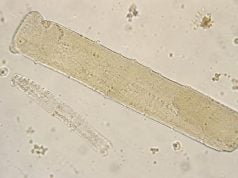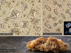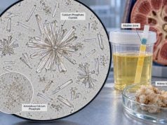
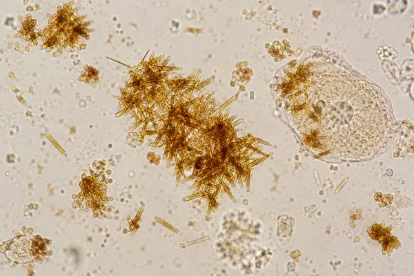
Bilirubin crystals in urine are an uncommon abnormal sediment finding that usually appears when conjugated bilirubin is being excreted in substantial amounts. Under the microscope, they may look like fine yellow, orange, or reddish-brown needles, granules, or clustered deposits, often in acidic urine. Their presence points toward bilirubinuria rather than identifying a specific liver disease. Bilirubin can enter urine when the water-soluble conjugated form builds up in blood, as may occur with hepatocellular injury or impaired bile flow. In practice, the urine bilirubin dipstick and blood liver tests are more important for routine evaluation than the crystal finding itself. Bilirubin crystals should also be distinguished from bile casts in kidney tissue, which are associated with severe hyperbilirubinemia and bile cast nephropathy. A crystal report is therefore best treated as a clue that needs confirmation and clinical context, especially when jaundice, dark urine, abnormal liver tests, or kidney dysfunction is present.
- Bilirubin crystals are abnormal and usually reflect bilirubinuria: they are most often associated with conjugated hyperbilirubinemia from liver or biliary disease.
- They are often yellow-brown needles or granules: acidic urine and high bilirubin concentration can favor crystal formation.
- The urine bilirubin dipstick is more useful than crystal quantity: crystals are not a standardized measure of bilirubin severity.
- Bilirubinuria points to conjugated bilirubin: unconjugated bilirubin is not water-soluble enough to be excreted in urine.
- Jaundice plus worsening kidney function needs prompt evaluation: severe hyperbilirubinemia can be associated with structural kidney injury, including bile cast nephropathy.
Table of Contents
- What Bilirubin Crystals in Urine Mean
- How Bilirubin Crystals Form and Look Under the Microscope
- Liver and Biliary Conditions Linked to Bilirubinuria
- How to Interpret Bilirubin Crystals With Other Tests
- Bilirubin Crystals vs Bile Cast Nephropathy
- Follow-Up and Repeat Testing
- When to Seek Prompt Medical Care
What Bilirubin Crystals in Urine Mean
Bilirubin crystals indicate that bilirubin-containing material has precipitated in the urine sediment. They are not considered a normal finding. The underlying signal is bilirubinuria: the presence of bilirubin in urine.
Bilirubin comes from the breakdown of heme, much of it from aging red blood cells. The initial unconjugated form travels in blood bound to albumin and is not water-soluble, so it does not normally pass into urine. The liver converts it to conjugated bilirubin, which is water-soluble and is normally secreted into bile. If conjugated bilirubin accumulates in the bloodstream because liver cells cannot process or excrete it normally, or because bile flow is blocked, the kidneys can filter some of it into urine.
This distinction explains why dark urine can be an early clue to conjugated hyperbilirubinemia. A person with predominantly unconjugated hyperbilirubinemia, such as many cases of hemolysis or Gilbert syndrome, may be jaundiced without urine bilirubin. A positive urine bilirubin test instead points toward a conjugated component and directs attention to hepatobiliary causes.
Blood testing separates total bilirubin into direct and indirect fractions. The direct fraction largely reflects conjugated bilirubin, although laboratory methods are not chemically perfect. When urine bilirubin is positive, a concurrent rise in direct serum bilirubin usually makes the result more coherent. If the urine is positive but serum bilirubin is normal, clinicians may repeat both tests, review possible interference, and consider whether the urine result was transient or falsely positive. That comparison is generally more clinically useful than assigning severity from the crystal count itself.
Bilirubin crystals are less common than a positive dipstick. They generally occur when bilirubin concentration and urine conditions allow pigment to precipitate. Because crystal formation depends on pH, concentration, temperature, and timing, the number of crystals does not provide a reliable measure of how high the blood bilirubin is.
The practical meaning is therefore not “the liver disease is severe because crystals are present.” It is “the urine contains enough conjugated bilirubin for visible sediment precipitation, so the bilirubinuria and its cause should be interpreted with liver tests, symptoms, and the rest of the urinalysis.”
How Bilirubin Crystals Form and Look Under the Microscope
Bilirubin crystals can appear as small yellow to reddish-brown needles, clusters of needles, granular deposits, or pigment coating cells and casts. They are more likely to be seen in acidic urine. Their strong yellow-brown color can help distinguish them from colorless crystals, but morphology is not always textbook-perfect.
Fresh specimen handling matters. As urine stands, cells degrade, pH can change, and crystals can precipitate after collection. That means a crystal observed in an older specimen may not exactly reflect what was present in the urinary tract. Laboratories should interpret pigment crystals with the urine pH, chemical bilirubin result, and specimen quality in mind.
Microscopy can also be challenging because several pigments and crystals can appear yellow-brown. Uric acid crystals may be yellow or amber, while drug-related crystals and cellular debris can create confusing shapes. Bilirubin is more convincing when the urine dipstick is positive for bilirubin and the clinical picture includes jaundice or abnormal liver chemistry.
A complete urine sediment examination can show whether bilirubin pigment is accompanied by red or white blood cells, casts, renal tubular epithelial cells, or other crystals. These additional findings may point toward a second process, such as kidney injury, infection, or hematuria, rather than being caused by bilirubin alone.
Automated urine analyzers can identify many particles, but unusual pigmented crystals may still require manual review. When the laboratory is uncertain, correlation with dipstick bilirubin and clinical chemistry is often more useful than attempting to classify every pigment granule solely by shape.
One reason morphology is difficult is that urine can contain several endogenous pigments. Hemoglobin and myoglobin can darken urine but do not create a positive bilirubin pad. Uric acid crystals may be orange-brown yet usually have more defined geometric forms. Drug metabolites can color urine red, orange, or brown without indicating liver disease. A microscopic pigment should therefore be checked against urine chemistry instead of identified from color alone. If bilirubin is chemically negative and the sediment is ambiguous, the laboratory may repeat the preparation or request a fresh sample before assigning clinical significance.
The urine pH also changes how easily pigment precipitates. Bilirubin is more likely to crystallize in acidic conditions, while prolonged standing can alter both pH and particle appearance. This is another reason that a fresh sample is preferable when an unusual crystal is being used as a diagnostic clue. A report from a delayed specimen should be interpreted cautiously if it conflicts with serum bilirubin or the patient’s symptoms.
Liver and Biliary Conditions Linked to Bilirubinuria
Bilirubinuria can occur when conjugated bilirubin backs up into the bloodstream. The broad causes fall into two groups: hepatocellular disease and cholestatic or obstructive disease.
Hepatocellular injury includes conditions in which liver cells are inflamed or damaged and cannot efficiently excrete bilirubin into bile. Examples include acute viral hepatitis, drug-induced liver injury, alcoholic hepatitis, severe metabolic liver disease, autoimmune hepatitis, and decompensated cirrhosis. The degree of bilirubinuria varies with the pattern and severity of injury.
Cholestasis or bile-duct obstruction occurs when bile cannot flow normally. Gallstones in the common bile duct, strictures, pancreatic or biliary tumors, primary sclerosing cholangitis, and other obstructive processes can increase conjugated bilirubin and produce dark urine. Pale stools and itching may accompany more pronounced cholestasis.
Bilirubinuria can occasionally precede obvious jaundice because filtered conjugated bilirubin darkens urine before skin color changes become striking. However, urine color is not specific. Dehydration, blood, myoglobin, medications, and foods can also darken urine, so visual appearance should not replace testing.
The finding is different from urine urobilinogen. Urobilinogen is produced downstream when intestinal bacteria metabolize bilirubin. Its pattern can differ in hepatocellular disease, hemolysis, and complete biliary obstruction. Bilirubin and urobilinogen can therefore provide complementary clues but neither test identifies a diagnosis by itself.
A bilirubin crystal report should prompt attention to the full liver panel, especially total and direct bilirubin, alanine aminotransferase, aspartate aminotransferase, alkaline phosphatase, and sometimes gamma-glutamyl transferase. The relative pattern helps clinicians distinguish hepatocellular injury from cholestatic disease and decide whether imaging is needed.
Pattern recognition is more useful than any single enzyme value. A predominantly cholestatic pattern, in which alkaline phosphatase is elevated out of proportion to aminotransferases, increases concern for impaired bile flow. A hepatocellular pattern, with much larger AST or ALT elevations, points more toward injury within liver cells, although mixed patterns occur. Direct bilirubin can rise in both groups. Imaging becomes especially important when obstruction is plausible because a blocked duct may require a procedure rather than medication alone.
The clinical history can narrow the differential quickly. Sudden right-upper-quadrant pain after meals may raise suspicion for gallstone disease, while a new medication or supplement can suggest drug-induced liver injury. Fever and rigors with jaundice raise concern for ascending cholangitis. Long-standing alcohol use, viral hepatitis exposure, autoimmune symptoms, or known chronic liver disease changes the probability of other causes. Bilirubin crystals do not distinguish among these possibilities; they simply reinforce that conjugated pigment is reaching the urine.
How to Interpret Bilirubin Crystals With Other Tests
The most useful interpretation combines urine chemistry, blood tests, and symptoms. A bilirubin crystal is a qualitative clue; it is not a substitute for measuring bilirubin in blood.
| Finding | What it suggests |
|---|---|
| Positive urine bilirubin | Supports urinary excretion of conjugated bilirubin |
| High direct serum bilirubin | Supports conjugated hyperbilirubinemia |
| Marked alkaline phosphatase elevation | Raises concern for cholestasis or biliary obstruction |
| Marked AST/ALT elevation | Supports hepatocellular injury, depending on the pattern |
| Rising creatinine | Signals kidney dysfunction that needs separate assessment |
| Protein, blood, or cellular casts | May indicate concurrent renal disease rather than isolated bilirubinuria |
Urine dipstick bilirubin testing also has limitations. Exposure to light can degrade bilirubin in the sample and contribute to false-negative results. Some medications or strongly colored urine can interfere with color interpretation. A negative dipstick in an old specimen is therefore less reassuring than a properly collected fresh sample when jaundice is clinically apparent.
A positive result may be discovered incidentally before a person knows they have liver or bile-duct disease. In that situation, repeating the urinalysis and checking blood liver tests is more useful than trying to count bilirubin crystals. If blood testing confirms conjugated hyperbilirubinemia, clinicians then investigate the cause based on the liver-enzyme pattern, medication history, alcohol exposure, infection risk, symptoms, and imaging when appropriate.
It is also important not to confuse bilirubin crystals with “bilirubin casts” on a routine urine report. Pigmented urinary casts can sometimes be seen in severe jaundice, but definitive bile cast nephropathy is a tissue-level diagnosis based on kidney injury and bile casts within tubules, often demonstrated on kidney biopsy. The terminology can overlap in casual use, so the laboratory wording and clinical setting matter.
The timing of abnormalities can also differ. Urine bilirubin may become positive before visible jaundice is obvious, while crystal formation is less predictable and depends on concentration and sediment conditions. That is why a bilirubin crystal report should trigger attention to the underlying bilirubinuria and liver-biliary evaluation rather than be treated as a stand-alone measure of disease severity.
Bilirubin Crystals vs Bile Cast Nephropathy
Bilirubin crystals in voided urine and bile cast nephropathy are related to excess bile pigments, but they are not the same diagnosis. Bilirubin crystals are a sediment finding. Bile cast nephropathy, also called cholemic nephropathy, refers to structural kidney injury associated with severe hyperbilirubinemia and bile material within renal tubules.
In bile cast nephropathy, several mechanisms may contribute to kidney dysfunction. High concentrations of bile acids and bilirubin can have direct tubular effects, while intratubular bile casts may obstruct flow and accompany acute tubular injury. The condition has been described in acute liver failure, severe cholestasis, cirrhosis, and obstructive jaundice.
A person can have bilirubin crystals without bile cast nephropathy. Conversely, the absence of reported urinary bilirubin crystals does not exclude bile-related kidney injury. Diagnosis of acute kidney injury still depends on serum creatinine trends and urine output, and the cause may be multifactorial. People with advanced liver disease can also develop volume-related kidney injury, infection-related AKI, medication toxicity, or hepatorenal physiology.
This distinction matters because a sediment finding should not be used to diagnose a serious renal complication by itself. When bilirubinuria occurs together with rising creatinine, oliguria, severe jaundice, or other evidence of acute kidney injury, clinicians evaluate both liver and kidney processes. A broader acute kidney injury blood test panel may help track creatinine, electrolytes, and other markers while the underlying hepatobiliary problem is addressed.
Follow-Up and Repeat Testing
If bilirubin crystals are reported unexpectedly, the first step is to confirm whether urine bilirubin is positive and whether the specimen was fresh. A repeat urinalysis may be appropriate when the result does not fit the clinical picture or sample handling was questionable.
Blood testing usually includes total and direct bilirubin plus liver enzymes. Depending on the pattern, clinicians may also check albumin, prothrombin time or INR, a complete blood count, viral hepatitis tests, or other targeted studies. Ultrasound is often used when biliary obstruction is possible because it can detect ductal dilation, gallstones, and some structural abnormalities.
Medication review is important. Prescription drugs, over-the-counter products, herbal supplements, and recent antibiotics can cause liver injury or change urine color. Alcohol use, recent illness, travel, transfusion history, autoimmune symptoms, and family history may also guide testing.
If kidney function is abnormal, the workup expands. Serum creatinine, eGFR, electrolytes, urine output, and the entire sediment become important. Proteinuria or hematuria may suggest concurrent kidney disease rather than a pure bilirubin effect. In severe liver disease, clinicians may need to distinguish cholemic nephropathy from other causes of acute kidney injury.
Bilirubin crystals themselves are not treated. Management focuses on the cause of conjugated hyperbilirubinemia. Resolving obstruction, stopping an offending drug when medically appropriate, treating hepatitis or infection, or managing advanced liver disease can reduce bilirubinuria as the underlying condition improves.
Serial urine microscopy is rarely used to monitor recovery because crystal formation is too dependent on specimen conditions. Trends in serum bilirubin, liver enzymes, symptoms, and kidney function are more meaningful. If the original crystal finding disappears while serum bilirubin remains high, that does not prove the hepatobiliary problem has resolved. Conversely, a repeat specimen may still show pigment for a short time after blood values begin improving.
When to Seek Prompt Medical Care
Prompt medical evaluation is appropriate for jaundice with fever, severe right-upper-abdominal pain, confusion, repeated vomiting, bleeding, marked weakness, or rapidly worsening illness. Fever and jaundice together can signal acute biliary infection, while confusion or bleeding in severe liver disease may indicate impaired liver function.
Dark urine with pale stools, intense itching, or progressive yellowing of the eyes also deserves timely assessment, especially when new. These symptoms can occur with significant cholestasis or obstruction and usually require blood tests rather than urine microscopy alone.
Kidney warning signs include a clear drop in urine output, swelling, shortness of breath, or rising creatinine. Severe jaundice plus acute kidney injury requires urgent evaluation because several potentially serious processes can occur at the same time. The urine crystal report can support the presence of bilirubinuria, but treatment decisions depend on the underlying liver, biliary, and kidney findings.
References
- Bilirubinuria 2026
- The Multifaceted Role of Bilirubin in Liver Disease: A Literature Review 2024 (Review)
- An updated approach to the evaluation of the urinary sediment 2025 (Review)
- The EFLM European Urinalysis Guideline 2023 2024 (Guideline)
- Bile cast nephropathy: A systematic review of case reports and case series 2025 (Systematic Review)
Disclaimer
Bilirubin crystals are an abnormal urine finding but cannot identify the cause or severity of liver disease by themselves. New jaundice, dark urine with systemic symptoms, or bilirubinuria accompanied by worsening kidney function should be evaluated by a qualified clinician with appropriate blood tests and, when needed, imaging.