Calcium phosphate crystals in urine usually mean that the urine environment favors precipitation of calcium and phosphate, especially when urine pH is high. A few crystals can appear without disease, but repeated or abundant calcium phosphate crystalluria matters more when there is a history of kidney stones, high urine calcium, low urine citrate, persistently alkaline urine, or a condition such as distal renal tubular acidosis. The two major calcium phosphate stone minerals are hydroxyapatite and brushite. They behave differently, but both are promoted by a combination of mineral concentration and urinary chemistry. Microscopy alone cannot tell whether a person has a stone or why the crystals formed. The useful next step is to interpret the sediment together with symptoms, urine pH, imaging, stone composition, blood tests, and—when stones recur—a 24-hour urine study. This distinction helps avoid both overreacting to an incidental crystal and missing a correctable stone risk.
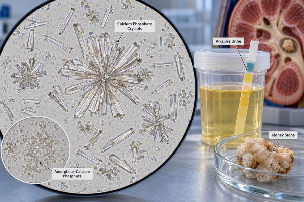
- Calcium phosphate crystals are most favored by alkaline urine, often when pH is above about 6.5.
- Crystals do not prove that a kidney stone is present; they are a clue to urine chemistry and stone risk.
- High urine calcium, low urine citrate, and high urine pH are major calcium phosphate stone risk factors.
- Persistently alkaline urine can occur with distal renal tubular acidosis, some medicines, or urease-producing urinary infections.
- Recurrent stones usually warrant stone analysis and a 24-hour urine evaluation rather than treatment based on microscopy alone.
Table of Contents
- What Calcium Phosphate Crystals Mean
- Appearance and Major Calcium Phosphate Mineral Types
- How Urine pH Drives Calcium Phosphate Crystal Formation
- Causes and Risk Factors for Calcium Phosphate Crystals
- How Crystals Relate to Calcium Phosphate Kidney Stones
- Testing and Follow-Up After Calcium Phosphate Crystals
- Prevention and When to Seek Care
What Calcium Phosphate Crystals Mean
Calcium phosphate crystals are a microscopic sign that calcium and phosphate have come out of solution in the urine. That process is called crystallization. It becomes more likely when the urine is alkaline, calcium concentration is high, citrate is low, or urine is concentrated enough for calcium phosphate to become supersaturated.
The finding is not the same as a diagnosis of kidney stones. People without stones may have occasional crystals, and people with stones may have no crystals in the particular sample examined. Timing matters because crystals can appear after urine sits, cools, or changes pH outside the body. A fresh specimen analyzed promptly gives the most useful picture of what was happening in the urinary tract.
The report also needs context. A few calcium phosphate crystals in an otherwise normal urinalysis may have little clinical importance. Repeated heavy crystalluria is more meaningful when it accompanies flank pain, microscopic blood, recurrent stones, nephrocalcinosis, or abnormal chemistry. A broader urine microscopy assessment can show whether red blood cells, white blood cells, casts, or bacteria point toward another process.
The most useful question is not simply, “Are crystals present?” It is, “What urine conditions allowed them to form?” That shifts attention to factors that can often be measured and modified.
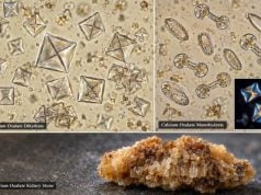
Calcium phosphate crystals also differ from calcium oxalate crystals. Calcium oxalate can form over a wider pH range, while calcium phosphate is much more strongly linked to alkaline urine. This is why the same patient may switch stone composition over time if urine chemistry changes.
Appearance and Major Calcium Phosphate Mineral Types
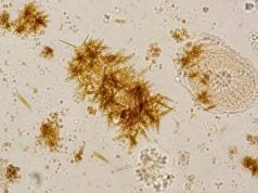
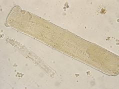
Calcium phosphate is not one single crystal form. Urine microscopy may show amorphous phosphates, wedge-shaped or prism-like crystals, rosettes, or other forms depending on the mineral phase and sample conditions. Some calcium phosphate material is difficult to identify with certainty by routine light microscopy, especially when crystals are tiny or amorphous.
Two mineral types are especially important in kidney stone disease:
- Hydroxyapatite or carbonate apatite: Apatite is a common calcium phosphate component in urinary stones. It can occur alone or mixed with calcium oxalate. Carbonate apatite tends to be favored at higher urine pH and may occur in infection-related settings, although its presence does not by itself prove infection.
- Brushite: Brushite is dicalcium phosphate dihydrate. It often forms in urine that is alkaline but not extremely alkaline. Brushite stones can be dense, hard, recurrent, and clinically challenging.
Routine microscopy cannot reliably tell the full mineral composition of a kidney stone. If a person passes a stone or has one removed, formal kidney stone analysis is far more definitive. Infrared spectroscopy or X-ray diffraction can identify mineral components and show whether calcium phosphate is dominant or mixed with calcium oxalate, uric acid, struvite, or another material.
Amorphous phosphate is especially easy to overinterpret. It may appear as fine granular material in alkaline urine and can sometimes resemble bacteria or nonspecific debris. The laboratory may use specimen pH, crystal pattern, solubility behavior, polarization, or repeat microscopy to clarify an uncertain finding.
The phrase “calcium phosphate crystals” therefore describes a laboratory observation, not a complete stone diagnosis. The morphology is useful, but it becomes clinically meaningful only when paired with chemistry, symptoms, and stone history.
How Urine pH Drives Calcium Phosphate Crystal Formation
Urine pH is one of the strongest drivers of calcium phosphate crystallization. As urine becomes more alkaline, phosphate shifts into chemical forms that bind calcium more readily. That raises calcium phosphate supersaturation and makes nucleation and crystal growth easier.
A spot urine pH test for kidney stone risk can be helpful, but a single reading is only a snapshot. Urine pH normally changes with meals, hydration, medications, and time of day. Persistently high values across several measurements are more informative than one alkaline result.
The exact pH at which a crystal forms depends on urine calcium, phosphate, citrate, ionic strength, and other factors. In general, brushite formation is favored around the mid-to-upper 6 range, while carbonate apatite becomes increasingly favored as pH rises further. This is why a pH of 6.8 has a different meaning in a person with high urinary calcium than in someone with low calcium and no stone history.
Citrate adds another layer. Citrate binds calcium and can reduce the amount of free calcium available to form crystals. Low urinary citrate, called hypocitraturia, therefore increases stone risk. Yet citrate treatment is often given as an alkali salt, which can raise urine pH. In calcium phosphate stone formers, clinicians may need to balance the benefit of more citrate against the possibility that a higher pH could increase calcium phosphate supersaturation. A urine citrate test helps show whether low citrate is actually present.
This pH dependence explains an important clinical point: trying to make urine “more alkaline” is not universally beneficial for stone prevention. Alkalinization is central for uric acid and cystine stones, but calcium phosphate stones require more individualized targets.
Causes and Risk Factors for Calcium Phosphate Crystals
The common risk pattern is high urine pH plus enough urinary calcium to create supersaturation. Several medical, dietary, and medication-related factors can produce that combination.
Important causes and associations include:
- Hypercalciuria: High urinary calcium is a major calcium-stone risk factor even when blood calcium is normal. A urine calcium test, usually as part of a 24-hour collection, helps quantify the problem.
- Distal renal tubular acidosis: In distal RTA, the kidney has difficulty acidifying urine. Urine can remain inappropriately alkaline while blood bicarbonate is low, and hypocitraturia is common. This combination strongly favors calcium phosphate stones and nephrocalcinosis.
- Primary hyperparathyroidism or other hypercalcemic states: Excess parathyroid hormone can raise blood calcium and urinary calcium. Persistent hypercalcemia calls for evaluation rather than dietary calcium restriction alone.
- Carbonic anhydrase inhibitor medicines: Topiramate and acetazolamide can raise urine pH and lower urine citrate, creating a characteristic calcium phosphate stone risk pattern.
- High sodium intake: More sodium delivered to the kidney can increase urinary calcium excretion in susceptible people.
- Low urine volume: Concentrated urine raises mineral activity and supersaturation even when the total daily excretion is not extreme.
- Urease-producing urinary infection: Some bacteria split urea and raise urine pH. This is classically associated with struvite but can also promote carbonate apatite. The presence of calcium phosphate alone does not establish a UTI.
Phosphate excretion can matter as well. A urine phosphorus test may be useful in selected metabolic evaluations, but phosphate is rarely interpreted alone.
Dietary calcium deserves nuance. Normal food-based calcium intake is generally not the same thing as pathologic hypercalciuria. Severe calcium restriction can have unintended effects, including more intestinal oxalate absorption. Stone prevention therefore focuses on the measured metabolic pattern rather than simply eliminating calcium.
How Crystals Relate to Calcium Phosphate Kidney Stones
Crystals are building blocks of stones, but a crystal in urine does not automatically become a stone. Stone formation requires crystals to nucleate, grow, aggregate, and remain in the kidney long enough to enlarge. Urine flow, crystal inhibitors, renal anatomy, and tissue surfaces all influence that process.
Calcium phosphate plays two roles in stone disease. It can form a stone itself, especially as apatite or brushite, and it can act as a surface on which calcium oxalate later grows. Microscopic calcium phosphate deposits in renal papillae can participate in plaque formation, creating a site where other crystals attach.
The composition matters because calcium phosphate stones are not managed exactly like calcium oxalate stones. Current research shows that people with calcium phosphate stones often have higher urine pH, hypercalciuria, and hypocitraturia. Brushite stone formers can have particularly recurrent disease and may require repeated procedures.
A urine supersaturation test can integrate several chemistry values into an estimate of how strongly the urine favors a given crystal phase. It is more informative than looking at crystal count alone because supersaturation reflects the interaction of calcium, phosphate, citrate, pH, volume, and other ions.
Stone composition can also change over time. A person who previously formed calcium oxalate stones may later form stones with more calcium phosphate if urine pH rises. Medication changes, alkali therapy, distal RTA, or evolving metabolic factors can contribute. Reanalyzing a new stone can therefore be worthwhile instead of assuming that every future stone is identical to the first.
One practical mistake is to label every alkaline-stone pattern as “infection stones.” True infection stones are most strongly associated with urease-producing organisms and often contain struvite. Calcium phosphate may coexist, but a urine culture and clinical picture are needed before infection is blamed.
Testing and Follow-Up After Calcium Phosphate Crystals
Follow-up should match the level of risk. A small incidental crystal finding in a symptom-free person often needs only clinical context and, if appropriate, a repeat fresh urinalysis. Recurrent stones, persistent crystalluria, nephrocalcinosis, or abnormal kidney function justify a more complete evaluation.
A typical workup may include:
- Repeat urinalysis and urine pH. This confirms whether alkaline urine and crystals persist and checks for blood, protein, white blood cells, and bacteria.
- Urine culture when infection is plausible. Symptoms such as burning, urgency, fever, or pyuria increase the value of culture. Culture is more reliable than crystal type for deciding whether bacteria are causing an infection.
- Stone analysis. A passed or removed stone should be analyzed when possible because composition guides prevention.
- Blood chemistry. Creatinine, electrolytes, bicarbonate, calcium, and sometimes phosphorus and parathyroid hormone can reveal kidney dysfunction, distal RTA, or hyperparathyroidism.
- 24-hour urine testing. Recurrent or high-risk stone formers often benefit from measurement of volume, calcium, citrate, sodium, oxalate, uric acid, phosphorus, creatinine, pH, and supersaturation.
A 24-hour urine stone risk profile is especially useful because values interact. For example, a urine calcium level that is only modestly high may become important when pH is 7.0 and citrate is low. The same calcium level can carry less calcium phosphate risk when urine volume is high and pH is lower.
Imaging may be needed when symptoms suggest a stone or obstruction. Ultrasound can detect hydronephrosis and many stones without radiation. Noncontrast CT is more sensitive and may be used when the diagnosis is uncertain or complications are a concern.
Testing should answer a management question. Repeating microscopy alone, without investigating persistent high pH or recurrent stones, may document crystals without explaining why they keep forming.
A complete metabolic evaluation is especially valuable after recurrent stones, bilateral stones, stones at a young age, nephrocalcinosis, or an unusual calcium phosphate composition such as brushite. These features raise the chance that a persistent metabolic driver is present. Two well-collected 24-hour urine samples may be used in some stone programs because day-to-day diet and fluid intake can shift pH, calcium, citrate, and volume enough to change interpretation.
Prevention and When to Seek Care
Prevention usually targets urine volume, sodium, urinary calcium, citrate, and the cause of persistently high pH. The right plan depends on the metabolic profile rather than the crystal name alone.
For many calcium stone formers, clinicians recommend enough fluid to produce roughly 2 to 2.5 liters or more of urine per day, unless a medical condition requires fluid restriction. Spreading fluid across the day and including some intake later in the evening can reduce long periods of concentrated urine.
Lowering excess sodium often helps because sodium and calcium handling in the kidney are linked. Normal dietary calcium from food is generally maintained rather than severely restricted. If hypercalciuria persists, a clinician may consider a thiazide-type diuretic depending on blood pressure, kidney function, and other factors.
Low citrate can be treated with dietary changes or potassium citrate in selected patients, but calcium phosphate stone formers need individualized monitoring because raising urine pH too far may increase calcium phosphate supersaturation. Follow-up urine testing can show whether citrate improved without creating an unfavorable pH shift.
If distal RTA, hyperparathyroidism, or a medication such as topiramate is contributing, treating or adjusting the underlying cause can be more effective than generic “stone diets.” Never stop a prescribed medicine solely because crystals appear; the prescribing clinician can weigh stone risk against the drug’s benefit and possible alternatives.
Seek prompt medical care for severe flank pain, persistent vomiting, visible blood with significant pain, fever or chills with urinary symptoms, very low urine output, or pain in someone with one functioning kidney. An obstructing stone plus infection can become an emergency because infected urine may be trapped above the blockage.
For a nonurgent crystal finding, the most useful next step is to ask whether the result was from a fresh sample, what the urine pH was, whether stones have occurred before, and whether there are metabolic risk factors. That turns an isolated microscopy report into a focused plan.
Follow-up should also check whether the prevention plan changes the calcium phosphate supersaturation, not just one isolated number. For example, a higher citrate value may look favorable, but if urine pH rises substantially at the same time, the net effect on calcium phosphate risk may be less favorable. Reviewing the full profile keeps treatment focused on the actual chemistry that drives crystal growth.
References
- Calcium Phosphate Nephrolithiasis: A Comprehensive Review. 2026 (Review)
- Metabolic Evaluation and Recurrence Prevention for Urinary Stone Patients: An EAU Guidelines Update 2024 (Guideline Update)
- Urinary Crystals Identification and Analysis 2024 (Review)
- Pathophysiology and Main Molecular Mechanisms of Urinary Stone Formation and Recurrence 2024 (Review)
- Differential identification of urine crystals with morphologic characteristics and solubility test 2022
Disclaimer
This article explains calcium phosphate crystals and stone risk for general education and does not diagnose the cause of an individual urine result. Crystal interpretation depends on specimen handling, urine chemistry, symptoms, medical history, and other tests. Seek urgent medical care for fever with flank pain, inability to pass urine, or other signs of an obstructed or infected urinary tract.