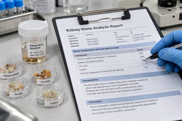
A kidney stone analysis test identifies the minerals and other substances that make up a stone after it has been passed or removed. The result can distinguish calcium oxalate, calcium phosphate, uric acid, cystine, struvite, drug-related material, and mixed stones. That information matters because stone composition can point toward different causes and prevention strategies. A uric acid stone, for example, often directs attention to persistently acidic urine and may be dissolvable with alkalinization, while a cystine stone suggests inherited cystinuria and lifelong high-risk follow-up. Reliable analysis is usually performed with infrared spectroscopy or X-ray diffraction rather than older chemical-only methods. Stone analysis does not replace blood tests or a 24-hour urine study, because the composition shows what formed, while metabolic testing helps explain why it formed and which risk factors are active now.
- Stone analysis directly identifies stone composition and is recommended for first-time stone formers when a stone sample is available.
- Infrared spectroscopy and X-ray diffraction are preferred methods because they can identify specific crystalline compounds more accurately than older wet-chemical analysis.
- Mixed stones are common, so a report may list several components and their approximate percentages rather than one stone type.
- Composition changes prevention: uric acid, cystine, infection, calcium oxalate, and calcium phosphate stones have different metabolic clues and treatment priorities.
- A stone result should be combined with urine and blood testing, especially after recurrent, bilateral, multiple, early-onset, or otherwise high-risk stones.
Table of Contents
- What a Kidney Stone Analysis Test Is
- How Stone Composition Is Tested
- Common Kidney Stone Types and What They Suggest
- How to Read a Stone Analysis Report
- How Results Change Follow-Up Testing and Prevention
- How to Collect and Submit a Stone
- Limitations and When Repeat Stone Analysis Matters
What a Kidney Stone Analysis Test Is
Kidney stone analysis is a laboratory examination of the solid material that formed in the urinary tract. The sample may be a stone passed naturally, a fragment captured during urination, or material removed during ureteroscopy, shock-wave treatment, or percutaneous stone surgery.
The purpose is to identify the stone’s chemical and crystalline composition. Current European guidance recommends stone analysis for first-time stone formers when material is available, using a validated method such as infrared spectroscopy or X-ray diffraction.
Composition is clinically useful because different crystals form under different conditions. Calcium oxalate often reflects combinations of high urine calcium, high oxalate, low citrate, or low volume. Uric acid points toward acidic urine. Cystine indicates cystinuria. Struvite suggests infection with urease-producing organisms. Calcium phosphate can raise questions about high urine pH, hypercalciuria, distal renal tubular acidosis, or hyperparathyroidism.
Stone analysis is not the same as imaging. CT can estimate stone density and sometimes suggest composition, but Hounsfield units overlap between stone types. Plain X-ray can show whether a stone is radiopaque, yet that also does not provide a precise chemical diagnosis.
It is also different from a 24-hour urine kidney stone risk test. Stone analysis tells you what crystallized in the past; urine chemistry measures the current environment that may cause the next stone.
How Stone Composition Is Tested
Modern stone laboratories mainly use Fourier transform infrared spectroscopy (FTIR) or X-ray diffraction (XRD). Both methods identify compounds by their physical molecular or crystal properties rather than by simple chemical reactions.
FTIR shines infrared light through or onto prepared stone material and measures the characteristic absorption pattern. Different minerals have different spectral “fingerprints.” A reference library is then used to identify calcium oxalate monohydrate, calcium oxalate dihydrate, uric acid, apatite, brushite, cystine, struvite, and other compounds.
XRD exposes crystalline material to X-rays and measures how the crystal lattice diffracts them. The diffraction pattern can identify specific crystalline phases. It is particularly useful when distinguishing compounds with similar elemental composition but different crystal structures.
Older wet-chemical analysis detects broad chemical groups such as calcium, phosphate, oxalate, or uric acid. It can be less accurate because it may not distinguish the exact crystalline compound and can misclassify mixed stones. Current guidelines and expert recommendations therefore favor FTIR or XRD.
Some specialized centers also examine stone morphology under stereomicroscopy or use Raman spectroscopy, electron microscopy, or elemental analysis. These methods can reveal layers, nuclei, growth patterns, and minor components. They are valuable for research and selected diagnostic questions but are not necessary for every routine stone.
The quality of the result depends on both the method and the sample. A large layered stone may contain different materials in the core and outer shell. If only one tiny fragment is tested, the report may miss another component. Detailed laboratories may analyze several regions when the sample allows.
Laboratory quality control also matters. Spectral identification depends on good preparation, a reliable reference library, and experienced interpretation. Two laboratories can occasionally report different percentages from the same heterogeneous stone if they analyze different fragments or use different classification rules. A major mismatch—such as one report calling a stone uric acid and another calling it calcium phosphate—deserves review, while small percentage differences in mixed stones may simply reflect sampling.
Some reports also include morphology. The color, surface, internal layers, and nucleus can provide clues about how a stone grew, especially when combined with spectroscopy. Morphology is not a substitute for chemical identification, but it can help distinguish a stone that grew around an infection-related core from one that accumulated layer by layer under changing metabolic conditions.
Common Kidney Stone Types and What They Suggest
Most stones are calcium based, but “calcium stone” is not a complete diagnosis. The exact mineral matters.
Calcium oxalate
Calcium oxalate is the most common stone material. It appears mainly as calcium oxalate monohydrate and calcium oxalate dihydrate. Risk factors include high urinary calcium, high oxalate, low citrate, and low urine volume.
A calcium oxalate result usually leads to review of a calcium oxalate stone risk profile, including urine calcium, oxalate, citrate, sodium, and volume. Normal dietary calcium is generally maintained because calcium eaten with meals helps bind oxalate in the intestine.
Calcium phosphate
Calcium phosphate stones may contain carbonate apatite, hydroxyapatite, or brushite. They often form in urine that is more alkaline than the environment favoring uric acid stones.
A substantial calcium phosphate component can prompt evaluation for hypercalciuria, high urine pH, distal renal tubular acidosis, hyperparathyroidism, or infection depending on the exact mineral and clinical context. Brushite stones can be particularly recurrent and may be harder than some other stones.
Uric acid
Uric acid stones are strongly associated with persistently acidic urine, often with low urine volume. Hyperuricosuria can contribute, but low pH is usually the central chemical driver.
This composition is clinically important because uric acid stones can sometimes be dissolved with supervised urine alkalinization. A uric acid stone risk evaluation focuses on urine pH, volume, and uric acid excretion.
Cystine
Cystine in a stone is strong evidence of cystinuria, an inherited amino acid transport disorder. Cystine stones often begin at a young age and recur frequently.
The finding usually leads to quantitative urine cystine testing, very high fluid targets, urine alkalinization, sodium reduction, family assessment, and sometimes cystine-binding thiol therapy. A cystine stone evaluation may also include genetic testing in selected cases.
Struvite and infection stones
Struvite is magnesium ammonium phosphate. It forms in alkaline urine associated with urease-producing bacteria. Infection stones can grow rapidly and may form large branching calculi.
A struvite component makes infection control and complete stone clearance especially important. Urine culture and the clinical infection history matter because not every alkaline urine or phosphate-containing stone is struvite.
Drug and rare stones
Some medications or their metabolites can crystallize directly in urine or promote unusual stone components. Rare stones can also contain 2,8-dihydroxyadenine, xanthine, ammonium urate, or other substances. Accurate physical analysis is particularly important because these uncommon findings may reveal a genetic disease, medication effect, or specific metabolic disorder that would be missed by generic prevention advice.
The percentage breakdown also matters. A small secondary component may be a clue rather than the main driver, while a dominant unusual mineral can change the work-up substantially. Clinicians therefore interpret the full composition report together with urine chemistry, blood tests, imaging, medications, and the patient’s age at first stone.
How to Read a Stone Analysis Report
A report may list one component or several, often with estimated percentages. For example, a stone might be reported as 80% calcium oxalate monohydrate and 20% carbonate apatite. That is a mixed stone, not two unrelated diagnoses.
The major component usually describes the dominant chemistry, while a minor component can still matter. A small calcium phosphate fraction in a mostly calcium oxalate stone may be common, whereas cystine or struvite as a component can carry more specific implications.
| Reported component | Common interpretation | Typical next focus |
|---|---|---|
| Calcium oxalate | Most common metabolic stone | Urine calcium, oxalate, citrate, volume, sodium |
| Calcium phosphate | Often linked with higher urine pH and hypercalciuria | Urine pH, calcium, citrate; blood calcium and bicarbonate when indicated |
| Uric acid | Acidic urine is usually central | Urine pH, volume, uric acid, metabolic health |
| Struvite | Suggests urease-producing infection | Culture, infection control, complete stone removal |
| Cystine | Indicates inherited cystinuria | Urine cystine, pH, volume, sodium, family evaluation |
| Rare/drug material | May indicate medication or genetic cause | Targeted medication, metabolic, or genetic workup |
The percentages are usually semiquantitative rather than a perfect molecular inventory. Small differences between 70% and 80% of a component may not change treatment. The clinically important issue is often which minerals are present and which one dominates.
The order of components can matter too. Some laboratories list the major mineral first, while others provide a formal percentage breakdown. If the report says only “calcium stone,” ask whether the laboratory can provide the specific compound. Calcium oxalate and calcium phosphate have overlapping but not identical causes, and the distinction can influence how aggressively urine pH, citrate, and endocrine causes are evaluated.
A small amount of a highly specific component may be clinically meaningful. Even a minor cystine component can raise concern for cystinuria, and struvite can suggest an infection contribution. By contrast, a small carbonate apatite fraction is common in many mixed calcium stones and does not automatically prove infection. Interpretation should match the mineral with urine culture, pH, history, and the rest of the metabolic workup.
A report may also use mineral names unfamiliar to patients. Whewellite is calcium oxalate monohydrate; weddellite is calcium oxalate dihydrate. Carbonate apatite is a calcium phosphate. Brushite is calcium hydrogen phosphate dihydrate. Struvite is magnesium ammonium phosphate.
If the report contains an unfamiliar rare compound, ask the ordering clinician or laboratory for interpretation rather than assuming it behaves like a common calcium stone.
How Results Change Follow-Up Testing and Prevention
Stone composition helps determine the next tests. In a first-time, low-risk calcium oxalate stone former, basic blood and urine screening plus general prevention may be enough. A high-risk stone type such as cystine, uric acid, or infection stone usually justifies more specific evaluation.
For recurrent or high-risk disease, guidelines commonly use one or two 24-hour urine collections. Measurements may include volume, pH, calcium, oxalate, citrate, uric acid, sodium, potassium, magnesium, and creatinine. Blood tests often include creatinine, electrolytes, calcium, uric acid, and bicarbonate, with PTH added when calcium is high or otherwise suspicious.
A calcium phosphate stone can make a blood calcium test especially relevant because primary hyperparathyroidism is a treatable cause. Persistently high urine pH with low citrate and low serum bicarbonate may raise concern for distal renal tubular acidosis.
Uric acid composition changes prevention toward alkalinization rather than simply “lowering uric acid.” Cystine composition triggers lifelong high-volume and alkalinization targets. Struvite shifts attention toward bacteria and stone clearance rather than metabolic diet advice alone.
Composition can also prevent unnecessary restriction. A patient with calcium oxalate stones may assume that eating less calcium is the obvious solution, but normal calcium intake with meals is usually preferred. The stone result starts the conversation, while urine testing identifies the actual modifiable abnormality.
It can also help prioritize specialist referral. A routine calcium oxalate stone with mild hypercalciuria may be managed with standard metabolic prevention, whereas cystine, 2,8-dihydroxyadenine, xanthine, or an unusual drug-related stone can justify nephrology, genetics, metabolic, or pharmacology input. Rare stone composition is often the first clue to a disorder that would otherwise be missed.
How to Collect and Submit a Stone
If a clinician asks you to capture a stone, a urine strainer is often the easiest method. These small mesh devices catch solid fragments while urine passes through.
When a fragment is found, follow the laboratory’s instructions. In many cases the stone is allowed to dry and placed in a clean specimen container. Do not add bleach, household chemicals, or unrequested preservatives because they can contaminate or alter the sample.
If several fragments pass, save all of them if practical. Different fragments may represent different layers or stones. Label the container as instructed, especially if samples came from different dates, kidneys, or surgical procedures.
A stone does not need to be large. Modern spectroscopy can analyze very small amounts of material. Even a sand-like fragment may be useful if it is truly stone material.
If the sample came from surgery, the urology team can usually send material directly to pathology or a stone laboratory. Ask whether compositional analysis was ordered; a generic pathology description is not always the same as a formal stone composition report.
Limitations and When Repeat Stone Analysis Matters
Stone analysis has limitations. A small fragment may not represent the whole stone, mixed stones can be heterogeneous, and laboratory methods vary. Old chemical analysis can be less precise than FTIR or XRD.
Composition can also change over time. A person who once formed calcium oxalate may later form more calcium phosphate after changes in urine pH, medication, anatomy, infection, or metabolic disease. A past report should therefore not be treated as permanent proof of every future stone type.
Repeat analysis is particularly useful when stones recur despite prevention, recurrence happens after a long stone-free period, the appearance or imaging characteristics change, or a new medical condition could alter stone chemistry.
Repeat analysis can also show whether treatment has shifted the mineral environment. For example, aggressive alkali therapy may reduce uric acid crystallization but, if urine pH becomes too high in a susceptible patient, later stones may contain more calcium phosphate. That does not mean the original treatment was necessarily wrong; it means prevention needs to be retuned using the new stone composition and current urine profile.
Stone analysis cannot show whether a stone is currently obstructing the ureter, whether infection is present, or whether kidney function is threatened. Those questions require symptoms, urinalysis, culture, blood tests, and imaging.
The strongest stone evaluation combines three kinds of information: composition tells what formed, metabolic testing explains the urine and blood environment, and imaging shows the current stone burden. Together, those data allow prevention to be tailored to the actual disease rather than to a generic idea of kidney stones.
References
- Guidelines – EAU Guidelines on Urolithiasis – Uroweb 2026 (Guideline)
- The Impact of Stone Composition on Treatment Strategies for Patients with Urolithiasis: A Narrative Review 2025 (Review)
- Determination of the kidney stone composition using infrared spectroscopy in Iran at a national referral center during 2019-2023 2025
- 2022 Recommendations of the AFU Lithiasis Committee: Epidemiology, stone analysis and composition. 2023 (Guideline)
- Kidney Stone Pathophysiology, Evaluation and Management: Core Curriculum 2023 2023 (Review)
- Metabolic Evaluation and Recurrence Prevention for Urinary Stone Patients: An EAU Guidelines Update 2024 (Review)
Disclaimer
This article is for general education and does not replace medical diagnosis or treatment. Stone analysis methods and reports vary by laboratory, and composition should be interpreted with imaging, blood tests, urine testing, symptoms, and medical history. Seek urgent care for stone symptoms with fever, inability to urinate, severe uncontrolled pain, or other signs of serious illness.