A blood calcium test helps identify medical conditions that can raise kidney stone risk, especially primary hyperparathyroidism. Most people who form calcium stones do not have high blood calcium, so this test is not a direct measure of how much calcium is in a stone or in the urine. Its main value is finding an abnormal calcium level that changes the evaluation. A persistently high total or ionized calcium level can point toward excess parathyroid hormone activity, certain cancers, excess vitamin D, medication effects, or other disorders that need further testing. A normal blood calcium level also does not rule out high urine calcium, which is a common stone risk factor. Clinicians therefore interpret serum calcium alongside albumin, kidney function, parathyroid hormone, 24-hour urine calcium, stone composition, and the broader metabolic profile rather than using one result alone.
- Blood calcium is mainly a screening test for systemic causes of stones, especially primary hyperparathyroidism; it does not measure urine calcium excretion.
- Typical adult total calcium reference ranges are roughly 8.5–10.5 mg/dL, but the laboratory’s own range should be used because methods differ.
- High or high-normal calcium may prompt a parathyroid hormone test, particularly in a person with recurrent calcium stones or hypercalciuria.
- Normal blood calcium does not rule out hypercalciuria; a 24-hour urine calcium test is needed to measure daily urinary calcium loss.
- Marked hypercalcemia with confusion, dehydration, vomiting, weakness, or heart rhythm symptoms needs prompt medical assessment, especially when calcium is around or above 14 mg/dL.
Table of Contents
- What a Blood Calcium Test Shows in Kidney Stone Evaluation
- Total Calcium, Albumin, and Ionized Calcium
- High Calcium, PTH, and Primary Hyperparathyroidism
- Other Causes of High Blood Calcium
- Why Normal Blood Calcium Can Still Occur With Calcium Stones
- Follow-Up Tests After an Abnormal Result
- Preparation, Repeat Testing, and What to Do Next
What a Blood Calcium Test Shows in Kidney Stone Evaluation
A blood calcium test asks a different question from a urine calcium test. Blood calcium shows whether calcium regulation in the body is abnormal; urine calcium shows how much calcium the kidneys excrete over time. Both can matter for stones, but they are not interchangeable.
Calcium is tightly regulated because it is needed for nerve signaling, muscle contraction, heart function, blood clotting, and bone structure. The parathyroid glands, kidneys, intestines, bones, and vitamin D system all help keep blood calcium within a narrow range. When serum calcium rises persistently, that can be a clue to a systemic disorder rather than simply a dietary issue.
In stone evaluation, serum calcium is commonly part of the basic metabolic workup. Current kidney stone guidelines recommend checking blood calcium because primary hyperparathyroidism is a treatable cause of recurrent calcium stones. The test is particularly important when stones are recurrent, bilateral, numerous, associated with nephrocalcinosis, or accompanied by high urinary calcium.
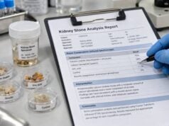
A blood calcium result cannot identify stone composition. Calcium oxalate and calcium phosphate stones contain calcium, but the calcium incorporated into a stone does not necessarily make the serum calcium level high. If a passed or removed stone is available, a kidney stone analysis is the direct way to determine its mineral makeup.
Total Calcium, Albumin, and Ionized Calcium
Most routine blood panels report total calcium. This includes calcium that is bound to proteins, mainly albumin, plus calcium that is free in the blood. The free fraction is called ionized calcium, and it is the biologically active form.
A common adult total calcium reference interval is about 8.5–10.5 mg/dL, or approximately 2.12–2.62 mmol/L. The exact limits vary by laboratory, age, method, and local population, so the printed reference interval should guide interpretation.
Albumin matters because a substantial portion of circulating calcium is protein-bound. A low albumin concentration can make total calcium look low even when ionized calcium is normal. A high albumin level, dehydration, or hemoconcentration can make total calcium look higher. For that reason, clinicians often review calcium together with albumin.
“Corrected calcium” formulas attempt to estimate what total calcium would be at a standard albumin level. They can be convenient, but they are imperfect, especially in people with kidney disease, critical illness, abnormal proteins, acid-base disorders, or substantial hypoalbuminemia. When the distinction matters, directly measured ionized calcium can be more informative.
Ionized calcium commonly has a reference range near 1.15–1.30 mmol/L, but laboratory methods differ. The sample is more sensitive to collection and handling conditions because blood pH changes the amount of calcium bound to proteins. That is why an ionized calcium test may be reserved for situations in which total calcium is difficult to interpret or the diagnosis remains uncertain.
One isolated result slightly above the reference range is usually not enough to diagnose a disorder. Dehydration, laboratory variation, albumin changes, supplements, and temporary illness can affect results. Persistent hypercalcemia generally deserves confirmation and a structured evaluation.
A low blood calcium level is usually not a direct kidney stone signal. It can occur with low albumin, vitamin D deficiency, hypoparathyroidism, advanced kidney disease, magnesium disorders, or other illnesses. In a stone former, low total calcium should therefore be interpreted with albumin and, when needed, ionized calcium rather than treated automatically with calcium supplements. Taking large calcium doses without knowing the cause can be unhelpful and may complicate stone prevention. Dietary calcium is different from indiscriminate supplementation: for many calcium oxalate stone formers, maintaining normal calcium intake with meals is preferred because calcium in the gut binds oxalate and can reduce its absorption.
High Calcium, PTH, and Primary Hyperparathyroidism
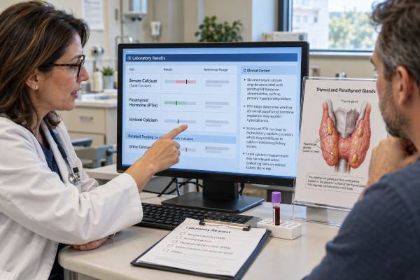
When a person with kidney stones has repeatedly high blood calcium, primary hyperparathyroidism is one of the most important causes to consider. In this condition, one or more parathyroid glands secrete too much parathyroid hormone, or PTH, relative to the calcium level.
PTH raises blood calcium through several pathways. It promotes calcium release from bone, increases kidney calcium reabsorption, alters phosphate handling, and stimulates activation of vitamin D, which increases intestinal calcium absorption. Even though PTH promotes renal calcium reabsorption, the overall calcium load delivered to the kidneys can rise enough to cause hypercalciuria.
The classic biochemical pattern is high calcium with a PTH level that is elevated or inappropriately normal. “Inappropriately normal” means the PTH falls within the laboratory range but is not suppressed as it should be when calcium is high. A very low PTH in the setting of hypercalcemia points clinicians toward non-parathyroid causes instead.
For stone formers, a parathyroid hormone test for kidney stones is commonly ordered when calcium is high or repeatedly high-normal and the clinical context raises suspicion. Some guidelines also recommend PTH testing in idiopathic hypercalciuria even when serum calcium is normal, because normocalcemic primary hyperparathyroidism and secondary causes can complicate interpretation.
Kidney stones are a recognized kidney manifestation of primary hyperparathyroidism. Modern international guidelines consider nephrolithiasis or nephrocalcinosis important evidence of renal involvement. In established primary hyperparathyroidism, evaluation commonly includes kidney function, 24-hour urine calcium, and imaging for stones or nephrocalcinosis when appropriate.
Parathyroid imaging is not usually the first step in diagnosis. Ultrasound, sestamibi scanning, or 4D CT is mainly used to localize abnormal glands after the biochemical diagnosis has been established and surgery is being considered. A high calcium result should therefore lead first to biochemical confirmation, not directly to imaging.
Other Causes of High Blood Calcium
Not every high calcium result in a stone former is caused by primary hyperparathyroidism. The PTH level helps divide the evaluation into PTH-dependent and PTH-independent causes.
If PTH is suppressed, clinicians may consider causes such as malignancy-related hypercalcemia, excess vitamin D activity, granulomatous diseases, prolonged immobilization, hyperthyroidism, adrenal disorders, or medication and supplement effects. The likely causes depend strongly on the person’s symptoms, age, medical history, and degree of calcium elevation.
Important medication and supplement contributors can include:
- thiazide diuretics, which reduce urinary calcium but can mildly raise serum calcium in some people;
- lithium, which can alter parathyroid regulation;
- excessive calcium supplements or large amounts of absorbable alkali;
- excessive vitamin D intake or unusually high vitamin D activity;
- high-dose vitamin A in some circumstances.
These associations do not mean that a person should stop prescribed medicine without guidance. For example, thiazide-type drugs are also used to reduce urinary calcium and prevent recurrent calcium stones in selected patients. If serum calcium rises, the prescriber needs to weigh the possible cause, dose, benefits, and alternatives.
Another important diagnosis is familial hypocalciuric hypercalcemia (FHH), an inherited condition in which blood calcium is usually mildly elevated but urinary calcium excretion tends to be low. FHH can resemble primary hyperparathyroidism on blood tests, yet it generally does not benefit from parathyroid surgery. A 24-hour urine calcium measurement and calcium-to-creatinine clearance ratio may help distinguish them, although the values overlap and genetic testing is sometimes needed.
Severe hypercalcemia is a different clinical situation from a mildly abnormal screening test. Calcium around or above 14 mg/dL, particularly with dehydration, confusion, weakness, constipation, vomiting, kidney injury, or arrhythmia symptoms, may require urgent treatment. A person who feels acutely ill should not wait for a routine stone clinic appointment.
Why Normal Blood Calcium Can Still Occur With Calcium Stones
Most calcium stone formers have normal serum calcium. That is because stone formation depends heavily on urine chemistry, not just blood calcium concentration.
Idiopathic hypercalciuria is a common example. In this condition, urinary calcium excretion is elevated despite a normal blood calcium level and no obvious systemic cause. Genetic tendency, sodium intake, intestinal absorption, bone mineral handling, and kidney calcium transport can all contribute.
This is why the urine calcium test for kidney stones answers an important question that serum calcium cannot. A 24-hour urine collection may show high calcium even when the blood value has always been normal.
Calcium stone risk also depends on several other factors. Low urine volume concentrates calcium and oxalate. High urine oxalate can increase calcium oxalate supersaturation even if urine calcium is only moderate. Low citrate removes a natural inhibitor of calcium crystal formation. High urine pH can favor calcium phosphate crystallization. High sodium intake can increase urinary calcium.
That interaction explains why a person should not interpret a normal blood calcium test as “no calcium stone risk.” The result is reassuring against persistent hypercalcemia, but it does not replace a full stone-risk profile.
The reverse is also true: a mildly high blood calcium level does not prove that a current stone is made of calcium or that hypercalcemia caused it. Stone composition, urinary risk factors, and repeated blood testing are needed to connect the findings.
Follow-Up Tests After an Abnormal Result
A high calcium result is usually evaluated step by step. The exact sequence varies, but common follow-up includes repeating the calcium measurement, reviewing albumin, and checking PTH. Depending on the situation, the clinician may also measure creatinine or estimated glomerular filtration rate, phosphate, magnesium, bicarbonate, 25-hydroxyvitamin D, and ionized calcium.
For a kidney stone patient, urine testing adds critical information. A 24-hour urine kidney stone risk test commonly measures volume, calcium, oxalate, citrate, uric acid, sodium, pH, and creatinine. If hyperparathyroidism is suspected, 24-hour urine calcium can help characterize renal calcium handling and may help distinguish primary hyperparathyroidism from FHH.
| Finding | Common next question | Possible follow-up |
|---|---|---|
| High calcium + high or non-suppressed PTH | Is primary hyperparathyroidism present? | Repeat calcium/PTH, kidney function, phosphate, vitamin D, urine calcium, renal and bone assessment |
| High calcium + suppressed PTH | Is there a non-parathyroid cause? | Medication review and targeted testing based on symptoms and history |
| Normal serum calcium + high urine calcium | Is this idiopathic hypercalciuria or another renal/metabolic cause? | Full 24-hour urine profile, sodium assessment, PTH when indicated |
| Mild lifelong hypercalcemia + low urine calcium | Could this be FHH? | Calcium clearance assessment and sometimes genetic evaluation |
If primary hyperparathyroidism is confirmed, surgery is the only definitive treatment. International guidelines consider kidney stones and nephrocalcinosis important renal indications when deciding on parathyroidectomy. The decision still requires an individualized endocrine and surgical assessment.
The kidney evaluation in primary hyperparathyroidism often includes more than a calcium number. Clinicians may look at estimated glomerular filtration rate, imaging evidence of nephrolithiasis or nephrocalcinosis, and 24-hour urinary calcium. A high urine calcium level supports a substantial filtered calcium load, but it is not required for stones to occur. Conversely, very low urinary calcium in someone with mild hypercalcemia raises the possibility of FHH or an incomplete collection. This is another reason to avoid making a diagnosis from one laboratory value.
Bone evaluation may also matter because the same parathyroid disorder that contributes to stones can affect skeletal mineral density. Depending on age, symptoms, and confirmed diagnosis, dual-energy X-ray absorptiometry and vertebral assessment may be part of the broader workup. These tests do not diagnose the kidney stone itself; they measure the systemic consequences of abnormal calcium and PTH regulation and help guide treatment decisions.
After treatment of a systemic cause, stone risk does not necessarily disappear completely. Some people continue to have hypercalciuria or other urine abnormalities and may need ongoing metabolic stone prevention.
Preparation, Repeat Testing, and What to Do Next
A routine blood calcium test usually requires little preparation. Fasting is not always necessary, although calcium may be included in a larger panel that has its own instructions. Follow the laboratory or clinician’s directions rather than fasting automatically.
Before testing, provide an accurate list of prescription medicines, over-the-counter products, calcium supplements, vitamin D, antacids, and other supplements. Do not stop them on your own just to produce a different result. The goal is to understand your real calcium physiology and identify anything that may need adjustment.
If the result is mildly high, a common next step is repeat testing under stable conditions. Good hydration can reduce the effect of hemoconcentration, but deliberately overdrinking is not necessary. Repeated abnormal results are more meaningful than a single borderline number.
The pattern over time can be especially useful. A calcium value that has been just above the upper limit on several tests is more informative than one isolated spike during dehydration. If historical laboratory results are available, clinicians may compare them with PTH, creatinine, albumin, phosphate, vitamin D, and medication changes. The combination can show whether hypercalcemia is persistent, newly developed, or linked to a specific treatment.
If your calcium is normal but you have recurrent calcium stones, ask whether a complete metabolic evaluation is appropriate. That may include blood tests, stone analysis, and one or two 24-hour urine collections. If blood calcium is high, ask whether PTH and vitamin D should be checked and whether urine calcium is needed.
The most useful interpretation is not “calcium is high, therefore calcium caused the stone.” It is a chain of questions: Is hypercalcemia real and persistent? Is PTH appropriately suppressed? Is urine calcium high or low? What type of stone formed? Are there other urine abnormalities? Answering those questions can reveal a treatable endocrine disorder or show that the main prevention target lies elsewhere.
References
- Metabolic Evaluation and Recurrence Prevention – EAU Guidelines on Urolithiasis – Uroweb 2026 (Guideline)
- Evaluation and Management of Primary Hyperparathyroidism: Summary Statement and Guidelines from the Fifth International Workshop 2022 (Guideline)
- Primary hyperparathyroidism: clinical manifestations, diagnosis and evaluation according to the Fifth International Workshop guidelines 2023 (Review)
- Screening for asymptomatic nephrolithiasis in primary hyperparathyroidism patients is warranted 2024
- Kidney Stone Pathophysiology, Evaluation and Management: Core Curriculum 2023 2023 (Review)
- UPDATE – Canadian Urological Association guideline: Evaluation and medical management of kidney stones 2022 (Guideline)
Disclaimer
This article is for general education and is not a substitute for medical diagnosis or treatment. Calcium reference ranges and urgency depend on the laboratory, symptoms, kidney function, albumin, medications, and other clinical factors. Seek prompt care for marked hypercalcemia or symptoms such as confusion, severe weakness, dehydration, persistent vomiting, or heart rhythm problems.