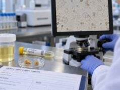
A parathyroid hormone (PTH) blood test helps determine whether a calcium disorder is contributing to kidney stones. The test is most useful when it is interpreted with a blood calcium level drawn at about the same time. In primary hyperparathyroidism, one or more parathyroid glands release too much PTH, causing blood calcium to rise and often increasing the amount of calcium filtered into the urine. That combination can promote calcium-based kidney stones. A high PTH result by itself does not prove primary hyperparathyroidism, however. Kidney disease, vitamin D deficiency, low calcium intake or absorption, and some medications can raise PTH for different reasons. Likewise, a PTH value inside the laboratory range can still be abnormal if calcium is high, because PTH should normally be suppressed in that situation. For stone formers, the key question is whether the calcium-PTH pattern points to a treatable hormonal cause.
- PTH should be interpreted with calcium: high calcium plus elevated or “inappropriately normal” PTH strongly suggests primary hyperparathyroidism after other causes are considered.
- High PTH with normal or low calcium is more often secondary: common causes include vitamin D deficiency and chronic kidney disease.
- Kidney stones are a recognized complication of primary hyperparathyroidism: confirmed nephrolithiasis can be an indication for parathyroid surgery in an appropriate patient.
- A 24-hour urine calcium test often follows abnormal calcium-PTH results: it helps measure hypercalciuria and can help distinguish primary hyperparathyroidism from some mimicking conditions.
- PTH reference ranges vary by assay: the relationship between PTH, calcium, kidney function, vitamin D, and phosphorus is more important than one isolated number.
Table of Contents
- Why PTH is tested in people with kidney stones
- How to read PTH and calcium together
- Primary hyperparathyroidism and kidney stone risk
- Other causes of a high PTH result
- Tests that usually accompany PTH
- What different result patterns may mean
- Follow-up, treatment, and when to seek care
Why PTH is tested in people with kidney stones
PTH is a hormone made by four small parathyroid glands in the neck. Its job is to keep blood calcium within a narrow range. When blood calcium falls, PTH rises and acts on bone, kidneys, and vitamin D metabolism to restore calcium. When blood calcium rises, normal parathyroid glands should reduce PTH secretion.
That feedback loop is why the test matters in kidney stone evaluation. If a stone former has an elevated blood calcium result, the next question is whether PTH is appropriately suppressed. If it is not, primary hyperparathyroidism becomes an important possibility.
Primary hyperparathyroidism can increase stone risk through several pathways. Higher PTH increases calcium release from bone and calcium reabsorption in the kidney while also increasing active vitamin D production, which can raise intestinal calcium absorption. The net effect in many patients is more calcium entering the bloodstream and, despite PTH’s renal effects, enough filtered calcium reaching the urine to cause hypercalciuria.
Most stones associated with primary hyperparathyroidism are calcium stones, commonly calcium oxalate and sometimes calcium phosphate. Not every person with the disorder develops stones, and not every person with stones has dramatic hypercalciuria. Stone formation also depends on urine volume, citrate, oxalate, pH, sodium intake, and other factors.
A clinician may order PTH when a stone former has high or high-normal serum calcium, repeated calcium stones, calcium phosphate stones, nephrocalcinosis, unexplained hypercalciuria, low serum phosphorus, osteoporosis, or another feature that raises concern for a parathyroid disorder. It commonly accompanies a blood calcium test for kidney stones rather than being used as a stand-alone screening test.
How to read PTH and calcium together
The PTH number is not interpreted the same way at every calcium level. The key principle is physiologic appropriateness.
If calcium is high, PTH should be low. Therefore, a PTH value that falls in the middle or upper part of the laboratory’s “normal” range may still be inappropriate. In a person with confirmed hypercalcemia, an elevated or non-suppressed PTH points toward PTH-dependent hypercalcemia, most commonly primary hyperparathyroidism.
If calcium is low, a rise in PTH is often an appropriate response. The body is trying to preserve calcium. That pattern occurs with vitamin D deficiency, calcium malabsorption, chronic kidney disease, and other forms of secondary hyperparathyroidism.
If calcium is normal but PTH is repeatedly high, clinicians consider normocalcemic primary hyperparathyroidism only after excluding common secondary causes. That usually requires repeat total and sometimes ionized calcium measurements, adequate vitamin D assessment, acceptable kidney function, and review of diet, gastrointestinal disease, and medications.
A useful framework is:
| Calcium | PTH | Typical interpretation |
|---|---|---|
| High | High or not suppressed | PTH-dependent hypercalcemia; primary hyperparathyroidism is a leading cause |
| High | Low | Look for non-PTH causes of hypercalcemia |
| Normal | High | Often secondary hyperparathyroidism; normocalcemic primary hyperparathyroidism is a diagnosis of exclusion |
| Low | High | Usually an appropriate secondary PTH response |
| Low | Low or inappropriately normal | Consider hypoparathyroidism or another failure of PTH response |
Total calcium is affected by albumin, so clinicians may use albumin-adjusted calcium or directly measure ionized calcium when the result is uncertain. Dehydration can also temporarily concentrate serum calcium. A mildly abnormal result is therefore often repeated before a major diagnosis is made.
The timing of paired tests can matter when results are borderline. Calcium and PTH are easiest to interpret when measured under similar conditions and before major changes in supplements or treatment. If calcium repeatedly sits at the upper limit while PTH is not suppressed, the pattern deserves more attention than either value viewed alone. If albumin is unusually high or low, ionized calcium can clarify whether the biologically active calcium is truly elevated. This step can prevent a patient from being labeled with primary hyperparathyroidism on the basis of a transient or protein-related total calcium change.
PTH assays have laboratory-specific reference intervals. Do not compare a value from one laboratory with another laboratory’s range. High-dose biotin and certain assay interferences can also affect some hormone tests, so tell the laboratory and clinician about supplements and follow any pretest instructions.
Primary hyperparathyroidism and kidney stone risk
Primary hyperparathyroidism is usually caused by a single benign parathyroid adenoma, although more than one gland can be involved. The condition ranges from mild laboratory abnormalities to symptomatic disease with kidney stones, bone loss, fractures, marked hypercalcemia, or kidney impairment.
Kidney stones are considered a classic renal manifestation. A person with primary hyperparathyroidism may have overt stones that cause renal colic, or silent stones and nephrocalcinosis found only on imaging. Because stones can recur, identifying the endocrine cause can change management more substantially than treating each stone episode separately.
The 2022 Fifth International Workshop guidelines list renal findings that can support surgery in otherwise asymptomatic primary hyperparathyroidism. These include kidney stones or nephrocalcinosis on imaging, an estimated glomerular filtration rate below 60 mL/min, and elevated 24-hour urine calcium above 250 mg/day in women or 300 mg/day in men. These thresholds are not universal definitions of stone risk; they are criteria used in a specific primary hyperparathyroidism management framework.
The amount of calcium in a 24-hour urine collection is useful because stone risk rises progressively as urine calcium increases. A patient with hyperparathyroidism can still form stones below a single cutoff if urine volume is low, citrate is low, or oxalate is high. Conversely, a high urine calcium level does not prove the parathyroid glands are the cause. Idiopathic hypercalciuria, high sodium intake, vitamin D excess, and other conditions can produce the same urine finding.
That is why the endocrine evaluation and the stone evaluation overlap but are not identical. The urine calcium test measures one important risk factor, while a broader urine profile shows whether calcium is actually creating high calcium oxalate or calcium phosphate supersaturation in that person’s urine.
Primary hyperparathyroidism should not be confused with secondary hyperparathyroidism related to chronic kidney disease. In kidney disease, PTH may rise because phosphorus retention, lower active vitamin D, and altered calcium balance stimulate the glands. The separate PTH test in kidney disease is interpreted according to a different clinical context.
Other causes of a high PTH result
A high PTH result is common and does not automatically mean a parathyroid adenoma. The most important alternative causes are conditions that lower calcium availability or alter kidney handling of calcium and phosphorus.
Vitamin D deficiency
Low vitamin D reduces intestinal calcium absorption. The parathyroid glands respond by releasing more PTH to maintain blood calcium. Serum calcium may remain normal even while PTH rises. Correcting vitamin D deficiency can normalize PTH in some people, although vitamin D replacement in a stone former may require follow-up of serum and urine calcium.
Chronic kidney disease
As kidney function declines, phosphate handling and vitamin D activation change. PTH often rises as part of chronic kidney disease-mineral and bone disorder. In this setting, the PTH level must be interpreted with eGFR, calcium, phosphorus, vitamin D status, and the stage of kidney disease. A kidney filtration test such as eGFR is therefore highly relevant when PTH is unexpectedly high.
Low calcium intake or poor absorption
Very low calcium intake, celiac disease, inflammatory bowel disease, pancreatic or biliary disorders, and bariatric surgery can reduce calcium absorption and stimulate PTH. Some of these conditions can paradoxically increase kidney stone risk through enteric hyperoxaluria even when urine calcium is not high.
Medications
Lithium can alter the calcium-sensing set point and produce a pattern resembling primary hyperparathyroidism. Thiazide diuretics can raise serum calcium in some people and lower urinary calcium. Antiresorptive osteoporosis medicines, some anticonvulsants, and other drugs can affect PTH indirectly. Medication history should be reviewed before interpreting an unexpected result.
Familial hypocalciuric hypercalcemia
Familial hypocalciuric hypercalcemia, or FHH, is an inherited condition that can resemble mild primary hyperparathyroidism because calcium is high and PTH may be normal or mildly elevated. Urinary calcium is typically low relative to serum calcium. Clinicians may calculate a calcium-to-creatinine clearance ratio and consider genetic testing when the pattern fits, especially in younger patients or those with a family history. This distinction matters because parathyroid surgery usually does not correct FHH.
Tests that usually accompany PTH
When kidney stones and an abnormal calcium-PTH pattern occur together, the next step is usually a small group of complementary tests rather than repeating PTH alone.
Common tests include:
- Repeat total calcium and albumin: confirms that hypercalcemia is persistent and helps interpret protein-bound calcium.
- Ionized calcium: useful when total calcium is borderline or albumin makes interpretation uncertain.
- 25-hydroxyvitamin D: identifies deficiency that can cause secondary hyperparathyroidism and may influence treatment.
- Creatinine and eGFR: assess kidney function, which strongly affects PTH interpretation and treatment decisions.
- Serum phosphorus: often low or low-normal in primary hyperparathyroidism because PTH increases renal phosphate excretion.
- 24-hour urine calcium and creatinine: quantify calcium loss and help assess collection quality and the differential diagnosis.
- A full 24-hour stone-risk profile: measures volume, calcium, oxalate, citrate, uric acid, sodium, pH, and supersaturation.
- Kidney imaging: looks for current stones or nephrocalcinosis.
- Stone analysis: confirms the mineral composition when a stone is available.
If recurrent calcium stones are the main issue, a 24-hour urine kidney stone risk test can show whether the parathyroid abnormality is accompanied by other modifiable urinary risks. This matters because parathyroid treatment does not eliminate every cause of calcium stones.
Imaging of the parathyroid glands is not the first test used to diagnose primary hyperparathyroidism. Ultrasound, sestamibi scanning, 4D CT, or other localization studies are generally performed after the biochemical diagnosis is established and surgery is planned. A scan cannot substitute for the calcium-PTH pattern.
Does the PTH blood test require fasting or special preparation?
PTH itself often can be measured without fasting, but the ordering clinician or laboratory may request a morning fasting sample when PTH is being paired with calcium, phosphorus, vitamin D, or other metabolic tests. Follow the specific instructions on the order rather than assuming every PTH test is prepared the same way.
Before the blood draw, provide a complete list of prescription medicines, over-the-counter products, calcium or vitamin D supplements, and high-dose biotin. Do not stop a prescribed drug unless the clinician tells you to. A medication may be part of the reason the test was ordered, and abruptly changing it can make the result less representative or create other risks.
PTH also varies biologically and analytically. A borderline result that conflicts with the calcium level may be repeated, ideally with the same laboratory method so the values can be compared more cleanly. Calcium should be confirmed as well, because a single mildly high result can reflect dehydration, albumin changes, or laboratory variation. In a stone evaluation, the trend across several measurements often matters more than one isolated number.
The practical preparation goal is consistency: obtain the blood tests under the conditions requested, avoid unplanned supplement changes, and make sure the clinician knows about factors that could alter calcium or the assay.
What different result patterns may mean
The following examples illustrate why PTH is a context test rather than a simple high-versus-low test.
High calcium, high PTH, recurrent calcium stones
This is a classic pattern for primary hyperparathyroidism. Confirmation usually includes repeat calcium, kidney function, vitamin D, phosphorus, urine calcium, and imaging for renal involvement. If the diagnosis is confirmed, recurrent stones make the condition clinically significant and often strengthen the case for parathyroid surgery.
High calcium, “normal” PTH
A normal-range PTH may still be abnormal when calcium is high. For example, a PTH of 50 pg/mL might fall within a laboratory range, but if calcium is clearly elevated, normal physiology should suppress PTH much lower. The clinician considers primary hyperparathyroidism, FHH, medications, and assay factors.
Normal calcium, high PTH, stones
Secondary causes should be excluded first. Vitamin D deficiency, reduced kidney function, low calcium intake, malabsorption, and medications are common. Normocalcemic primary hyperparathyroidism requires repeated evidence over time and careful exclusion of those causes. The stone itself does not establish the diagnosis.
High urine calcium, normal blood calcium and normal PTH
This is common in idiopathic hypercalciuria. The treatment approach often emphasizes sodium reduction, normal dietary calcium, adequate urine volume, and selected medication rather than parathyroid surgery.
High blood calcium, low urine calcium
This pattern raises the possibility of FHH, particularly when hypercalcemia is mild and long-standing. It can also occur with low calcium intake, vitamin D deficiency, reduced kidney function, or thiazide use, so interpretation still requires context.
Follow-up, treatment, and when to seek care
Treatment depends on the diagnosis, not on the PTH value alone. If primary hyperparathyroidism is confirmed and the patient has kidney stones, parathyroidectomy is often the definitive treatment. Surgery removes the overactive gland or glands and can correct the hormonal driver of hypercalcemia. The decision still considers age, surgical risk, kidney function, bone health, calcium severity, and patient preference.
If the PTH rise is secondary, treatment targets the cause. That may mean correcting vitamin D deficiency, improving calcium intake or absorption, managing chronic kidney disease, or reviewing medications. In stone formers, changes should be made with awareness of urine calcium and the overall stone profile.
After successful parathyroid surgery, some people remain at risk for stones because they also have low urine volume, high sodium intake, hyperoxaluria, hypocitraturia, or another independent factor. A repeat metabolic stone evaluation may therefore be useful when stones were recurrent or the preoperative urine profile showed multiple abnormalities.
Do not start or stop calcium, vitamin D, citrate, or thiazide therapy solely from an online interpretation of PTH. These treatments can shift serum and urine calcium in different directions and may be inappropriate in certain kidney or endocrine conditions.
Urgent medical assessment is appropriate for symptoms of significant hypercalcemia such as marked weakness, confusion, severe dehydration, repeated vomiting, or worsening kidney function, especially when calcium is substantially elevated. A stone emergency also requires urgent care when severe flank pain occurs with fever or chills, when urine output stops, or when vomiting prevents hydration. Those symptoms may indicate an infected obstruction or another complication that cannot wait for routine endocrine follow-up.
For most stone formers, however, an abnormal PTH result is a reason for structured outpatient evaluation. The practical goal is to determine whether the parathyroid glands are driving calcium excess, identify any kidney consequences, and treat the underlying cause rather than chasing the hormone number by itself.
References
- Evaluation and Management of Primary Hyperparathyroidism: Summary Statement and Guidelines from the Fifth International Workshop 2022 (Guideline)
- Primary hyperparathyroidism: clinical manifestations, diagnosis and evaluation according to the Fifth International Workshop guidelines 2023 (Review)
- Metabolic Evaluation and Recurrence Prevention – EAU Guidelines on Urolithiasis – Uroweb 2026 (Guideline)
- Metabolic evaluation for prevention of kidney stones – CARI Guidelines 2025 (Guideline)
- Metabolic evaluation of urolithiasis: a narrative review. 2025 (Review)
Disclaimer
This article is for general education and does not diagnose hyperparathyroidism or determine whether surgery is appropriate. PTH must be interpreted with calcium, kidney function, vitamin D status, medications, and the individual’s stone history. Seek urgent medical care for severe symptoms of hypercalcemia or a kidney stone accompanied by fever, inability to urinate, or persistent vomiting.