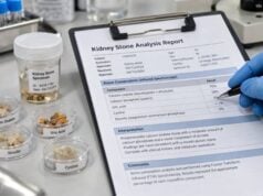

A calcium oxalate stone risk test uses urine chemistry to show why calcium oxalate crystals may be forming and which risk factors are most practical to change. The most important measurements usually include urine oxalate, calcium, citrate, volume, pH, sodium, uric acid, and creatinine. These results work together: a modest rise in oxalate can matter much more when urine volume is low, while good citrate and high urine volume can reduce risk even when one marker is mildly abnormal. Calcium oxalate is the most common kidney stone mineral, but the cause differs from person to person. Some people mainly have high urine calcium, others have excess oxalate absorption, low citrate, dehydration, high sodium intake, or a combination. The test is therefore most useful when interpreted as a pattern alongside stone analysis, diet, medications, medical history, and kidney function.
- The core calcium oxalate risk pattern is high urine calcium and/or oxalate combined with low urine volume or low citrate.
- Urine oxalate is especially important because small increases can substantially raise calcium oxalate supersaturation.
- Normal dietary calcium is usually preferred over calcium restriction because calcium eaten with meals binds oxalate in the intestine.
- A common prevention goal is more than 2.5 L of urine per day, with extra fluid needed during heat, exercise, or heavy sweating.
- Severe or unexplained hyperoxaluria needs medical evaluation because bowel disease, bariatric surgery, or primary hyperoxaluria may require targeted treatment.
Table of Contents
- What a Calcium Oxalate Stone Risk Test Measures
- Urine Oxalate and Hyperoxaluria
- Urine Calcium and Hypercalciuria
- Urine Citrate and Volume as Protective Factors
- How the Results Work Together
- Causes of Abnormal Results and Follow-Up Tests
- Prevention Based on the Urine Pattern
What a Calcium Oxalate Stone Risk Test Measures
A calcium oxalate risk evaluation is usually performed with one or two 24-hour urine collections. The test measures how much stone-forming material enters the urine over a full day and how concentrated that material becomes in the final urine volume.
The most important measurements are urine volume, calcium, oxalate, citrate, sodium, uric acid, pH, and creatinine. Many laboratories also calculate calcium oxalate supersaturation, a summary measure of how strongly the urine favors calcium oxalate crystal formation.
A urine supersaturation test can be helpful because stone risk is not determined by one value alone. Calcium and oxalate interact chemically, citrate binds calcium and inhibits crystals, and urine volume changes the concentration of everything in the sample.
Stone composition should ideally be confirmed when material is available. A kidney stone analysis can distinguish calcium oxalate from calcium phosphate, uric acid, cystine, struvite, and mixed stones. This matters because a prevention strategy that helps one stone type can be less useful—or occasionally counterproductive—for another.
Calcium oxalate itself can appear in different crystalline forms, mainly calcium oxalate monohydrate and calcium oxalate dihydrate. These forms do not require completely separate home treatment plans, but they can provide clues about the urine environment and stone history. Mixed stones are common, so a report that says “80% calcium oxalate, 20% calcium phosphate” is often more useful than a generic label of “calcium stone.” The urine profile should be interpreted against that actual composition.
Timing also matters. A baseline metabolic collection is best performed when the person is stable, back on their usual diet, and not intentionally overhydrating after a recent emergency visit. European guidance suggests waiting until the acute episode has settled and the patient has returned to normal habits. Collecting during vomiting, infection, or a temporary crash diet can create values that do not represent everyday risk.
The test is most often used after recurrent stones, multiple stones, stones in both kidneys, a strong family history, stone disease at a young age, bowel disease, bariatric surgery, or other features that suggest higher recurrence risk. It can also be useful after a first stone when the patient wants individualized prevention.
Urine Oxalate and Hyperoxaluria
Oxalate is a small organic molecule that comes from both food and normal metabolism. It has a strong attraction to calcium. When urine contains enough free calcium and oxalate, the two can combine into calcium oxalate crystals.
Many laboratories consider roughly 20–40 mg/day a typical adult range, but cutoffs vary by method, sex, body size, and laboratory. A result above the reference range is called hyperoxaluria. The degree of elevation matters: mild dietary hyperoxaluria is very different from the marked values seen in some intestinal disorders or primary hyperoxaluria.
Urine oxalate is important because calcium oxalate supersaturation is highly sensitive to oxalate concentration. A relatively small increase may have a meaningful effect, particularly when urine volume is low. This is one reason a urine oxalate test is a central part of calcium oxalate stone evaluation.
Common causes of high urine oxalate include:
- frequent intake of very high-oxalate foods in susceptible people;
- low calcium intake with meals, which leaves more oxalate free for intestinal absorption;
- fat malabsorption from inflammatory bowel disease, chronic pancreatic disease, short bowel, or some bariatric procedures;
- high-dose vitamin C, which can be metabolized to oxalate;
- rare genetic primary hyperoxaluria, in which the liver overproduces oxalate.
Enteric hyperoxaluria deserves special attention. When fat is poorly absorbed, fatty acids bind intestinal calcium. That leaves less calcium available to bind oxalate, so more free oxalate is absorbed into the bloodstream and then excreted by the kidneys. Chronic diarrhea can compound the problem by lowering urine volume and citrate.
A very high or unexplained oxalate result should not be treated simply by eliminating a long list of foods. The clinician may need to investigate gastrointestinal disease, medication and supplement use, kidney function, or a genetic disorder.
Collection quality should also be checked before assuming that a surprising result reflects biology. A missed urine sample can make daily oxalate excretion look falsely low, while collecting beyond 24 hours can make it look falsely high. Creatinine excretion and the recorded start and stop times help the laboratory and clinician judge whether the sample is plausible. If the result does not fit the clinical picture, repeating the collection is often more informative than acting on a questionable number.
Urine Calcium and Hypercalciuria
High urinary calcium, or hypercalciuria, is one of the most common findings in calcium stone formers. It can occur even when blood calcium is completely normal.
Common adult thresholds used in practice are around 250 mg/day for women and 300 mg/day for men, although laboratories and guidelines vary. Some clinicians interpret urinary calcium continuously rather than treating one cutoff as a sharp boundary because stone risk rises progressively with higher excretion.
Idiopathic hypercalciuria is common. “Idiopathic” means no single secondary disease explains the finding. Genetics, sodium intake, intestinal calcium absorption, bone mineral handling, and kidney calcium transport can all contribute.
High sodium intake is especially relevant. Sodium and calcium handling in the kidney are linked, so higher sodium excretion often accompanies higher urine calcium. A urine sodium test for kidney stones can therefore show whether salt intake may be undermining calcium-stone prevention.
Less commonly, high urine calcium may be related to primary hyperparathyroidism, excessive vitamin D activity, certain medicines, renal phosphate-wasting disorders, or other systemic conditions. Blood calcium is useful for screening because persistent hypercalcemia changes the differential diagnosis. The blood calcium test for kidney stones complements the urine calcium result rather than replacing it.
One common mistake is sharply restricting dietary calcium after a calcium oxalate stone. In most adults, normal calcium intake—roughly 1,000–1,200 mg/day from food depending on age and guideline—is preferred. Calcium eaten with meals binds oxalate in the gut, reducing oxalate absorption. Very low calcium intake can therefore increase urinary oxalate even if it slightly lowers urinary calcium.
Urine Citrate and Volume as Protective Factors
Citrate is one of the urine’s natural defenses against calcium stones. It binds calcium, reducing the amount available to pair with oxalate or phosphate, and it can inhibit crystal growth and aggregation.
Low urine citrate is called hypocitraturia. A value below roughly 320 mg/day is often considered low in adults, but laboratories and clinical approaches vary. Some clinicians also consider citrate relative to urine volume or calcium rather than relying on one absolute cutoff.
Hypocitraturia can occur with chronic metabolic acidosis, diarrhea, distal renal tubular acidosis, high animal-protein intake, low fruit and vegetable intake, low potassium intake, or certain medications. It may also be present without an obvious cause.
A urine citrate test is especially useful because low citrate is treatable. Diet can help in some people, while potassium citrate or another alkali may be prescribed when appropriate. Treatment requires attention to kidney function, blood potassium, urine pH, and stone composition.
Urine volume is equally important. Low volume does not add a new stone-forming chemical; instead, it concentrates calcium, oxalate, uric acid, and other solutes already present. That can sharply increase supersaturation.
Many kidney stone guidelines advise enough fluid to produce more than about 2.5 L of urine per day in adults. The needed fluid intake is usually higher than the urine target because water is also lost through breathing, skin, sweat, and stool. Someone working outdoors in summer may need far more fluid than the same person during a cool, sedentary day.
How the Results Work Together
The key to interpretation is interaction. Consider two people who each excrete 280 mg of urine calcium per day. If one produces 3.0 L of urine with good citrate and normal oxalate, the calcium oxalate supersaturation may be modest. If the other produces 1.2 L with high oxalate and low citrate, the crystal-driving environment is much stronger.
That is why the most useful report often includes supersaturation rather than only individual analytes.
| Pattern | Likely effect on calcium oxalate risk | Typical focus |
|---|---|---|
| Low volume | Concentrates calcium and oxalate | Increase fluid intake and spread it through the day |
| High calcium + high sodium | Raises calcium oxalate supersaturation | Reduce sodium; consider targeted medication if needed |
| High oxalate + low dietary calcium | More intestinal oxalate absorption | Normalize calcium with meals and moderate high-oxalate foods |
| Low citrate | Removes calcium-binding and crystal-inhibiting protection | Dietary alkali or prescribed citrate when appropriate |
| High oxalate + low volume + low citrate | Strong combined risk pattern | Evaluate cause and address several factors together |
Urine pH also helps distinguish calcium oxalate from calcium phosphate tendencies. Calcium oxalate can form across a broad pH range. Calcium phosphate becomes more favored as urine pH rises, particularly when urinary calcium is high. If a mixed stone contains substantial calcium phosphate, aggressively raising pH may need more careful monitoring.
Creatinine helps assess collection quality. A surprisingly low 24-hour creatinine may suggest missed urine, although age, sex, body size, muscle mass, diet, and kidney function all influence expected excretion. If the collection is incomplete, all daily excretion values can appear falsely low.
Causes of Abnormal Results and Follow-Up Tests
When the pattern is abnormal, the next step is to identify whether it reflects everyday diet, a medication, a gastrointestinal problem, an endocrine disorder, or a genetic condition.
Blood testing commonly includes creatinine, electrolytes, bicarbonate, calcium, and uric acid. If serum calcium is high or suspiciously high-normal, PTH may be checked for primary hyperparathyroidism. Low bicarbonate plus alkaline urine and low citrate can raise concern for distal renal tubular acidosis.
Marked hyperoxaluria may lead to a more focused gastrointestinal and genetic evaluation. Primary hyperoxaluria is rare, but it is important because untreated disease can cause recurrent calcium oxalate stones, nephrocalcinosis, and progressive kidney damage. Severe oxalate elevation, childhood onset, family history, or kidney failure out of proportion to ordinary stone disease should lower the threshold for specialist evaluation.
Medication and supplement review matters. High-dose vitamin C can raise oxalate. Topiramate and some other carbonic anhydrase-inhibiting drugs can lower citrate and raise urine pH, more often promoting calcium phosphate stones. Loop diuretics can increase calcium excretion, while thiazide-type drugs can reduce it.
Diet history should focus on patterns rather than one “bad food.” Important questions include total fluid intake, salt, calcium with meals, animal protein, vitamin C supplements, high-oxalate foods, and whether diarrhea or fat malabsorption is present.
If the first 24-hour urine was collected during unusual circumstances—illness, travel, extreme diet change, or heat exposure—a second collection under normal conditions may be more representative. Many guidelines favor two collections for a detailed baseline evaluation.
Prevention Based on the Urine Pattern
Calcium oxalate prevention works best when each intervention has a measurable target. General advice is useful, but a personalized urine profile can show which changes matter most.
For low urine volume, the goal is enough fluid to reach the urine output target. Spreading fluid from morning through bedtime is often more effective than drinking a large amount at once. Extra intake is needed during heat, sports, sauna use, vomiting, or diarrhea.
For high urine calcium, sodium reduction is often one of the first practical steps. Normal dietary calcium should usually be maintained. If hypercalciuria remains important and stones continue, a clinician may consider a thiazide or thiazide-like medication, balancing potential benefits against effects on blood pressure, potassium, glucose, and other factors.
For high urine oxalate, avoid extreme restriction unless a clinician or dietitian recommends it. The most useful strategies often include normal calcium with meals, reducing the highest-oxalate foods that are consumed frequently, avoiding excessive vitamin C, and treating malabsorption when present. Enteric hyperoxaluria may require additional measures such as calcium taken with meals under medical guidance.
For low citrate, more fruits and vegetables and less excessive animal protein may help. Potassium citrate is effective for many hypocitraturic calcium stone formers, but urine pH should be monitored because excessive alkalinization can shift risk toward calcium phosphate.
For people with several abnormalities, treatment priorities can be staged. Correcting low urine volume often comes first because it lowers the concentration of every lithogenic solute. Sodium reduction may then lower urinary calcium, while normal calcium with meals can reduce oxalate absorption. This kind of sequencing helps avoid an overly restrictive diet and makes it easier to identify which changes produced measurable improvement. A registered dietitian familiar with kidney stones can be useful when the plan involves sodium, calcium, oxalate, protein, and gastrointestinal disease at the same time.
A repeat 24-hour urine after several weeks or months can confirm whether the intervention changed the intended marker. This feedback is valuable: it turns prevention from guesswork into a cycle of measurement, targeted change, and remeasurement.
The best result is not necessarily a completely “normal” panel. The practical aim is to lower supersaturation, correct major abnormalities, and reduce actual stone growth or recurrence while keeping the plan safe and sustainable. Imaging and stone events still matter: a better urine profile is encouraging, but prevention is ultimately judged by fewer new stones, less stone growth, and fewer procedures over time, while preserving good nutrition and overall kidney health.
Small changes are worth measuring because several modest improvements can add up. For example, raising urine volume, lowering sodium enough to reduce calciuria, and pairing normal dietary calcium with meals may collectively lower calcium oxalate supersaturation even when no single urine value moves dramatically.
References
- Metabolic Evaluation and Recurrence Prevention – EAU Guidelines on Urolithiasis – Uroweb 2026 (Guideline)
- Metabolic Evaluation and Recurrence Prevention for Urinary Stone Patients: An EAU Guidelines Update 2024 (Review)
- Kidney Stone Pathophysiology, Evaluation and Management: Core Curriculum 2023 2023 (Review)
- Kidney Stone Prevention 2023 (Review)
- Metabolic changes in kidney stone disease 2023 (Review)
- UPDATE – Canadian Urological Association guideline: Evaluation and medical management of kidney stones 2022 (Guideline)
Disclaimer
This article is for general education and does not replace individualized medical care. Urine reference ranges, treatment thresholds, and prevention targets vary by laboratory, stone type, kidney function, medical history, and medication use. Discuss major abnormalities—especially marked hyperoxaluria, recurrent stones, or reduced kidney function—with a qualified clinician.