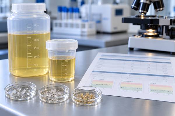
A urine supersaturation test estimates how strongly your urine favors crystal formation for specific stone minerals, most often calcium oxalate, calcium phosphate, and uric acid. It is usually calculated from a 24-hour urine collection rather than measured directly. The laboratory combines urine volume, mineral concentrations, pH, and chemical interactions to estimate whether the urine is below, near, or above the solubility limit for each compound. A higher supersaturation generally means a more favorable environment for crystals to form and grow, but the number is not a direct prediction that a stone will appear. Results differ by stone type and laboratory calculation method, so trends are often more useful than one isolated value. Supersaturation is especially helpful when individual urine tests look only mildly abnormal: low volume, modestly high calcium, borderline oxalate, or low citrate can combine into a clearly high calcium oxalate supersaturation even when no single result looks dramatic.
- Supersaturation summarizes the combined effect of urine volume, minerals, pH, and inhibitors rather than one isolated risk factor.
- Calcium oxalate supersaturation rises with higher calcium or oxalate and lower urine volume; citrate can reduce calcium available for crystallization.
- Calcium phosphate supersaturation is highly sensitive to urine pH and often rises as pH moves above about 6.0.
- Uric acid supersaturation rises sharply in acidic urine, especially when urine volume is low or uric acid concentration is high.
- There is no single universal “normal” supersaturation target; use the laboratory range, stone type, and change over time.
Table of Contents
- What urine supersaturation means
- How the test is calculated
- Calcium oxalate supersaturation
- Calcium phosphate supersaturation
- Uric acid supersaturation
- How to interpret high, low, and changing results
- How to lower supersaturation and use follow-up testing
What urine supersaturation means
Supersaturation describes whether urine contains more dissolved stone-forming material than would be expected to remain comfortably in solution under those chemical conditions. In simplified terms, a laboratory compares the ion activity product of a mineral with its solubility product. The resulting value is often called relative supersaturation, saturation index, or simply supersaturation.
When the solution is undersaturated, crystals tend to dissolve rather than grow. When it is supersaturated, crystal formation becomes thermodynamically possible. That does not mean a stone is guaranteed. Urine contains inhibitors and promoters of crystallization, and stones also depend on nucleation sites, crystal retention, anatomy, and time.
The value is useful because kidney stones are a systems problem. A person may have urine calcium only slightly above target and oxalate near the upper end of normal, but if urine volume is low the concentrations of both rise. The combined calcium oxalate supersaturation may therefore be high. Conversely, increasing urine volume can reduce supersaturation even when the total amount of calcium excreted per day changes very little.
Most comprehensive 24-hour urine stone risk profiles report at least calcium oxalate, calcium phosphate, and uric acid supersaturation. Some laboratories calculate additional indices for brushite or other minerals.
Supersaturation is not the same as stone composition. A high calcium phosphate value does not prove the stone is calcium phosphate. A kidney stone analysis remains the best way to identify what a recovered stone actually contains. The two tests work together: stone analysis shows what formed, while urine supersaturation helps explain why the current urine environment may favor recurrence.
How the test is calculated
A supersaturation value is calculated from several measured urine variables. The exact algorithm varies by laboratory, but common inputs include calcium, oxalate, phosphate, uric acid, citrate, magnesium, sodium, potassium, ammonium, chloride, sulfate, urine pH, and total volume.
The laboratory does more than multiply concentrations. Minerals in urine exist in different ionic forms and bind to other molecules. Citrate, for example, complexes with calcium and reduces free calcium activity. Magnesium can bind oxalate. Phosphate changes charge as pH changes. Uric acid shifts between soluble urate and less soluble uric acid depending strongly on pH. Computer programs account for these interactions to estimate the concentration of the chemically active species.
That is why two people with the same urine calcium can have different calcium stone supersaturation. If one has higher citrate, greater volume, or a different pH, the amount of free calcium available to crystallize may be lower.
The test depends on a complete 24-hour collection. Missing urine can distort both total excretion and concentration. Overcollection can do the opposite. A collection performed during unusual dieting, illness, or extreme fluid intake may not represent ordinary risk. Initial testing is usually most useful when a person follows their usual eating and drinking pattern.
The numerical scale is not standardized across every laboratory. Some systems report values where 1.0 corresponds conceptually to the solubility threshold, while others use proprietary indexes or reference ranges. Even when 1.0 has a thermodynamic meaning, a clinical target may be lower or higher depending on the mineral and the laboratory model. Do not compare a value from one laboratory directly with a different laboratory’s range without checking the method.
This is one reason within-person trends are valuable. If the same laboratory shows calcium oxalate supersaturation falling from 9 to 5 after more fluid and less sodium, that direction is informative even if another lab uses a somewhat different scale.
Supersaturation also differs from a simple concentration. A urine calcium concentration can look lower because someone produced more urine, even though the total amount of calcium excreted over 24 hours stayed high. The supersaturation calculation uses the complete chemical environment, so it can show whether dilution was enough to reduce crystallization pressure. Conversely, a normal daily calcium amount can contribute to high supersaturation when volume is very low or oxalate is elevated.
A single collection is a snapshot of one day. Meals, exercise, weather, work conditions, fluid access, and medications can change urine chemistry from day to day. For people with recurrent stones or a high-risk history, two baseline 24-hour collections are often used because they can reveal whether an apparent abnormality is persistent. Creatinine excretion and collection timing also help clinicians judge whether the specimen was probably complete.
Calcium oxalate supersaturation
Calcium oxalate is the most common kidney stone mineral. Its supersaturation is driven mainly by urine calcium, urine oxalate, and urine concentration, with citrate and other complexing substances modifying the chemistry.
Higher calcium raises the amount available to pair with oxalate. Higher oxalate has a strong effect because oxalate is present at much lower concentrations than calcium; relatively small changes can meaningfully alter the calcium oxalate ion product. Low urine volume concentrates both.
Common patterns that increase calcium oxalate supersaturation include:
- hypercalciuria;
- hyperoxaluria;
- low urine volume;
- low citrate;
- high sodium intake that increases urinary calcium;
- enteric hyperoxaluria after malabsorption; and
- combinations of several borderline abnormalities.
A urine oxalate test and urine calcium test help identify the dominant driver. If oxalate is high, treatment may focus on normal calcium intake with meals, removal of major oxalate sources, vitamin C supplements, or malabsorption. If calcium is high, sodium reduction, evaluation for hyperparathyroidism, or medication may be more relevant.
Citrate is important because it binds calcium and inhibits crystal growth. A person with normal calcium and oxalate can still have high supersaturation if urine is concentrated and citrate is low. Increasing urine volume is therefore one of the most reliable ways to lower calcium oxalate supersaturation across different causes.
The goal is not to force every individual component into the middle of a reference range. It is to reduce the overall crystallization pressure while keeping treatment nutritionally sound and medically safe.
Calcium phosphate supersaturation
Calcium phosphate supersaturation is especially sensitive to urine pH. As pH rises, phosphate shifts toward the divalent form that combines readily with calcium. This is why calcium phosphate supersaturation can rise even when total phosphate excretion barely changes.
Key contributors include:
- high urine pH;
- high urine calcium;
- low citrate;
- low urine volume; and
- enough urinary phosphate to support crystallization.
Calcium phosphate supersaturation often begins to rise rapidly as urine pH moves above about 6.0. Brushite and hydroxyapatite stones are particularly associated with higher pH. The urine pH result therefore deserves close attention when calcium phosphate supersaturation is elevated.
Causes of persistently alkaline urine include distal renal tubular acidosis, medications such as topiramate or acetazolamide, alkali therapy, and some urinary infections. The correct response depends on the cause. For example, potassium citrate may be helpful when citrate is low, but it also raises pH. In a calcium phosphate stone former, the clinician may need to balance the citrate benefit against a rise in calcium phosphate supersaturation rather than simply increasing alkali until pH reaches a generic target.
A high calcium phosphate supersaturation is not automatically an instruction to restrict dietary phosphorus. Modern calcium phosphate stone management focuses more on urine pH, calcium, citrate, sodium, volume, and underlying disorders than on severe phosphate restriction.
When calcium phosphate supersaturation is high despite apparently normal individual values, the calculated index can reveal that several modest changes are adding together. That is one of its main practical advantages.
Uric acid supersaturation
Uric acid supersaturation behaves differently because pH is often the dominant factor. Uric acid is far less soluble in acidic urine. As pH falls, soluble urate is converted to uncharged uric acid, which can precipitate.
A person can therefore have high uric acid supersaturation even when 24-hour uric acid excretion is not markedly elevated. Persistently low urine pH below about 5.5, low urine volume, and higher uric acid concentration work together to increase risk.
This explains why uric acid stones are common in people with type 2 diabetes, insulin resistance, obesity, metabolic syndrome, and gout. Many of these patients have impaired urinary ammonium buffering and persistently acidic urine.
The uric acid stone risk profile therefore emphasizes pH and volume in addition to the absolute uric acid amount. Treatment often includes urine alkalinization, commonly with potassium citrate when appropriate, because raising pH can sharply reduce uric acid supersaturation and can even dissolve existing pure uric acid stones in selected patients.
Volume still matters. If urine output is low, the uric acid concentration rises even if total daily excretion stays the same. Increasing fluid intake can lower supersaturation without changing purine metabolism.
A high uric acid supersaturation result should not be interpreted as proof of gout. Gout is a clinical disorder related to urate crystal deposition in joints and tissues and is evaluated with symptoms and serum uric acid, while urinary supersaturation describes the chemistry inside the urinary tract.
How to interpret high, low, and changing results
A high supersaturation means the urine chemistry is favorable for that mineral to crystallize. The next question is which measured variables are causing it. The treatment should target the driver rather than the index alone.
A low supersaturation is generally favorable, but it does not guarantee that no stones will form. A previously formed stone can remain in the kidney, anatomy can promote crystal retention, and day-to-day urine chemistry varies. Imaging and stone history still matter.
The most informative comparison is often before versus after intervention. If calcium oxalate supersaturation falls after urine volume rises and sodium falls, the treatment is working chemically. If uric acid supersaturation remains high despite lower uric acid excretion, pH may still be too low. If calcium phosphate supersaturation rises after citrate therapy, the increase in pH may need to be weighed against the citrate benefit.
| High index | Common drivers to review | Typical focus |
|---|---|---|
| Calcium oxalate | Calcium, oxalate, volume, citrate | Fluid, sodium/calcium handling, oxalate source, citrate |
| Calcium phosphate | pH, calcium, citrate, volume, phosphate | Cause of alkaline urine, hypercalciuria, safe citrate strategy |
| Uric acid | pH, uric acid, volume | Alkalinization, fluid, purine/metabolic factors |
Recent research has explored using serial relative supersaturation patterns to predict recurrence. Some studies have found that persistently high values are associated with more recurrent stones, but specific thresholds from small cohorts should not be treated as universal clinical cutoffs. Laboratory reference ranges and established guideline-based risk factors remain more appropriate for individual care.
Context matters when a result seems inconsistent. A high value collected during vomiting, fasting, heavy sweating, or unusually low fluid intake may overstate a person’s typical exposure, while deliberate overhydration on collection day may make the profile look safer than usual. Fever, diarrhea, temporary diet changes, and recently started medicines can have similar effects. If the collection was not representative, repeating it under ordinary conditions is often more useful than making a major treatment decision from the isolated number.
Stone composition provides another check. Persistently high uric acid supersaturation is more compelling when a prior stone contains uric acid, while an unexpectedly high calcium phosphate value in a person known to form calcium oxalate stones should prompt review of urine pH, medication changes, and whether stone composition has changed. The result should fit the broader clinical picture rather than replace it.
How to lower supersaturation and use follow-up testing
The most broadly effective way to lower supersaturation is to increase urine volume. More water dilutes calcium, oxalate, phosphate, uric acid, and other solutes. Current guidelines commonly target at least 2.0–2.5 L of urine per day, with some recommending 2.5 L or more for most stone formers.
After volume, treatment becomes stone-specific:
- For high calcium oxalate supersaturation: reduce excess sodium, maintain normal dietary calcium, address high oxalate, correct low citrate, and treat hypercalciuria when indicated.
- For high calcium phosphate supersaturation: investigate persistently high pH, lower high urine calcium, improve volume, and use alkali thoughtfully when citrate is low.
- For high uric acid supersaturation: raise urine pH to the clinician-directed target, increase volume, and address hyperuricosuria or excessive purine intake when present.
The urine volume test is especially useful because a higher volume can improve all three major supersaturation indexes at once.
Repeat 24-hour urine testing is commonly performed about 8–12 weeks after starting targeted prevention. The repeat panel answers a practical question: did the intervention lower the relevant supersaturation without worsening another one? For example, aggressive alkalinization may reduce uric acid supersaturation but raise calcium phosphate supersaturation. That tradeoff is visible only when the whole profile is reviewed.
Two baseline collections can improve confidence because diet and hydration vary day to day. Once values are stable, follow-up can be less frequent. Using the same laboratory when possible makes trends easier to interpret.
Supersaturation is most useful as a decision aid, not a score to chase in isolation. It turns a long list of urine measurements into a chemical summary, but the individual components still show what can be changed. When stone composition, supersaturation, and the rest of the 24-hour urine profile point in the same direction, prevention can be much more targeted. The most useful follow-up is therefore a pattern: a complete collection, a lower relevant supersaturation, improvement in the variables being treated, and a plan that the person can maintain on ordinary days rather than only during testing.
References
- Metabolic evaluation for prevention of kidney stones – CARI Guidelines 2025 (Guideline)
- Diagnosis and risk factors of kidney stones – CARI Guidelines 2025 (Guideline)
- Metabolic Evaluation and Recurrence Prevention for Urinary Stone Patients: An EAU Guidelines Update 2024 (Review)
- 24-Hour Urinary Chemistries and Kidney Stone Risk 2024
- Study on changes in urinary relative supersaturation and risk of stone recurrence in patients after urolithiasis surgery 2025
Disclaimer
This article provides general information about urine supersaturation testing and does not replace individualized kidney stone evaluation. Supersaturation calculations and reference ranges vary by laboratory, and treatment targets depend on stone composition, kidney function, medications, and the full urine profile. Discuss major diet or medication changes with a qualified clinician.