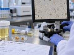
A uric acid stone risk test looks mainly at urine pH, urine volume, and uric acid excretion to determine whether the urine is favorable for uric acid crystallization. Of these factors, persistently acidic urine is usually the most important. Uric acid becomes much less soluble as pH falls, so stones can form even when the total amount of uric acid in a 24-hour collection is not dramatically high. That is why a normal blood uric acid level or a modest urine uric acid result does not rule out uric acid stones. A complete evaluation also considers stone analysis, kidney function, gout, diet, metabolic syndrome, diabetes, chronic diarrhea, and medications. The purpose of testing is practical: identify whether the main problem is low pH, low urine volume, excessive uric acid excretion, or a combination, then choose a prevention plan that changes the chemistry actually driving the stone.
- Low urine pH is the strongest uric acid stone marker: a persistently acidic pH below about 5.5 strongly favors uric acid crystallization.
- High urine uric acid is not required: many people with pure uric acid stones have low pH without marked hyperuricosuria.
- Low urine volume raises risk by concentrating uric acid: prevention commonly aims for at least about 2–2.5 L of urine daily, individualized to the patient.
- A 24-hour urine test is more informative than one spot sample: it measures uric acid excretion and volume while showing the overall daily chemistry.
- Metabolic syndrome, obesity, type 2 diabetes, gout, high animal-protein intake, and chronic gastrointestinal alkali loss can contribute to acidic urine or uric acid risk.
Table of Contents
- What a uric acid stone risk test measures
- Why acidic urine pH matters most
- Urine uric acid and urine volume
- Common causes of a uric acid stone pattern
- How to interpret common result patterns
- Testing, collection, and pH monitoring
- Follow-up and prevention based on results
What a uric acid stone risk test measures
A uric acid stone evaluation is usually part of a broader metabolic kidney stone work-up rather than a single named laboratory test. The essential measurements are urine pH, urine volume, and urinary uric acid. A comprehensive 24-hour urine panel often adds creatinine, sodium, urea nitrogen, calcium, oxalate, citrate, potassium, magnesium, and supersaturation calculations.
The main components are:
- Urine pH: shows how acidic or alkaline the urine is. Uric acid is poorly soluble in acidic urine.
- Urine volume: determines how concentrated uric acid and other solutes become.
- 24-hour urine uric acid: measures how much uric acid is excreted over the day.
- Uric acid supersaturation: some laboratories calculate the chemical tendency for uric acid to crystallize from pH, concentration, and other variables.
- Creatinine: helps judge whether the 24-hour collection was reasonably complete.
Blood tests often include serum uric acid and creatinine/eGFR. Serum uric acid is useful for gout and systemic urate metabolism, but it does not substitute for urine testing. A person can have uric acid stones with a normal blood uric acid level.
Stone composition is also important. A kidney stone analysis test can confirm whether a passed or removed stone is uric acid, calcium oxalate, calcium phosphate, cystine, struvite, or mixed. Without composition, the clinician may infer uric acid risk from urine pH and imaging, but direct analysis is stronger evidence.
The risk profile is therefore a combination of chemistry and context. The urine pH measurement and the urine uric acid test answer different parts of the same question.
Why acidic urine pH matters most
Uric acid chemistry changes sharply with pH. At lower pH, more urate is converted to poorly soluble uric acid. Once the concentration exceeds its solubility, crystals can nucleate, grow, and aggregate into stones.
This is why persistently low urine pH is often more important than the total amount of uric acid excreted. European guidance notes that urine pH below about 5.5 is typical of uric acid stone formation. A patient may excrete an ordinary amount of uric acid but still create high uric acid supersaturation if the urine is both acidic and concentrated.
Urine pH also varies through the day. Meals, fasting, exercise, medications, and acid-base status can shift it. A single office dipstick result therefore cannot fully characterize a patient’s pattern. The most meaningful finding is repeated or sustained acidity, especially when it appears in a 24-hour profile or in fresh home measurements obtained as instructed.
The pH relationship also helps distinguish stone types. Calcium oxalate stones can form across a broader pH range. Calcium phosphate stones become more favorable as urine becomes more alkaline. Uric acid, by contrast, is strongly associated with low pH.
That does not mean higher pH is always better. Excessive alkalinization can raise calcium phosphate supersaturation. Treatment targets must therefore be matched to the stone type and monitored rather than pursued without limits.
When a clinician prescribes alkali for uric acid stones, fresh urine pH measurements at different times of day can help adjust the dose. This is different from simply relying on the average pH reported from a stored 24-hour collection.
The timing of pH checks can reveal a problem that a daily average hides. Urine may be adequately alkaline after a potassium citrate dose but become strongly acidic overnight or before the next dose. For a patient trying to dissolve a known uric acid stone, those low-pH periods can slow dissolution even when the 24-hour average looks acceptable. A clinician may therefore ask for several fresh measurements across the day and use them to adjust dose timing as well as total dose.
Treatment should avoid overshooting. Raising pH improves uric acid solubility, but urine that is kept unnecessarily alkaline can increase calcium phosphate supersaturation in susceptible patients. The aim is a pH range appropriate to the treatment goal—prevention versus active dissolution—while continuing to monitor the rest of the urine chemistry.
Urine uric acid and urine volume
Hyperuricosuria means excessive uric acid excretion in urine. European guidance defines it as more than 4 mmol/day in adult women and more than 5 mmol/day in adult men, although laboratories may use different mass-based reference ranges. In U.S. practice, values around 750 mg/day for women and 800 mg/day for men are also commonly used as reference points. These cutoffs should be interpreted with the laboratory’s own range and the patient’s stone type.
High urine uric acid can result from high purine intake, gout or increased urate production, rapid cell turnover, some blood disorders, chemotherapy-related cell breakdown, and rare enzyme disorders. It can increase the concentration of uric acid available to crystallize.
However, a “normal” 24-hour uric acid result does not make a low pH safe. The risk is driven by the interaction of amount, pH, and volume.
Urine volume is equally practical. A 24-hour uric acid excretion of 700 mg behaves differently when dissolved in 1.0 L of urine versus 2.5 L. Higher volume lowers concentration and supersaturation. General stone guidelines commonly aim for urine output above 2.0–2.5 L/day, while the exact fluid intake needed depends on sweating, climate, exercise, body size, and gastrointestinal losses.
The urine volume test for kidney stones can help explain why someone who “drinks a lot” may still produce insufficient urine. Hot environments, heavy exercise, fever, or diarrhea can consume much of the fluid before it reaches the bladder.
A useful interpretation asks three questions together: Is the urine too acidic? Is the uric acid load high? Is the urine too concentrated? Treating only one factor may leave substantial residual risk.
Common causes of a uric acid stone pattern
Several medical and lifestyle patterns can push urine toward uric acid crystallization.
Metabolic syndrome, obesity, and type 2 diabetes
Uric acid stones are strongly associated with insulin resistance and metabolic syndrome. One important mechanism is impaired renal ammonium buffering, which can leave urine more acidic. Studies of stone formers show that as metabolic syndrome features accumulate, urine pH tends to fall and uric acid supersaturation tends to rise.
This does not mean every person with obesity or diabetes will form uric acid stones. It means a low pH result in that context has a plausible metabolic explanation and should not be dismissed as a random urine finding.
Gout and high uric acid production
Gout reflects systemic urate excess and can coexist with uric acid stones. Some patients have both high urinary uric acid and low pH. Others mainly have acidic urine. Serum and urine uric acid therefore provide complementary information.
A blood uric acid test for kidney stones can help assess gout and hyperuricemia, but it cannot determine urine pH or urine concentration.
High animal-protein or purine intake
Large amounts of meat and other purine-rich foods can increase uric acid production and dietary acid load. Excess animal protein can also lower citrate and urine pH in susceptible people. The goal is usually moderation rather than eliminating all protein.
Chronic diarrhea or intestinal losses
Diarrhea can cause bicarbonate and fluid loss. The kidneys compensate by conserving alkali and excreting more acidic urine, while dehydration lowers urine volume. That combination can produce a strong uric acid risk pattern even without gout.
Other causes
Low urine pH can occur with some forms of chronic kidney disease, autosomal dominant polycystic kidney disease, high endogenous acid production, and other metabolic conditions. Very high uric acid excretion may also occur with myeloproliferative disorders or rapid cell turnover. The broader medical history determines whether specialized testing is needed.
Some patients have more than one mechanism at the same time. A person with type 2 diabetes may have persistently acidic urine, eat a high-purine diet, and produce only 1.3 L of urine each day. In that situation, correcting pH alone may reduce risk but leave the urine unnecessarily concentrated, while increasing fluids without addressing the acidity may not lower uric acid supersaturation enough. This is one reason a complete 24-hour urine profile is more useful than a single pH strip or serum uric acid value.
The pattern can also change over time. Weight loss, improved diabetes control, a new medication, dietary change, chronic diarrhea, or a change in kidney function may alter pH or uric acid excretion. A patient who previously formed uric acid stones may therefore need repeat testing rather than assuming that the original risk profile is permanent.
How to interpret common result patterns
The most useful interpretation focuses on combinations rather than one red value on the report.
| Result pattern | Likely meaning | Typical next focus |
|---|---|---|
| pH <5.5, normal urine uric acid, low or normal volume | Acidic urine is the dominant risk | Alkalinization and adequate urine volume |
| pH <5.5, high urine uric acid | Both acidity and uric acid load contribute | Address pH, fluid, purine exposure, and cause of hyperuricosuria |
| Low urine volume with borderline pH | Concentration may be amplifying risk | Increase urine output and reassess supersaturation |
| High serum uric acid but urine pH not acidic | Gout risk may be present without a classic uric acid stone pattern | Confirm stone type and complete urine chemistry |
| High uric acid excretion but pH >5.5 | Hyperuricosuria may contribute to calcium oxalate risk instead of pure uric acid stones | Review stone analysis and calcium oxalate supersaturation |
Supersaturation can clarify these patterns. A urine supersaturation test combines the chemical effects of pH and concentration into a more direct estimate of crystallization tendency.
Imaging can provide another clue because uric acid stones are radiolucent on plain X-rays, although they are visible on CT. Dual-energy CT can sometimes help distinguish uric acid from non-uric-acid stones. Imaging is supportive; stone analysis remains preferable when material is available.
Testing, collection, and pH monitoring
A 24-hour urine collection should represent a normal day unless the clinician gives different instructions. Deliberately drinking far more water than usual or avoiding normal foods can make the baseline result less useful.
The basic collection method is to discard the first urine at the start time, then collect every void for the next 24 hours, including the final void at the end time. Missing urine can falsely lower volume and total uric acid, sodium, citrate, and other daily measurements. Creatinine excretion helps assess whether the collection is plausible, but it cannot detect every error.
For a recurrent or high-risk stone former, two 24-hour collections may be preferred because pH, volume, sodium, and uric acid can vary substantially from day to day. Testing is generally most informative after an acute stone episode has settled and the person has returned to the usual diet and environment.
Spot pH testing serves a different purpose. During alkalinization therapy, the clinician may ask for fresh urine pH measurements several times during the day. This helps detect whether the medicine is working between doses. pH should be measured promptly after voiding because standing urine can change chemically or bacteriologically.
Dipsticks are convenient but have limited precision. A pH meter or higher-quality strips may be recommended when small differences matter for treatment. Do not change alkali dosing solely from one unexpected reading without following the prescribing plan.
Follow-up and prevention based on results
Uric acid stones are unusual in that changing urine chemistry can sometimes dissolve existing stones as well as prevent new ones. The main medical strategy is urine alkalinization, commonly with potassium citrate when appropriate. Sodium bicarbonate is another option in selected patients, but sodium load can be undesirable for some stone formers and people with hypertension or fluid-sensitive conditions.
The exact pH target depends on whether the goal is prevention or active dissolution and on the risk of calcium phosphate crystallization. Clinicians often use a somewhat higher pH during dissolution than during routine prevention, while avoiding sustained over-alkalinization. Home pH monitoring can help maintain the chosen range.
High fluid intake is the other foundation. The relevant goal is enough intake to produce the target urine volume, not a fixed number of glasses. People who sweat heavily may need substantially more fluid than people in a cool sedentary environment.
Dietary treatment may include moderating excessive purine-rich animal protein, reducing excess sodium, maintaining a balanced diet rich in fruits and vegetables when medically appropriate, and addressing weight, insulin resistance, or diabetes. Dietary change is most effective when it targets the pattern actually seen on testing.
Allopurinol is not automatically required for every uric acid stone. It is most useful when excessive uric acid production or hyperuricosuria remains an important contributor, including selected patients with gout. If low pH is the main abnormality, alkalinization may be the more direct therapy.
Follow-up 24-hour urine testing can show whether urine pH, volume, uric acid excretion, and supersaturation improved. European guidance suggests reassessment around 8–12 weeks after starting pharmacologic prevention, then periodic monitoring once the pattern is stable. Stone imaging may also be repeated when dissolution is being attempted or recurrence risk is high.
The main safety concern with alkalinization is that treatment affects potassium, sodium, kidney function, and calcium phosphate risk. Potassium citrate may not be appropriate in people with significant hyperkalemia risk or certain kidney conditions. Medication should therefore be prescribed and monitored rather than self-started from an abnormal urine pH.
Seek urgent care for fever or chills with flank pain, inability to urinate, severe uncontrolled pain, or persistent vomiting. Those symptoms may indicate obstruction or infection and are not problems that can be managed by adjusting urine pH at home.
A good uric acid stone risk test ultimately identifies the dominant mechanism. If urine is persistently acidic, correcting pH is central. If uric acid excretion is high, its cause also matters. If volume is low, dilution is essential. Measuring all three prevents treatment from focusing on the wrong target.
References
- Metabolic Evaluation and Recurrence Prevention – EAU Guidelines on Urolithiasis – Uroweb 2026 (Guideline)
- Type 4 renal tubular acidosis and uric acid nephrolithiasis: two faces of the same coin? 2023 (Review)
- Pharmacologic treatment of kidney stones: Current medication and pH monitoring 2023 (Review)
- Association of Urine Findings with Metabolic Syndrome Traits in a Population of Patients with Nephrolithiasis. 2022
- Obesity, Metabolic Syndrome, Diabetes and Kidney Stones: Strengthening Links Over Time? 2026 (Review)
Disclaimer
This article is for general education and does not diagnose stone composition or provide an individualized alkalinization dose. Urine pH targets, potassium or bicarbonate treatment, and uric acid medicines depend on kidney function, blood electrolytes, stone type, and other medical conditions. Seek urgent care for a suspected stone with fever, inability to urinate, severe uncontrolled pain, or persistent vomiting.