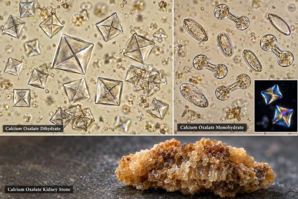
Calcium oxalate crystals are common urine crystals and are the mineral found in most kidney stones, but seeing them on microscopy does not mean a stone is present. They form when calcium and oxalate in urine become supersaturated and precipitate. Calcium oxalate dihydrate crystals often look like small envelopes, while calcium oxalate monohydrate can appear as dumbbells, ovals, or elongated picket-fence forms. Crystals may appear temporarily after dehydration, diet changes, or a concentrated urine sample, and some healthy people have them without ever developing stones. Their importance rises when crystalluria is persistent or occurs with recurrent renal colic, known stones, high urinary calcium or oxalate, low citrate, low urine volume, or kidney injury. A 24-hour urine study and stone analysis are more useful than crystal count alone for determining why a person forms stones and what prevention strategy fits the metabolic pattern.
- Calcium oxalate crystals are common and not automatically abnormal: they can appear in healthy urine without a kidney stone.
- Envelope-shaped crystals usually represent calcium oxalate dihydrate: monohydrate forms may look dumbbell-shaped, oval, or elongated.
- Stone risk rises with sustained supersaturation: low urine volume, hypercalciuria, hyperoxaluria, and low citrate are important contributors.
- Urine pH is less decisive than for uric acid or calcium phosphate stones: calcium oxalate can crystallize across a broad pH range.
- Persistent crystals with pain, stones, or kidney injury need follow-up: 24-hour urine chemistry and stone analysis can identify the driver.
Table of Contents
- What Calcium Oxalate Crystals Mean
- Appearance and Types Under the Microscope
- Why Calcium Oxalate Crystals Form
- How Crystals Relate to Kidney Stones
- Important Causes and Risk Factors
- Testing and Prevention After Recurrent Findings
- When Calcium Oxalate Crystals Need Prompt Care
What Calcium Oxalate Crystals Mean
Calcium oxalate crystals form when dissolved calcium and oxalate exceed their solubility in urine. This state is called supersaturation. Supersaturation is necessary for crystal formation, but it does not guarantee that a kidney stone will develop. Many crystals are flushed out before they grow or attach to the kidney.
This is why a microscopy report should be read as a snapshot of urine chemistry at the time the sample was collected. A dehydrated person may briefly have concentrated urine and visible crystals even if their average daily stone risk is low. Refrigeration or delayed examination can also increase precipitation after the urine leaves the body.
The finding becomes more meaningful when it is repeated in fresh samples or occurs in a person with known calcium oxalate stones. In that setting, microscopy supports a pattern already suggested by symptoms, imaging, or stone analysis. The broader urine microscopy test can also show blood, infection markers, tubular cells, or casts that change the urgency and differential diagnosis.
Calcium oxalate crystalluria alone does not tell whether the main problem is too much calcium, too much oxalate, too little urine, too little citrate, or a combination. Those drivers require quantitative testing.
One isolated report is therefore best interpreted as a risk clue, not a diagnosis. Crystal number also depends on when the specimen was collected, how concentrated it was, and how quickly it reached the microscope. A fresh sample that repeatedly shows abundant calcium oxalate crystals, especially alongside blood or stone symptoms, carries more weight than a few crystals in an old, concentrated specimen.
Appearance and Types Under the Microscope
Calcium oxalate commonly appears in two hydrated forms. Calcium oxalate dihydrate is called weddellite, while calcium oxalate monohydrate is called whewellite. The distinction is useful because the forms have different morphology and may occur under different biochemical conditions, although both can be present in the same person or stone.
Calcium oxalate dihydrate typically forms colorless, highly refractile crystals shaped like an envelope or an octahedron. Under ordinary microscopy, the crossed lines through the center create the familiar envelope appearance. These crystals may occur in healthy urine and are common in stone formers.
Calcium oxalate monohydrate has more variable shapes. It may look like a dumbbell, oval, elongated oval, or narrow picket fence. Some monohydrate forms can be confused with other crystals, so laboratory experience matters. Polarized light and solubility characteristics can help when morphology is atypical.
The quantity may be reported as few, moderate, or many crystals. These categories are not a standardized stone-risk scale. The number depends on urine concentration, centrifugation, specimen temperature, pH, and the laboratory method. A person with “many” crystals in one concentrated specimen may have a lower long-term risk than someone with fewer crystals but severe hyperoxaluria on repeated 24-hour testing.
Calcium oxalate crystals are generally strongly birefringent under polarized light. This can help distinguish them from amorphous material and some other sediment particles. A definitive stone composition, however, should be established by formal analysis of a passed or removed stone rather than inferred solely from urine microscopy.
Morphology can also hint at the urinary environment but cannot identify the cause by itself. Calcium oxalate crystals may appear across a broad pH range, so a single urine pH does not confirm or exclude them. When the shape is atypical, laboratory staff may use polarization, solubility behavior, repeat microscopy, or formal stone analysis rather than relying on appearance alone.
Why Calcium Oxalate Crystals Form
Crystal formation reflects the balance between promoters and inhibitors of crystallization. The major promoters are calcium and oxalate concentration, while urine volume strongly controls how concentrated these substances become. Citrate is an important inhibitor because it binds calcium and reduces the amount available to combine with oxalate.
Low urine volume is one of the most common and modifiable risk factors. When daily urine output is low, calcium and oxalate concentrations rise even if their total excretion is not unusually high. Hot weather, heavy sweating, inadequate fluid intake, diarrhea, and occupations that limit bathroom access can all contribute.
Hypercalciuria means excess calcium excretion in urine. It can occur with otherwise normal blood calcium and is common among calcium-stone formers. High sodium intake can increase urinary calcium, while hyperparathyroidism, excessive vitamin D activity, and some other disorders can also contribute. A urine calcium test for kidney stones helps quantify this risk rather than relying on microscopy.
Hyperoxaluria means excess urinary oxalate. It may come from high oxalate intake, increased intestinal absorption, reduced gut calcium binding, fat malabsorption, bariatric surgery, inflammatory bowel disease, or rare inherited disorders called primary hyperoxalurias. High-dose vitamin C can also increase oxalate production in susceptible people. A urine oxalate test is especially useful when calcium oxalate stones recur or enteric hyperoxaluria is suspected.
Hypocitraturia removes a natural inhibitor of calcium crystallization. It can occur with metabolic acidosis, chronic diarrhea, low potassium states, distal renal tubular acidosis, and other conditions. Measuring urine citrate helps determine whether citrate therapy or dietary changes are relevant.
The key concept is supersaturation: urine contains more dissolved calcium and oxalate than can remain comfortably in solution under the current conditions. Low volume raises both concentrations at once. Citrate and other inhibitors can reduce crystal growth and aggregation, so a low citrate level may matter even when calcium and oxalate are only moderately elevated.
How Crystals Relate to Kidney Stones
Calcium oxalate is the most common major component of urinary stones. Stone formation usually involves several steps: supersaturation, nucleation of microscopic crystals, growth, aggregation, and retention within the kidney. If crystals simply pass into the urine, no stone forms.
Many calcium oxalate stones begin on calcium phosphate deposits called Randall’s plaques on renal papillae. These subepithelial plaques can become exposed to urine and provide a surface on which calcium oxalate grows. Other stones may form through different pathways, including crystal plugging of tubules.
A crystal in voided urine therefore shows that precipitation occurred somewhere in the urinary system or specimen, but it does not show whether crystals were retained in the kidney. Imaging such as ultrasound or noncontrast CT is used to diagnose stones when symptoms or risk justify it. A kidney stone analysis is the best way to confirm composition when stone material is available.
Crystalluria may be more useful when it is persistent. Repeated calcium oxalate crystals in fresh urine, especially when abundant, can suggest ongoing supersaturation and may help monitor a known stone-forming tendency. Even then, 24-hour urine supersaturation measurements are more quantitative and actionable than microscopy alone.
Urine pH plays a smaller role in calcium oxalate than in uric acid or calcium phosphate stones. Calcium oxalate can form across a broad pH range. Very acidic urine may contribute to mixed uric acid and calcium oxalate processes, while higher pH may shift calcium precipitation toward phosphate, but there is no single calcium oxalate pH cutoff.
A stone can form through several steps—nucleation, crystal growth, aggregation, and retention in the kidney. Calcium oxalate may also grow on calcium-phosphate deposits in renal papillae, often called Randall’s plaque. This helps explain why a spot urine sample can show few or no crystals even in a person who has already formed a calcium oxalate stone.
Stone composition is often mixed. A report may describe mostly calcium oxalate with smaller amounts of calcium phosphate, and that mix can provide clues about urine pH and the surface on which the stone started. Repeating stone analysis after a later event can be useful when the composition changes, because prevention that fits one mineral pattern may not fit another as well.
Important Causes and Risk Factors
Most calcium oxalate crystalluria is related to ordinary stone-risk factors rather than poisoning or rare genetic disease. Still, some contexts deserve special attention.
Enteric hyperoxaluria occurs when fat malabsorption leaves free fatty acids in the intestine to bind calcium. With less calcium available to bind oxalate, more free oxalate is absorbed and later excreted in urine. This can occur after certain bariatric procedures, in Crohn disease, short bowel syndrome, chronic pancreatitis, and other malabsorptive states.
Primary hyperoxaluria is a rare inherited disorder in which the liver overproduces oxalate. It can cause recurrent stones, nephrocalcinosis, kidney injury, and systemic oxalate accumulation in severe disease. Early-onset recurrent calcium oxalate stones, very high urinary oxalate, nephrocalcinosis, or a strong family history may prompt specialized testing.
Ethylene glycol poisoning can produce calcium oxalate monohydrate crystals, acute kidney injury, and severe metabolic acidosis. However, crystals are neither sensitive nor specific enough to diagnose poisoning. The emergency diagnosis depends on exposure history, acid-base status, osmolar findings, kidney function, and toxicology testing. A person with possible antifreeze ingestion needs immediate emergency care rather than waiting for urine microscopy.
Dietary pattern influences risk but should be handled carefully. Very low dietary calcium can paradoxically raise oxalate absorption because less calcium is available in the gut to bind oxalate. For many calcium oxalate stone formers, normal dietary calcium taken with meals is preferred over severe calcium restriction. High sodium intake, excess animal protein in some patients, and inadequate fluids can also worsen stone risk.
The presence of crystals after eating a high-oxalate food does not establish chronic hyperoxaluria. The more important question is whether urine chemistry remains unfavorable across a full day and over repeated collections.
Dietary oxalate is only part of the story. Absorbing calcium and oxalate together in the intestine tends to bind oxalate before it reaches the urine, which is why severe calcium restriction can be counterproductive for some stone formers. High sodium intake can raise urinary calcium, while chronic diarrhea or fat malabsorption may simultaneously lower urine volume and citrate and increase oxalate absorption.
Testing and Prevention After Recurrent Findings
People with a single incidental crystal finding and no stone history often need no extensive evaluation. Hydration and repeat testing may be enough if the result was unexpected. Recurrent stones, bilateral stones, childhood stones, a solitary kidney, nephrocalcinosis, or repeated crystalluria raises the value of metabolic testing.
A typical 24-hour stone evaluation measures urine volume, calcium, oxalate, citrate, uric acid, sodium, creatinine, pH, and often calculated supersaturation. A 24-hour urine kidney stone risk test gives a much better picture of daily exposure than a spot urine sample.
Prevention is based on the abnormality found. Common strategies include producing a high daily urine volume, reducing excessive sodium, maintaining appropriate dietary calcium, moderating high-oxalate foods when oxalate is elevated, and increasing citrate when hypocitraturia is present. Some patients benefit from thiazide-type medication for hypercalciuria or potassium citrate for low citrate, but treatment should be individualized to the metabolic profile and medical history.
Fluid advice is often framed around urine output rather than a fixed number of glasses. Many stone guidelines aim for at least about 2.5 liters of urine per day in recurrent stone formers, which may require more than 2.5 liters of fluid intake depending on sweating, climate, diet, and activity. People with heart failure, advanced kidney disease, or other fluid restrictions need individualized advice.
Repeat 24-hour urine testing can show whether changes improved supersaturation. The goal is not necessarily to make every crystal disappear from microscopy; it is to lower the biochemical conditions that allow stones to grow and recur.
For recurrent stones, a 24-hour collection is more informative than a single microscopic field because it quantifies urine volume, calcium, oxalate, citrate, sodium, uric acid, creatinine, and pH over a full day. Results should be interpreted together: for example, high oxalate has different implications when urine volume is excellent versus very low. Repeat collections can also show whether prevention measures are actually changing risk.
Diet changes work best when they match the measured abnormality. People with high urine sodium may lower urinary calcium by reducing sodium; those with low citrate may benefit from more fruits and vegetables or prescribed citrate; and people with enteric hyperoxaluria may need specific strategies around fat malabsorption and calcium timing with meals. Generic advice to “avoid all oxalate” can make diets unnecessarily restrictive and may miss a more important driver such as low urine volume or high sodium.
When Calcium Oxalate Crystals Need Prompt Care
Calcium oxalate crystals alone rarely require urgent treatment. Prompt assessment is needed when they occur with severe flank pain, persistent vomiting, fever, chills, inability to urinate, or visible blood in the urine. These symptoms can indicate an obstructing stone, infection, or another urinary problem.
Fever plus suspected obstruction is particularly urgent because an infected obstructed kidney can deteriorate quickly and may require drainage in addition to antibiotics. A sudden drop in urine output or a rise in creatinine also deserves timely evaluation.
Emergency care is appropriate for suspected ethylene glycol exposure, severe confusion, rapid breathing, metabolic acidosis, or unexplained acute kidney injury with compatible history. In that situation, calcium oxalate crystals are only one clue in a time-sensitive toxicology emergency.
For stable patients with recurrent calcium oxalate crystalluria, the most useful next step is usually not repeated microscopy alone. Confirm stone composition when possible, quantify 24-hour urine risk factors, and target the specific abnormalities that keep urine supersaturated.
Prevention should be individualized rather than based on crystals alone. Some people mainly need more fluid; others have hypercalciuria, enteric hyperoxaluria, hypocitraturia, or a medication-related problem that requires targeted treatment. If a passed or removed stone is available, composition testing is valuable because it confirms whether calcium oxalate is truly the dominant stone material before long-term therapy is chosen.
References
- Urinary Crystals Identification and Analysis 2024 (Review)
- Differential identification of urine crystals with morphologic characteristics and solubility test 2022 (Review)
- Metabolic Evaluation and Recurrence Prevention for Urinary Stone Patients: An EAU Guidelines Update 2024 (Guideline)
- Pathophysiology and Main Molecular Mechanisms of Urinary Stone Formation and Recurrence 2024 (Review)
- Management of urinary stones: state of the art and future perspectives by experts in stone disease 2024 (Review)
Disclaimer
Calcium oxalate crystals do not by themselves diagnose a kidney stone or identify its cause. Recurrent stones, severe pain, fever, reduced urine output, kidney injury, or suspected toxic exposure should be evaluated by a qualified clinician using the appropriate imaging and metabolic or emergency testing.